Figure 1
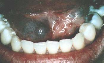
A 23-year-old woman presented with a slightly bluish, dome-shaped lesion on the right side of the mouth, just lateral to the frenulum and below the tongue (figure). She had no significant symptoms, although she occasionally experienced a “funny taste” in her mouth. The lesion seemed to vary in size.
Question
This lesion represents which one of the following?
- A blocked salivary duct.
- B. A thyroglossal duct cyst.
- C. A ranula.
- D. A cyst of the sublingual salivary gland.
- E. A hematoma of the floor of the mouth.
Figure 2
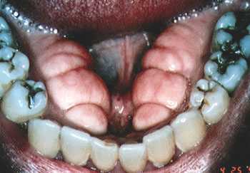
A 65-year-old man presented with firm nodules on the inside of the gums, medial to the mandible (figure). The nodules had been there for quite some time and were noted by his physician during a routine visit. They were completely asymptomatic and did not bother the patient.
Question
These nodules represent which one of the following conditions?
- A. Epulis.
- B. Gingival hyperplasia.
- C. Unerupted tertiary teeth.
- D. Torus mandibularis.
- E. Cysts of the gum.
Figure 1 Discussion
The answer is C: a ranula.1,2 A ranula is a mucocele of the minor salivary glands, located under the tongue on the floor of the mouth. When the small ducts of these glands become plugged, the secretions may build up into large, mucus-filled cystic structures that often partially rupture and discharge a foul-tasting material; these ruptures may cause the lesion to shrink in size. The lesions have a tendency to recur but do not pose any significant health risk to the patient, except for their nuisance value.
Figure 2 Discussion
The answer is D: torus mandibularis. Tori are bony extoses, usually seen on the center of the hard palate as torus palatini, or, as pictured here, attached to the mandible.1,3 They are usually asymptomatic but may cause dental problems, such as in fitting dentures or braces, and have no potential for malignancy. Epulis are soft, fleshy growths on the gums, whereas gingival hyperplasia usually occurs in the triangular area of gum between the teeth in patients taking drugs such as phenytoin.
