Exfoliative dermatitis, also known as erythroderma, is an uncommon but serious skin disorder that family physicians must be able to recognize and treat appropriately. Although the etiology is often unknown, exfoliative dermatitis may be the result of a drug reaction or an underlying malignancy. The approach to treatment should include discontinuation of any potentially causative medications and a search for any underlying malignancy. One of the most common malignancies associated with exfoliative dermatitis is cutaneous T-cell lymphoma, which may not manifest for months or even years after the onset of the skin condition. Hospitalization is usually necessary for initial evaluation and treatment. In the hospital, special attention must be given to maintaining temperature control, replacing lost fluids and electrolytes, and preventing and treating infection. The long-term prognosis is good in patients with drug-induced disease, although the course tends to be remitting and relapsing in idiopathic cases. The prognosis of cases associated with malignancy typically depends on the outcome of the underlying malignancy.
Exfoliative dermatitis is a disease process in which most, and sometimes all, of the skin is involved in erythematous inflammation resulting in massive scaling.1 A variety of diseases and other exogenous factors may cause exfoliative dermatitis. Unfortunately, the clinical picture does not contribute to an understanding of the underlying cause. Therefore, it is important to identify and treat any underlying disease whenever possible and to remove any contributing external factors.2
Incidence
Most published studies of exfoliative dermatitis have been retrospective and thus do not address the issue of overall incidence. Exfoliative dermatitis accounts for about 1 percent of all hospital admissions for dermatologic conditions.3
Although the disease affects both men and women, it is more common in men, with an average male-to-female ratio of 2.3:1. The average age at onset is 55 years, although exfoliative dermatitis may occur at any time.2
Pathogenesis
Exfoliative dermatitis is the result of a dramatic increase in the epidermal turnover rate. In patients with this disorder, the mitotic rate and the absolute number of germinative skin cells are higher than normal. Moreover, the time necessary for cells to mature and travel through the epidermis is decreased. This compressed maturation process results in an overall greater loss of epidermal material, which is manifested clinically as severe scaling and shedding. Normal epidermis undergoes some exfoliation every day, but the scales that are lost contain little, if any, important viable material, such as nucleic acids, soluble proteins and amino acids.4 In exfoliative dermatitis, however, protein and folate losses may be high.5
The pathogenesis of exfoliative dermatitis is a matter of debate. In recent years, clinicians have come to believe that this condition is secondary to a complicated interaction of cytokines and cellular adhesion molecules. Interleukin (IL)-1, IL-2, IL-8, intercellular adhesion molecule 1 (ICAM-1), tumor necrosis factor and interferon gamma are the cytokines that may have roles in the pathogenensis of exfoliative dermatitis.2
Etiology
The most common causes of exfoliative dermatitis are preexisting dermatoses, drug reactions, malignancies and other miscellaneous or idiopathic disorders.
PRIMARY DERMATOLOGIC DISORDERS
Dermatologic disorders occasionally present as exfoliative dermatitis. The most common of these are psoriasis, atopic dermatitis, seborrheic dermatitis, contact dermatitis and pityriasis rubra pilaris. Other dermatoses associated with erythroderma are listed in Table 1.2,3,6–8
TABLE 1 Dermatoses Associated with Erythroderma
| Atopic dermatitis |
| Candidiasis |
| Contact dermatitis |
| Dermatophytosis |
| Ichthyosis |
| Lichen planus |
| Mastocytosis |
| Nummular eczema |
| Pemphigus |
| Photosensitive eczema |
| Pityriasis rubra pilaris |
| Prurigo |
| Psoriasis |
| Reiter's syndrome |
| Scabies |
| Seborrheic dermatitis |
| Staphylococcus scalded skin syndrome |
| Stasis with autoeczematization |
| Subacute cutaneous lupus erythematosus |
| Vitamin deficiency |
MEDICATIONS
Since the earliest descriptions of exfoliative dermatitis, medications have been known to be important causative agents. Hence, the apparent increase in cases of exfoliative dermatitis may be related to the introduction of many new drugs. Drug eruptions that initially present as morbilliform, lichenoid or urticarial rashes may progress to generalized exfoliative dermatitis. Antiepileptic medications, antihypertensive medications, antibiotics, calcium channel blockers and a variety of topical agents (Table 2)2,3,6–9 can cause exfoliative dermatitis, but theoretically, any drug may cause exfoliative dermatitis. Therefore, the clinician should always consider drugs as a possible cause.
TABLE 2 Drugs Associated with Erythroderma
| Acetaminophen |
| Actinomycin D (Cosmegan) |
| Allopurinol (Zyloprim) |
| Aminoglycosides |
| Aminophylline |
| Amiodarone (Cordarone) |
| Arsenic |
| Aztreonam (Azactam) |
| Barbiturates |
| Calcium channel blockers |
| Captopril (Capoten) |
| Carbamazepine (Tegretol) |
| Cephalosporins |
| Chinese herbs |
| Chloroquine (Aralen) |
| Chlorothiazide (Diuril) |
| Chlorpromazine (Thorazine) |
| Chlorpropamide (Diabinese) |
| Cimetidine (Tagamet) |
| Cisplatin (Platinol) |
| Clofazimine (Lamprene) |
| Clotrimazole (Lotrimin) |
| Codeine |
| Cyclobenzaprine (Flexeril) |
| Dapsone |
| Dimercaprol (BAL in Oil) |
| Ethylenediamines |
| Gold |
| Hydantoins |
| Hydroxychloroquine (Plaquenil) |
| Interleukin-2 (Proleukin) |
| Interferon alfa (Roferon-A, Intron A, Alferon N) |
| Interferon beta (Avonex, Betaseron) |
| Iodine (Pima syrup) |
| Isoniazid (Laniazid, Nydrazid; also in Rifamate, Rimactane) |
| Isosorbide dinitrate (Isordil, Sorbitrate) |
| Isotretinoin (Accutane) |
| Lithium (Eskalith, Lithobid) |
| Mefloquine (Larium) |
| Mephyntoin (Mesantoin) |
| Mercurials |
| Mercury |
| Mexilitene (Mexitil) |
| Minocycline (Dynacin, Minocin, Vectrin) |
| Mitomycin-C (Mutamycin) |
| Neomycin (Neosporin) |
| Nitrofurantoin (Furadantin, Macrodantin) |
| Omeprazole (Prilosec) |
| Para-amino salicylic acid (Sodium P.A.S.) |
| Penicillins |
| Phenolphthalein (Agoral, Alophen, Modane) |
| Phenothiazines |
| Phenobarbital (Donnatal, Bellatal) |
| Phenytoin (Dilantin) |
| Quinacrine |
| Quinidine (Quinidex) |
| Ranitidine (Zantac) |
| Rifampin (Rifadin, Rimactane; also in Rifamate) |
| Streptomycin |
| Sulfadiazine |
| Sulfonamides |
| Sulfonylureas |
| Terbutaline (Brethine, Bricanyl) |
| Tetrachloroethylene |
| Tetracyclines |
| Thalidomide (Synovir) |
| Thiazide diuretics |
| Trimethoprim (Trimpex; also in Bactrim, Septra) |
| Tolbutamide (Orinase) |
| Vancomycin (Vancocin) |
A pseudolymphoma reaction with fever, arthralgias, lymphadenopathy, hepatosplenomegaly, anemia and erythroderma may develop as a result of hypersensitivity to dapsone or antiepileptic drugs. If cutaneous pathology also mimics cutaneous T-cell lymphoma, it can be very difficult to differentiate a drug-induced skin condition from exfoliative dermatitis associated with a malignancy.2,9
MALIGNANCIES
Malignancies are a major cause of exfoliative dermatitis. Reticuloendothelial neoplasms, as well as internal visceral malignancies, can produce erythroderma, with the former being the more predominant cause.
The cutaneous T-cell lymphomas are the lymphomas most commonly associated with exfoliative dermatitis. The most notable member of this group is mycosis fungoides. Studies indicate that mycosis fungoides may cause 25 to 40 percent of all cases of malignancy-related erythroderma.6,7 The erythroderma may arise as a progression from a previous cutaneous T-cell lymphoma lesion or appear simultaneously with the cutaneous T-cell lymphoma, or it may precede the appearance of the cutaneous T-cell lymphoma lesion. When it precedes cutaneous T-cell lymphoma lesions, exfoliative dermatitis becomes the presenting sign of the underlying malignancy.
The time interval between the appearance of exfoliative dermatitis and the appearance of cutaneous T-cell lymphoma lesions can vary from months to years or even decades. Sézary syndrome, the leukemic variant of mycosis fungoides, is also associated with exfoliative dermatitis. The erythrodermic form of mycosis fungoides and the Sézary syndrome may also be difficult to distinguish from benign erythroderma. Immunophenotypic studies with the use of advanced antibody panels may be useful in the differential diagnosis of these two forms.10 Reticulum cell sarcoma is another form of cutaneous T-cell lymphoma that may cause exfoliative dermatitis.
Acute and chronic leukemia may also cause exfoliative dermatitis. The relative risk of leukemia inducing erythroderma is highly variable, ranging from 11 to 50 percent.11
Internal (visceral) malignancies cause about 1 percent of all cases of exfoliative dermatitis.11 Frequently, erythroderma is the presenting sign of the malignancy. Patients with carcinoma of the colon, lung, prostate and thyroid have presented with erythroderma. More recently, carcinomas of the fallopian tube,12 larynx13 and esophagus14 have been reported as causes of exfoliative dermatitis. Insidious development of the erythroderma, progressive debilitation of the patient, absence of previous skin disease and resistance to standard therapy are features that may suggest an underlying malignancy.6,11
OTHER ASSOCIATED DISORDERS
Erythroderma is also associated with disorders that cannot easily be classified into groups. Exfoliative dermatitis has been reported in association with hepatitis, acquired immunodeficiency syndrome, congenital immunodeficiency syndrome (Omenn's syndrome) and graft-versus-host disease.2,15–17
In reviews of erythroderma, a significant percentage of patients (about 25 percent) do not receive a specific etiologic diagnosis. Some of these patients undergo spontaneous resolution. Other cases are ultimately classifiable as another dermatosis. A significant number of these patients eventually progress to cutaneous T-cell lymphoma.8
CLINICAL MANIFESTATIONS
Clinically, the first stage of exfoliative dermatitis is erythema, often beginning as single or multiple pruritic patches, involving especially the head, trunk and genital region. These patches tend to spread until, after a matter of days or weeks, most of the skin surface is covered with an erythematous, pruritic eruption. Usually, but not always, the palms of the hands, the soles of the feet and the mucous membranes are spared. In some studies, the nose and paranasal area are spared. This has been called the “nose sign.”18
Once the erythema is well established, scaling inevitably follows (Figure 1). The scales may be small or large, superficial or deep. Acute processes usually favor large scales, whereas chronic processes produce smaller ones. The exfoliative process also may involve the scalp, with 25 percent of patients developing alopecia.4 Nails can often become dystrophic, particularly in patients with preexisting psoriasis.4,6
FIGURE 1.
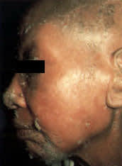
Elderly man with exfoliative dermatitis of the face caused by psoriasis.
The most frequently noted symptoms in patients with exfoliative dermatitis include malaise, pruritis and a chilly sensation. Both hyperthermia and hypothermia are reported. Other clinical findings include lymphadenopathy, hepatomegaly, splenomegaly, edema of the foot or ankle4,6 and gynecomastia.19
The scaling that occurs in exfoliative dermatitis can have severe metabolic consequences, depending on the intensity and the duration of the scaling. Since cutaneous function as a multiprotective barrier is so disrupted in exfoliative dermatitis, the body loses heat, water, protein and electrolytes, and renders itself much more vulnerable to infection. Exfoliative dermatitis is also a risk factor for epidemic spread of methicillin-resistant Staphylococcus aureus.6,20
Heat loss is another major concern that accompanies a defective skin barrier in patients with exfoliative dermatitis. Loss of normal vasoconstrictive function in the dermis, decreased sensitivity to the shivering reflex and extra cooling that comes from evaporation of the fluids leaking out of the weeping skin lesions all result in thermoregulatory dysfunction that can cause hypothermia or hyperthermia.6 The basal metabolic rate also is increased in patients with exfoliative dermatitis. A catabolic state thus ensues, which is often responsible for significant weight loss.
Each of these physiologic disruptions is potentially life-threatening. Hypothermia can result in ventricular flutter, decreased heart rate and hypotension. Increased peripheral blood flow can result in high-output cardiac failure. Hypervolemia can also occur in patients with exfoliative dermatitis, contributing to the likelihood of cardiac failure.21–24
HISTOPATHOLOGY
In most patients with erythroderma, skin biopsies show nonspecific histopathologic features, such as hyperkeratosis, parakeratosis, acanthosis and a chronic perivascular inflammatory infiltrate, with or without eosinophils. Even patients with clear histories of preexisting dermatoses tend to have biopsies that are not diagnostic when they present with erythroderma.2
Laboratory Findings
Laboratory evaluation of patients with erythroderma is generally not very helpful in determining a specific diagnosis. Typical laboratory values include mild anemia, leukocytosis, eosinophilia, elevated erythrocyte sedimentation rate, abnormal serum protein electrophoresis with a polyclonal elevation in the gamma globulin region, and elevated IgE levels.1–3,6–8
Blood counts and bone marrow studies may reveal an underlying leukemia. Analysis for circulating Sézary cells may be helpful, but only if the cells are identified in unequivocally large numbers.
Treatment
Hospitalization and dermatologic consultation are indicated in most cases to ensure that all of the necessary cutaneous, laboratory and radiologic investigations and monitoring are performed. The balance of fluids and electrolytes should be closely monitored, since dehydration or hypervolemia can be problems. Also, physicians should be vigilant about possible secondary infection, whether cutaneous, pulmonary or systemic.
In the acute phase, before determination of the etiology, treatment consists of measures to soothe the inflamed skin. These measures include bed rest, lukewarm soaks or baths, bland emollients and oral antihistamines.25–27
In patients with chronic idiopathic erythroderma, emollients and topical steroids may be effective. Other patients may warrant PUVA (psoralen plus ultraviolet A) phototherapy, systemic steroids (if psoriasis has been ruled out), retinoids (for exfoliative dermatitis secondary to psoriasis and pityriasis rubra pilaris), or immunosuppressive agents such as methotrexate (Rheumatrex) and azathioprine (Imuran).25–27
When used as adjunctive therapy, behavior modification designed to eliminate persistent scratching has been successful in reducing the rate of excoriation and increasing the rate of healing.28
No uniformity of opinion exists concerning the best treatment for cutaneous T-cell lymphoma. Options include use of PUVA light therapy, total-body electron beam irradiation, topical nitrogen mustard, systemic chemotherapy and extracorporeal photopheresis. Consultation with an oncologist who is well-versed in treatment of cutaneous T-cell lymphoma is advisable once the disease progresses to the tumor stage.
Clinical Course, Prognosis and Sequelae
Even though exfoliative dermatitis is a complex disorder involving many factors, the underlying disease is usually the key determinant of the course and prognosis. Drug-induced exfoliative dermatitis is usually short-lived once the inciting medication is withdrawn and appropriate therapy is administered. Patients with underlying skin disorders may respond much more slowly to therapy, but clearing almost always occurs eventually. The clinical course of patients with malignancies depends on the type of malignancy and the response to appropriate therapy. Patients who have exfoliative dermatitis of unknown cause tend to have an unpredictable course, usually replete with multiple remissions and exacerbations.4
In patients who develop complications (i.e., infection, fluid and electrolyte abnormalities, cardiac failure), the rate of mortality is often high. The most common causes of death in patients with exfoliative dermatitis are pneumonia, septicemia and heart failure.
Sequelae of exfoliative dermatitis are not widely reported. However, patchy, diffuse areas of postinflammatory hyperpigmentation and hypopigmentation may occur, especially in patients with darker skin.1,4 One case of posterythrodermic generalized vitiligo beginning six weeks after the onset of exfoliative dermatitis has been reported.29,30 Residual eruptive nevi and keloid formation are rare sequelae. Mild to severe alopecia and transient or permanent nail dystrophy also may be encountered.
