Transient ischemic attack is no longer considered a benign event but, rather, a critical harbinger of impending stroke. Failure to quickly recognize and evaluate this warning sign could mean missing an opportunity to prevent permanent disability or death. The 90-day risk of stroke after a transient ischemic attack has been estimated to be approximately 10 percent, with one half of strokes occurring within the first two days of the attack. The 90-day stroke risk is even higher when a transient ischemic attack results from internal carotid artery stenosis. Most patients reporting symptoms of transient ischemic attack should be sent to an emergency department. Patients who arrive at the emergency department within 180 minutes of symptom onset should undergo an expedited history and physical examination, as well as selected laboratory tests, to determine if they are candidates for thrombolytic therapy. Initial testing should include complete blood count with platelet count, prothrombin time, International Normalized Ratio, partial thromboplastin time, and electrolyte and glucose levels. Computed tomographic scanning of the head should be performed immediately to ensure that there is no evidence of brain hemorrhage or mass. A transient ischemic attack can be misdiagnosed as migraine, seizure, peripheral neuropathy, or anxiety.
Based on an increased understanding of brain ischemia and the introduction of new treatment options, a working group has proposed redefining transient ischemic attack (TIA) as “a brief episode of neurological dysfunction caused by focal brain or retinal ischemia, with clinical symptoms typically lasting less than one hour, and without evidence of acute infarction.”1(p1715) This definition underscores the urgency of recognizing TIA as an important warning of impending stroke and facilitating rapid evaluation and treatment of TIA to prevent permanent brain ischemia.
Epidemiology
An estimated 200,000 to 500,000 TIAs occur annually in the United States.2 One study2 found that 25 percent of patients who presented to an emergency department with TIA had adverse events within 90 days; 10 percent of the events were strokes, and the vast majority of the strokes were fatal or disabling.3 More than 50 percent of all adverse events occurred within the first four days after the TIA. Notably, of the patients with TIA who returned to the emergency department with stroke (10.5 percent), approximately one half had the stroke within the first 48 hours after the initial TIA. In 2.6 percent of patients with TIA, hospitalization was required for cardiac events, including congestive heart failure, unstable angina, cardiac arrest, and ventricular arrhythmia.
Clinical Presentation
The more common clinical presentations of TIA are described in Table 1. In general, a TIA presents as a syndrome rather than any one sign or symptom.
Pre-emergency Department Care
There is no reliable way to determine if the abrupt onset of neurologic deficits represents reversible ischemia without subsequent brain damage or if ischemia will result in permanent damage to the brain (e.g., stroke). Therefore, all patients with symptoms of TIA should receive an expedited evaluation.
Office staff should be trained to inform the family physician immediately if a patient calls or presents with symptoms that could represent a TIA. Neurologic symptoms that crescendo with increasing frequency, duration, or severity are particularly ominous signs of impending stroke.
Most patients with possible TIA should be sent immediately to the nearest emergency department. If they have had symptoms for fewer than 180 minutes, they should be sent to an emergency department that offers acute thrombolytic therapy. Patients should not drive themselves to the hospital. To speed evaluation, it is appropriate to activate the 9-1-1 Emergency Medical Service system for transport.2,3
TABLE 1 Common Clinical Presentations of TIA
| Affected area | Signs and symptoms | Implications |
|---|---|---|
| Cranial nerves | Visual loss in one or both eyes | Bilateral loss may indicate more ominous onset of brainstem ischemia. |
| Double vision | If double vision is subtle, the patient may describe it as “blurry” vision. | |
| Vestibular dysfunction | True vertigo is likely to be described as a spinning sensation rather than nonspecific lightheadedness. | |
| Difficulty swallowing | Trouble swallowing may indicate brainstem involvement; if the swallowing problem is severe, there may be an increased risk of aspiration. | |
| Motor function | Unilateral or bilateral weakness affecting the face, arm, or leg | Bilateral signs may indicate more ominous onset of brainstem ischemia. |
| Sensory function | Unilateral or bilateral: either decreased sensation (numbness) or increased sensation (tingling, pain) in the face, arm, leg, or trunk | If sensory dysfunction occurs without other signs or symptoms, the prognosis may be more benign, but recurrence is high. |
| Speech and language | Slurring of words or reduced verbal output; difficulty pronouncing, comprehending, or “finding” words | If speech is severely slurred or facial drooling is excessive, there is an increased risk of aspiration. |
| Writing and reading also may be impaired. | ||
| Coordination | Clumsy arms, legs, or trunk; loss of balance or falling (particularly to one side) with standing or walking | Incoordination of limbs, trunk, or gait may indicate cerebellar or brainstem ischemia. |
| Psychiatric or cognitive function | Apathy or inappropriate behavior | These symptoms can indicate frontal lobe involvement and frequently are misinterpreted as poor volitional cooperation. |
| Excessive somnolence | This symptom may indicate bilateral hemispheric or brainstem involvement. | |
| Agitation or psychosis | Rarely, these symptoms may indicate brainstem ischemia, particularly if they occur in association with cranial nerve or motor dysfunction. | |
| Confusion or memory changes | These rarely are isolated symptoms; more frequently, they are associated with language, motor, sensory, or visual changes. | |
| Inattention to surrounding environment, particularly to one side; if severe, patient may deny deficit or even his or her own body parts. | Depending on the severity of neglect, the physician may need to lift the patient's arm to check for strength, rather than rely on the patient to perform this task. |
TIA = transient ischemic attack.
On presentation to the emergency department, patients who have had symptoms for fewer than 180 minutes might be candidates for treatment with tissue-type plasminogen activator (tPA).4,5 If a patient is not a candidate for tPA treatment, antiplatelet therapy should be initiated as soon as it can be determined that there are no contraindications.4–6 [Reference6: SOR A, rating of benefits]
Inpatient or Outpatient Evaluation
Guidelines issued by the National Stroke Association7 recommend evaluation within hours of the onset of TIA symptoms, preferably in an emergency department. If appropriate imaging studies are not immediately available in the emergency department or out-patient setting, the patient should be hospitalized for observation.7 [SOR C, expert opinion] Relative indications for more extended inpatient evaluation for TIA or stroke are listed in Table 2.
TABLE 2 Relative Indications for Inpatient Evaluation of Possible TIA or Stroke*
| Condition | Implications |
|---|---|
| High-risk cardioembolic source: acute myocardial infarction (especially if large and significant wall-motion abnormality is present), mural thrombi, new-onset atrial fibrillation | Consider anticoagulation. |
| TIAs manifested by major symptoms such as dense paralysis or severe language disorder | Possible evolving large hemispheric stroke with increased risk of brain swelling |
| Increasing frequency or severity of TIAs (crescendo pattern) | Possible evolving thromboembolic stroke |
| Evidence of high-grade carotid artery stenosis | Carotid artery evaluation for possible emergency intervention (surgery, stent, or angioplasty) |
| Drooling, imbalance, decreased alertness, difficulty swallowing | Increased risk of falling, or of aspiration and other pulmonary complications |
| Severe headache, photophobia, stiff neck, recent syncope | Possible subarachnoid hemorrhage: obtain emergency computed tomographic scan of the head; if the scan is negative but clinical suspicion remains high, cerebrospinal fluid evaluation or possible cerebral angiography is needed. |
TIA = transient ischemic attack.
*—May require more than 23 hours of observation in the emergency department, or hospitalization for observation.
Patients with symptoms of acute TIA for fewer than 24 to 48 hours should undergo diagnostic testing in the emergency department.8 [SOR C, expert opinion] Patients whose symptoms have resolved for more than 48 hours should receive urgent inpatient or outpatient evaluation.
Initial Work-Up for Suspected TIA
The first step in evaluating a patient with symptoms of TIA is to confirm the diagnosis (Figure 1).
DIFFERENTIAL DIAGNOSIS
The most common imitators of TIA are glucose derangement, migraine, seizure, post-ictal states, and tumors (especially with acute hemorrhage).
TIA typically has a rapid onset, and maximal intensity usually is reached within minutes. Fleeting episodes lasting one or two seconds or nonspecific symptoms such as fatigue, lightheadedness (in the absence of other cerebellar or brainstem symptoms), and bilateral rhythmic shaking of the limbs are less likely presentations of acute cerebral ischemia.
Distinguishing TIA from migraine aura can be difficult. Younger age, previous history of migraine (with or without aura), and associated headache, nausea, or photophobia are more suggestive of migraine than TIA. In general, migraine aura tends to have a marching quality; for example, symptoms such as tingling may progress from the fingers to the forearm to the face. Migraine aura also is more likely to have a more gradual onset and resolution, with a longer duration of symptoms than in a typical TIA.
FIGURE 1. Initial Work-Up for Suspected TIA
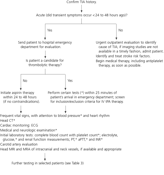
Initial work-up for the patient with possible transient ischemic attack (TIA). (IV = intravenous; tPA = tissue-type plasminogen activator; CT = computed tomography; ECG = electrocardiography; PT = prothrombin time; aPTT = activated partial thromboplastin time; INR = International Normalized Ratio; MRI = magnetic resonance imaging; MRA = magnetic resonance angiography)
If a patient has explosive onset of a severe headache, with or without photophobia, stiff neck, or syncope, acute subarachnoid hemorrhage is a possibility. Rarely, TIA is mistaken for the first presentation of multiple sclerosis in young patients or for amyotrophic lateral sclerosis in older patients.
HISTORY
A general medical history should be obtained in all patients with suspected TIA. Special emphasis should be given to possible symptoms of TIA (Table 1), and stroke risk factors should be identified to determine the likelihood that the symptoms are caused by TIA. Modifiable risk factors for stroke include hypertension, diabetes, cardiac disease, elevated blood lipid levels, carotid artery stenosis, smoking, sickle cell anemia, excessive alcohol use, obesity, and physical inactivity.7
Whether hypercholesterolemia is an independent primary risk factor for stroke remains uncertain.9 However, hypercholesterolemia is a significant risk factor for coronary heart disease (CHD) and therefore can be considered an important risk factor for ischemic stroke. There appears to be a stronger data relationship between total and low-density lipoprotein cholesterol levels, as well as a protective influence of high-density lipoprotein cholesterol levels, in cervical carotid artery atherosclerosis.10
Other important information includes a family history of stroke (including cerebral aneurysm or hypercoagulable state), the use of over-the-counter or illicit drugs, a history of migraine or “severe headaches,” recent head trauma, previous systemic clots and, in a woman of childbearing age, a history of spontaneous abortion. Certain findings may indicate the need for special diagnostic tests (Table 3).
PHYSICAL EXAMINATION
Vital signs should be evaluated, including blood pressures in both arms, to rule out stenosis of the subclavian artery, which may manifest as grossly asymmetric pressures. Auscultation of the heart and neck also should be performed. Carotid bruits, when present, are neither highly specific nor highly sensitive for carotid artery stenosis.
All patients with possible TIA should receive a detailed, documented neurologic examination, with emphasis on cognitive and language function, cranial nerve function, facial and limb strength, sensory function, deep tendon reflex symmetry, and coordination. This examination can be helpful in determining whether a patient previously had an unrecognized stroke. It also can serve as a baseline examination if the patient's neurologic status worsens or neurologic symptoms recur. Occasionally, the neurologic examination may identify a nonischemic cause for an acute neurologic deficit (e.g., acute radial nerve palsy, isolated third-nerve palsy in a patient with diabetes mellitus).
DIAGNOSTIC TESTS
Brain Imaging
Computed tomographic (CT) scanning of the head without contrast medium should be performed to identify sub-arachnoid hemorrhage, intracranial hemorrhage, or subdural hematoma. Urgent identification of these conditions is critical because neurosurgical intervention or special management may be required.
If hemorrhage is present, treatment with tPA or anticoagulants that may worsen central nervous system bleeding should be avoided. Special measures may be needed to manage blood pressure if the patient is found to have hypertension-mediated intracranial hematoma, and further testing may be required if the patient is found to have subarachnoid hemorrhage (e.g., cerebral angiography to rule out aneurysm).
CT scanning also can identify conditions that mimic TIA, including tumors and other masses (especially if hemorrhage occurs acutely within a mass), as well as conditions that are associated with seizures or auras. A head CT scan can identify signs of early brain damage or evidence of old strokes.11,12 Finally, CT scanning of the head with contrast medium should be performed in the febrile patient to rule out an infectious cause or in the patient with a suspected mass (e.g., metastatic carcinoma, abscess).
Because of increased bony artifact in the posterior fossa, CT scanning is not sensitive for evaluating disease in the brainstem or cerebellum. In these instances, magnetic resonance imaging (MRI) is the preferred study.
Electrophysiologic Testing
All patients should have a baseline electrocardiogram (ECG) with rhythm strip.6,12,13 If the ECG is abnormal or the patient has a history of cardiac disease, echocardiography should be performed. Atrial fibrillation and left ventricular hypertrophy (suggesting unrecognized chronic hypertension) are important risk factors for stroke. Recent data suggest that the 90-day risk for a cardiac event is seven times higher in patients with TIA and abnormal ECG findings than in those with a normal ECG (4.2 versus 0.6 percent).13
If the ECG is unrevealing, cardiac monitoring in selected patients could help diagnose paroxysmal atrial fibrillation (or other arrhythmias in patients with syncope or palpitations). In patients with untreated atrial fibrillation, echocardiography may identify a thromboembolic source or left ventricular systolic dysfunction, both of which are common predictors of ischemic stroke.14
TABLE 3 Further Diagnostic Testing Based on the History in Patients Undergoing Evaluation for Possible TIA*
| History | Implications | Tests |
|---|---|---|
| Headache in postpartum or dehydration setting | Venous thrombosis | MRI with venography or cerebral angiography |
| Fever | Subacute or acute bacterial endocarditis | Blood cultures, head CT scan with and without contrast medium; in selected patients with confirmed bacterial endocarditis, perform cerebral angiography to rule out a mycotic aneurysm. |
| Confusion, headache, seizure | CNS vasculitis | Cerebral angiography, ESR, lumbar puncture (to look for elevation of white blood cell counts in particular) |
| Hypertensive encephalopathy | Careful blood pressure monitoring in intensive care setting; consider MRI. | |
| Rheumatologic disease, sympathomimetic drug use | CNS vasculitis | Consider cerebral angiography, ESR, lumbar puncture (to look for elevation of white blood cell counts in particular). |
| Recent myocardial infarction | Cardioembolic source | Transthoracic or esophageal echocardiography |
| Head, neck, jaw pain, especially after trauma | Carotid or vertebral dissection | Consider cerebral angiography or other neck neuroimaging studies (see text). |
| Abrupt onset of severe headache with photophobia, or recent syncope | Subarachnoid hemorrhage | Emergency head CT scan; if the scan is negative, evaluate cerebrospinal fluid for elevated red blood cell count or perform cerebral angiography to rule out aneurysm or arteriovenous malformation. |
| Confusion, stupor, coma, other brainstem symptoms (poor prognosis) | Vertebrobasilar ischemia | Consider intracranial magnetic resonance angiography or cerebral angiography; if basilar artery is significantly thrombosed, consider intra-arterial thrombolytic therapy (if available). |
| Brain swelling, impending herniation | Immediate head CT scan; if the scan is positive, emergency neurosurgical intervention may be required. | |
| No obvious risk factors for stroke | “Cryptogenic stroke,” patent foramen ovale, intra-atrial septal aneurysm, valvular or aortic arch disease | Consider cerebral angiography, transesophageal echocardiography, and work-up for hypercoagulable state. |
TIA = transient ischemic attack; MRI = magnetic resonance imaging; CT = computed tomography; CNS = central nervous system; ESR = erythrocyte sedimentation rate.
*—The initial work-up for the patient with possible TIA is outlined in Figure 1.
[ corrected] Transesophageal echocardiography is superior to transthoracic echocardiography for evaluating possible dysfunction of the left atrium (including thrombus) or a patent foramen ovale (an etiology for paradoxical emboli), atrial septal defects (including aneurysm), and aortic plaque. Recent clinical trials15,16 suggest that transesophageal echocardiography should be considered in patients without an identifiable cause of TIA or known cardiac disease, because it may detect a condition requiring therapeutic intervention (e.g., anticoagulation for thrombus). Aortic plaque, which has been associated with stroke, can be visualized well on transesophageal echocardiography.
Laboratory Tests
A complete blood count with platelet count should be obtained to rule out polycythemia, thrombocytopenia, and thrombocytosis. It is helpful to know the pro-thrombin time (PT), activated partial thromboplastin time (aPTT), and International Normalized Ratio (INR) before antiplatelet or anticoagulation therapy is administered; the PT, aPTT, and INR can be elevated in some hypercoagulable states.
The glucose level should be determined to rule out hypoglycemia or hyperglycemia and to help diagnose occult diabetes. Blood urea nitrogen and creatinine levels are important, because poor renal status may prohibit the use of contrast media in imaging studies. An erythrocyte sedimentation rate (ESR) should be obtained to potentially rule out vasculitis. Finally, a drug of abuse screen, a pregnancy test, a homocystine level determination, or a blood alcohol level measurement should be performed in selected patients.
Follow-Up Evaluation
LIPID PROFILE
After the initial more abbreviated evaluation in the emergency department, risk factors for stroke can be reassessed thoroughly later in the evaluation. Recent data indicate that treatment with statins (3-hydroxy-3-methylglutaryl coenzyme A reductase inhibitors) reduces the risk of stroke by about 30 percent in patients with CHD.17,18 Therefore, a fasting lipid profile reflective of the patient's normal eating habits should be obtained, and statin therapy should be initiated if indicated.
HYPERCOAGULABLE STATES
Patients with known risk factors for stroke and those with a history of migraine, spontaneous abortion, pulmonary emboli, or deep venous thrombosis, or a family history of any of these conditions, should be evaluated for hypercoagulable states. Initial tests include ESR, antinuclear antibody test, rapid plasma reagent test, and antiphospholipid antibody tests. Referral to a hematologist or neurologist can ensure cost-effective evaluation of the multiple coagulation-factor abnormalities and conditions that can cause embolic stroke.
TESTING FOR ARTERIAL PATENCY AND BLOOD FLOW
Carotid duplex ultrasonography should be performed in a reliable laboratory, preferably one with validation against the results of cerebral angiography. Alternatively, cerebral and cervical vessels can be evaluated by magnetic resonance angiography (MRA) with contrast medium or by CT angiography. If the work-up demonstrates carotid or other large-vessel atherosclerotic disease in the patient with TIA and unrecognized CHD, coronary artery testing is recommended.19
MRI
Clear advantages of MRI of the brain over CT scanning of the head include better imaging of tissues (i.e., greater sensitivity for early edema), superior imaging within the posterior fossa (including the brainstem and cerebellum), additional planes of imaging (sagittal, coronal, and oblique), and no exposure to radiation.
A clear disadvantage of brain MRI is that it may or may not identify hemorrhage. For this reason, although MRI can be helpful, it should not replace urgent CT scanning of the head in the initial work-up of patients with possible TIA. When cerebrovascular malformation, aneurysm, cerebral venous thrombosis, or arteritis is suspected, MRI or MRA is preferred.
Diffusion-weighted imaging detects cellular edema as early as 10 to 15 minutes after symptom onset. However, this technique is not yet widely available.
MRA
This imaging modality is a noninvasive means of assessing intra- and extracranial vessels. Current MRA techniques use intravenously administered contrast medium (gadolinium) to visualize the vessels.
MRA with the administration of contrast medium also is effective in identifying vertebrobasilar stenosis, although recent data suggest that intracranial vertebral artery disease can be missed.20 Depending on the MRA acquisition technique, the percentage of intracranial vessel stenosis can be overestimated (sensitivity of approximately 85 percent compared with cerebral angiography).21 Therefore, if accuracy is therapeutically important, cerebral angiography is necessary.
When near occlusion of the carotid artery cannot be distinguished from complete occlusion on MRA or carotid Doppler ultrasound studies, cerebral angiography should be considered. Surgery generally cannot be performed on completely occluded vessels.
Special consideration should be given to patients who present with a history or symptoms that suggest arterial dissection. This condition can be diagnosed using neck MRI scans in certain sequences that can identify hemorrhage within the vessel wall (T1-weighted images with fat suppression).
Patients with carotid artery dissection can present with acute or subacute unilateral neck, head, or jaw pain. These symptoms may be associated with visual or language deficits, or with sensorimotor deficits, particularly in the opposite arm. More typically, patients with carotid artery dissection present with only some of these features, such as temporal headache with lateral neck pain and, possibly, transient visual obscuration (amaurosis fugax) because of thromboemboli in the ophthalmic artery.
Both carotid and vertebral artery dissections have been described after trauma, although spontaneous dissection also is common. Patients should be evaluated for connective tissue disease because of the associated increased risk of dissection.
If the MRI or MRA study is inconclusive, cerebral angiography should be used to rule out arterial dissection or better define the percentage of vessel narrowing.
CT Angiography
This modality is another state-of-the-art technique for detecting blood flow to the brain. CT angiography also is becoming a useful imaging modality for identifying carotid or vertebral artery dissection. Because the technique requires venous injection of contrast dye, the patient's renal status should be considered before the test is performed.
Conventional CT scanning in combination with CT angiography currently is being evaluated as an addition to the diagnostic imaging tools for use in patients with TIA or stroke. This combination can provide useful information about vascular anatomy (in the form of three-dimensional reconstructions) and the extent and location of infarction. It may allow rapid evaluation of patients with TIA or stroke in hospitals or institutions that do not have MRI capability.
Cerebral Angiography
This technique continues to be the gold standard for complete evaluation of intracranial and extracranial vessels. With cerebral angiography, both arterial and venous phases of cerebral blood flow can be visualized (dynamic study). However, cerebral angiography is an invasive technique that can result in neurologic complications (total incidence rate: 1.3 to 4.6 percent),22,23 including major stroke or death in 0.1 to 1.3 percent of patients, depending on the study.24,25
Relative indications for cerebral angiography include suspected carotid dissection unconfirmed on a noninvasive neuroimaging study, subarachnoid hemorrhage (to identify bleeding source), intracerebral hemorrhage in the absence of hypertension, and vasculitis. If one of these conditions is suspected, referral to a neurologist can be helpful in obtaining and interpreting the angiogram.
Special Considerations
VERTEBROBASILAR ISCHEMIA
Typical signs and symptoms of ischemic syndromes involving the anterior and posterior circulations are listed in Table 4. The brainstem and cerebellum are confined within the posterior fossa, a bony cavity with poor tolerance of brain swelling or mass effects (e.g., from hemorrhage). Because brainstem structures are essential for preserving critical respiratory function and arousal states, patients with vertebrobasilar ischemia should be monitored closely. It also is crucial to search for life-threatening cerebrovascular disease, such as basilar artery stenosis or thrombosis or disease affecting multiple large vessels (e.g., bilateral, vertebral, or carotid artery stenosis).
TIA IN A YOUNG PATIENT
When a TIA occurs in a patient younger than 45 years, particularly if there are no clear risk factors for stroke, it is advisable to refer the patient to a neurologist for consideration of specialized testing. For example, it may be necessary to determine the utility of cerebral angiography to rule out vasculitis, carotid artery dissection, and other forms of nonath-erosclerotic vasculopathy, or lumbar spinal puncture with cerebrospinal fluid evaluation may be required to rule out chronic infection or inflammation.
TABLE 4 Typical Characteristics of Ischemic Syndromes Involving the Anterior and Posterior Circulations
| Ischemic syndrome: circulation involved | Signs | Symptoms |
|---|---|---|
| Anterior circulation* | Visual-field cut | Inability to see well (i.e., difficulty reading or driving |
| Language dysfunction (left hemisphere most often affected): aphasia | Difficulty finding or understanding words, inability to read, garbled or slurred speech | |
| Motor dysfunction: contralateral face, arm, or leg weakness | Dropping objects; depending on severity, inability to lift or move a body part or objects | |
| Sensory dysfunction: contralateral increased or decreased sensation to pain, heat, or cold | Tingling (paresthesias), numbness, or pain | |
| Behavior dysfunction (right hemisphere): inattention to surrounding environment, particularly to one side; if severe, patient may deny deficits or even his or her own body parts | The patient usually reports no symptoms, but family members or others report that the patient has difficulty dressing, ignores half of food on a plate, or has poor attention to one side of the room or to someone speaking to the patient on one side versus the other (most often, the left side is ignored). | |
| Posterior circulation† | Nystagmus | Vertigo (spinning sensation) |
| Disconjugate gaze | If subtle, blurry or double vision | |
| Homonymous visual-field cut | Inability to see well, especially to one side | |
| Contralateral weakness | Dropping objects, inability to fully lift or move the limb | |
| Incoordination of trunk or limbs (ataxia) | Clumsiness, falling, inability to coordinate an action (e.g., drink from a cup without spilling contents) | |
| Motor or sensory dysfunction on opposite side of cranial nerve deficits (crossed signs suggest brainstem involvement) | For example, the patient may report double vision, droopiness on the left side of the face, and dragging of the right leg (because of weakness). | |
| Bilateral signs | Abrupt weakness of both legs, falling | |
| Decreased mentation; stupor or coma | Family members or others report that the patient has poor responsiveness or that they are unable to arouse the patient. |
*—Includes the internal carotid artery, middle cerebral artery, and anterior cerebral artery, as well as the branches of these arteries.
†—Includes the vertebral arteries, basilar artery, and posterior cerebral artery, as well as the branches of these arteries.
Because cardiac abnormalities are among the most common causes of TIA in young patients, a baseline ECG with rhythm strip should be obtained, and transthoracic and transesophageal echocardiography should be considered. A toxicology screen for drugs of abuse (especially sympathomimetic compounds) usually is performed.
Several newly identified, genetically based metabolic and hematologic syndromes have been found to be associated with stroke. With some of these syndromes, initial symptoms occur in the younger years (late childhood, adolescence, or early adulthood). Diagnosis of these syndromes may require specialized tests. Such testing could be important to better define treatment options and prognosis, as well as to identify family members who may be at risk for TIA or stroke.
