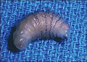
A 63-year-old woman presented with a two-month history of tender, pruritic papules on her left lower abdomen. The lesions were first noticed several days after returning from a trip to South America. Physical examination revealed three 6- to 8-mm firm nodules with central ulceration and mild erythema (Figure 1). On closer inspection, the nodules appeared to pulsate. The larva in Figure 2 was manually extracted from one of the lesions.
FIGURE 1.
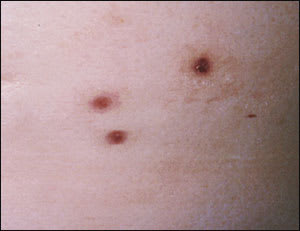
FIGURE 2.
Question
Which one of the following is the most likely cause of the patient's lesions?
A. Scabies.
B. Leishmaniasis.
C. Cutaneous myiasis.
D. Chigger bites.
E. Furunculosis.
Discussion
The answer is C: cutaneous myiasis (botfly infestation). Several different species of dipterous (two-winged) flies have life cycles that include parasitic larval stages that invade the skin or other tissue of host animals, especially domestic livestock and occasionally humans.1 Cutaneous myiasis occurs indirectly when adult flies deposit their eggs on the thorax of an unsuspecting vector (e.g., fleas, lice, mosquitoes, ticks, other flies). Larvae develop within the eggs over a span of five to 15 days. When the vector bites a host animal, the fly larvae drop to the skin surface and penetrate through the bite site or an adjacent hair follicle.2,3 Cutaneous infestation is manifested by development of inflamed, pruritic papules. A small punctum in the center of each lesion serves as a breathing hole and may drain serosanguineous fluid as the larva grows.4 The mature larva emerges from the wound in six to 12 weeks, falls to the ground, and pupates into adult flies in about 30 days.5
The fly species that cause cutaneous myiasis differ according to geographic location. Cordylobia anthropophaga, the tumbu fly, is indigenous to tropical Africa and is a common cause of myiasis in that region of the world. Dermatobia hominis, the human botfly, is distributed widely throughout Central and South America. Most cases of cutaneous myiasis in the United States occur in persons returning from these endemic areas.6
Treatment of cutaneous myiasis consists of manual removal of the larvae. Occlusion of the breathing punctum (e.g., petrolatum, adhesive tape) is advocated by some as a removal method.7 When the larva backs out of the occluded punctum, which may take up to 24 hours, then it may be removed manually. Others have found this to be ineffective and prefer surgical excision. Care must be taken to avoid lacerating the larva, as retained larval parts may precipitate a foreign body reaction.8,9
Scabies is caused by the mite Sarcoptes scabiei, which burrows into the stratum corneum and deposits its eggs. Characteristically, it produces intensely pruritic papules with linear burrows. This infestation often is transmitted by sexual or other close contact, not by travel to tropical locales. Scabies lesions typically would be smaller and more numerous than the lesions seen with cutaneous myiasis. Suspected scabies may be confirmed by microscopic identification of the mite or its feces in skin scrapings.
Leishmaniasis is a parasitic disease caused by protozoa of the family, Leishmania. The disease is transmitted to humans by the bite of female sandflies (Phlebotomus or Lutzomyia). The condition is endemic to many areas of the developing world, including Central and South America, Africa, the Middle East, and Asia. Skin lesions from leishmaniasis may vary from multiple small crusted lesions to a single large ulcerating sore. The organisms may be identified in skin biopsy specimens using Wright's stain.
Chigger bites are caused by the harvest mite or red bug, which commonly is found in grasslands of the southeastern United States. The larval form of the mite attaches to the skin, obtains a blood meal, and then falls off. Pruritic erythematous papules develop at bite sites, most commonly on the lower legs. No punctum would be seen in these lesions, and the inflammation would not persist for weeks to months, as occurs in myiasis.
Furunculosis is an acute, tender, perifollicular abscess that generally is caused by Staphylococcus aureus infection. The infection typically would respond rapidly to antibiotics, with incision and drainage for more advanced lesions.
Selected Differential Diagnosis of Persistent Pruritic Papules
| Condition | Characteristics |
|---|---|
| Scabies | Excoriated papules and plaques with burrows |
| Leishmaniasis | Indurated, ulcerated papules and plaques with scars |
| Cutaneous myiasis | Tender, ulcerated papules with central punctum |
| Chigger bites | Hemorrhagic, erythematous papules and pustules |
| Furunculosis | Erythematous, circumscribed, tender perifollicular nodules |
