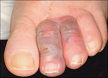
A 68-year-old man presented to the emergency department complaining of painful swelling in the second and third toes of his left foot, which had started four days earlier. The patient denied trauma or fever. His medical history was significant for hypertension and atrial fibrillation. Medications included lisinopril, sotalol, and warfarin. During the past six months, his anticoagulation consistently had been in the therapeutic range. Progression of pain and discoloration motivated him to seek care. Physical examination revealed the left second and third toes were tender, edematous, and purplish with sharp demarcation at the base of both digits. Lymphangitic streaking was absent (see accompanying figure). Peripheral pulses were normal in both legs, and the anklebrachial index also was normal bilaterally (left index was 1.2, right was 1.3). Left foot radiographs showed swelling, but no evidence of osteomyelitis or fracture. The patient was admitted and received empiric antibiotic treatment (levofloxacin) along with hydrocodone for pain.
Question
Based on the patient’s history and physical examination, which one of the following is the correct diagnosis?
A. Cellulitis.
B. Cryoglobulinemia.
C. Trauma.
D. Warfarin skin necrosis.
E. Blue toe syndrome.
Discussion
The answer is E: blue toe syndrome. Also known as “trashfoot,” blue toe syndrome is an arterial embolic disorder. Thrombogenic and atherogenic sources for emboli need to be considered in the diagnostic work-up. Transthoracic echocardiogram was performed in this patient and was negative for vegetations or clots. Computed tomography scanning revealed no evidence of abdominal aortic aneurysm or atheromatous plaque.
A thrombogenic etiology initially seemed more likely in this patient, given his known atrial fibrillation, the distal location of the embolic event, and its simultaneous onset in adjacent toes. However, when review of laboratory values confirmed consistently therapeutic anticoagulation and no cardioembolic source was found on transthoracic echocardiogram, attention shifted toward a possible atherogenic source.
Aortic atherosclerotic plaques are recognized as an important cause of lower extremity embolic disease.1 Plaques may become dislodged and embolize during intravascular catheter procedures, but this patient had no such history. Warfarin use for prevention of embolic disease is well known, but some studies have noted that warfarin use also may be a cause of emboli.2 It has been postulated that warfarin prevents clots from stabilizing injured atherosclerotic plaque surfaces, which then allows cholesterol contents to embolize.3
When the diagnostic work-up in this patient revealed no obvious thrombogenic etiology, the presumptive diagnosis was an atheromatous embolic event. Antibiotic therapy was stopped during hospitalization given the lack of apparent benefit. The patient was discharged with oral hydrocodone for pain relief. During the weeks after discharge, the patient’s pain slowly improved, but the purplish color of his toes persisted. A vascular surgery consult recommended continued outpatient pain management, with observation to see if toe amputation eventually would be required.
The differential diagnosis of the toe findings in this patient includes cellulitis, cryoglobulinemia, trauma, and warfarin skin necrosis. Cellulitis was unlikely, given the sharply demarcated border to the skin change, the absence of fever, and lack of response to antibiotic therapy. Cryoglobulinemia may lead to purpuric or gangrenous lesions in the distal extremities, and its incidence is increasing recently because of its association with hepatitis C infection. Skin lesions from cryoglobulinemia typically would be more widespread and be unlikely to affect only two adjacent toes. Trauma was unlikely given a negative history in a reliable patient and unremarkable radiographs. Warfarin skin necrosis typically occurs during the first few days of anticoagulation therapy and commonly appears in areas of adipose accumulation (i.e., breast, buttock, thigh). The patient had been receiving warfarin therapy for many months without previous complications.
Selected Differential Diagnosis of Swollen, Discolored Toes
| Condition | Characteristics |
|---|---|
| Cellulitis | Erythema, edema, warmth, pain with or without fever |
| Cryoglobulinemia | Recurrent palpable purpura on lower extremities with or without arthralgias and renal disease |
| Trauma | Positive history with corresponding contusion pattern |
| Warfarin skin necrosis | Onset during first week of warfarin use in areas of adipose accumulation |
| Blue toe syndrome | Acute onset of painful cyanotic discoloration caused by embolism in a vascular distribution pattern |
