
Am Fam Physician. 2004;70(9):1713-1720
Tympanometry provides useful quantitative information about the presence of fluid in the middle ear, mobility of the middle ear system, and ear canal volume. Its use has been recommended in conjunction with more qualitative information (e.g., history, appearance, and mobility of the tympanic membrane) in the evaluation of otitis media with effusion and to a lesser extent in acute otitis media. It also can provide useful information about the patency of tympanostomy tubes. Tympanometry is not reliable in infants younger than seven months because of the highly compliant ear canals of infants. Tympanogram tracings are classified as type A (normal), type B (flat, clearly abnormal), and type C (indicating a significantly negative pressure in the middle ear, possibly indicative of pathology). According to the Agency for Healthcare Research and Quality guidelines on otitis media with effusion, the positive predictive value of an abnormal (flat, type B) tympanogram is between 49 and 99 percent. A type C curve may be useful when correlated with other findings, but by itself it is an imprecise estimate of middle ear pressure and does not have high sensitivity or specificity for middle ear disorders.
Otitis media with effusion (OME) is defined as fluid in the middle ear without signs or symptoms of ear infection.1 Acute otitis media (AOM) is defined as the presence of middle ear effusion in conjunction with the recent, abrupt onset of one or more signs or symptoms of inflammation of the middle ear.2 AOM is the most frequently diagnosed disease in children, and its treatment results in more than 20 million antibiotic prescriptions annually in the United States.3,4 Frequently, AOM is overdiagnosed,5,6 and failure to differentiate AOM from OME may be the most common cause of unnecessary antibiotic prescriptions.3 Numerous studies have shown that some physical findings commonly used to predict AOM, such as redness or retraction of the tympanic membrane, have poor sensitivity and specificity.7–9 The definitions for AOM and OME require detecting the presence of middle ear effusion.1,2,10
| Key clinical recommendation | Label | References |
|---|---|---|
| Pneumatic otoscopy is the primary tool for diagnosing middle ear effusion in acute otitis media or otitis media with effusion. | A | 1 |
| Tympanometry is an optional tool that can be used to confirm suspected otitis media with effusion. | C | 1,13 |
| Tympanometry can be used in the evaluation of middle ear function in an infant with a suspected hearing disorder. | C | 1,16,17 |
The handheld tympanometer is a device that provides quantitative information on the function of structures and the presence of fluid in the middle ear. The graphic display of this data is the tympanogram. A pneumatic otoscopic examination of the tympanic membrane should be performed before tympanometry.6 Using pneumatic otoscopy with tympanometry improves the accuracy of diagnosis because many abnormalities of the eardrum and ear canal that might cause an abnormal tracing can be visualized. Determining the presence of obstructing cerumen in the canal, perforation or ventilation tubes in the tympanic membrane, and characteristics of the tympanic membrane (e.g., color, mobility, position, translucency) are helpful in correlating tympanometry findings with clinical disease.8,11,12 When comparing either test alone, pneumatic otoscopy has a better sensitivity and specificity than tympanometry for the diagnosis of OME.1 The two tests can be complementary, because pneumatic otoscopy provides a qualitative measure of tympanic membrane mobility (i.e., does the tympanic membrane move with insufflation?) and tympanometry produces more quantitative information (e.g., numeric and graphic data about generated positive and negative pressures, absorption of acoustic energy by the middle ear system, ear canal volume).11,13
The American Academy of Pediatrics (AAP)/American Academy of Family Physicians (AAFP)/Agency for Healthcare Research and Quality (AHRQ) guideline on OME recommends that performance of tympanometry be optional for confirming suspected OME.1,13 The guideline states that “the accuracy of pneumatic otoscopy in routine clinical practice may be less than that shown in published results because clinicians have varying training and experience. When the diagnosis of OME is uncertain, tympanometry or acoustic reflectometry should be considered as an adjunct to pneumatic otoscopy.” The Institute for Clinical System Improvement concluded that tympanometry may be useful in establishing the diagnosis of OME, but that it usually was not necessary for diagnosing (or documenting resolution of) AOM.14 The AAP/AAFP/AHRQ guidelines for AOM require the documentation of middle ear effusion for the diagnosis of AOM by tympanometry, pneumatic otoscopy, acoustic reflectometry, tympanocentesis, or the visualization of fluid in the external ear canal with tympanic membrane perforation.2 However, for OME and AOM, pneumatic otoscopy is recommended as the primary tool for diagnosis of middle ear effusion.
Other guidelines advise using tympanometry to evaluate middle ear function in infants suspected of having hearing disorders15 but not as a screening tool for periodic pediatric health examinations.1,16,17 Tympanometry is not reliable in infants younger than seven months because the ear canals of infants are highly compliant.17,18 In clinical practice, adherence to practice guidelines for use of pneumatic otoscopy and tympanometry for diagnosing OME is poor.19
Efficacy
Family physicians can use and interpret tympanograms for more accurate clinical decision making.8,20,21 The success rate for performing tympanometry (i.e., the ability to obtain a clinically useful tympanogram tracing) is between 74 and 94 percent (compared with a success rate of 85 to 91 percent for otoscopy).22
In a small Turkish study using confirmation of middle ear effusion by myringotomy as the gold standard, tympanometry had a positive predictive value and specificity of 96 and 92 percent, respectively; a negative predictive value and sensitivity of 96 percent each; and a false-positive rate of 8 percent in detecting the presence or absence of middle ear fluid in normal appearing ears.23 The predictive ability of tympanometry was lower, however, if the otoscopic examination showed retraction or other signs of effusion, but myringotomy demonstrated the absence of middle ear fluid.
In the 1994 AHRQ guidelines on OME, which we re reaffirmed in 1997, the positive predictive value of an abnormal (defined as a flat, type B tracing) tympanogram was between 49 and 99 percent.13 The AHRQ reexamined the evidence regarding the diagnosis and natural history of OME and published their findings in May 2002.24 Of the eight diagnostic studies reviewed (including portable tympanometry), the summary statement recommends pneumatic otoscopy as the preferred test (pooled sensitivity of 94 percent and specificity of 80 percent). Another analysis was performed of five studies using portable tympanometry, and 31 studies using professional tympanometry. Among the eight diagnostic methods, professional tympanometry (using a B or C2 curve as abnormal) tied with pneumatic otoscopy for the highest sensitivity at 93.8 percent (95 percent confidence interval [CI]: 91.1 to 96.4 percent), compared with myringotomy (Table 1).25 The diagnostic test with the highest specificity was professional tympanometry (using static compensated acoustic admittance at 0.1) at 94.1 percent (95 percent CI: 83.9 to 100 percent). The Canadian Task Force on Preventive Health Care reported that tympanometry has sensitivity and specificity greater than 80 percent in predicting fluid found in the middle ear at surgery.16 Studies combining tympanometry with clinical signs and symptoms have shown a sensitivity of 90 percent and a specificity of 75 percent in diagnosing OME.8
Using the Tympanometer
Portable tympanometers (resembling standard otoscopes) and desktop instruments are available. The probe is placed snugly in the external ear canal. A sound stimulus generator transmits acoustic energy into the canal while a vacuum pump introduces positive and negative pressures into the ear canal. A microphone in the instrument detects returning sound energy. Four useful pieces of objective data are obtained by the tympanometer (Table 2).26–28
| Estimated volume of air in front of the probe (equivalent ear canal volume [Vea]) |
| This volume is displayed on the tympanogram printout. The range of normal is age-dependent. |
| Maximum compliance (mobility) of the middle ear system (static admittance) |
| The mobility of the tympanic membrane is maximal when air pressures are equal on both sides of it.11 Static admittance is the greatest amount of acoustic energy absorbed by the middle ear system (the vertical peak of the tympanogram tracing). |
| Pressure at which the middle ear system has the greatest absorption of sound energy, or mobility (tympanometric peak pressure) |
| This value is an estimation of middle ear pressure and normally is around zero. It is the point on the horizontal axis (pressure axis) where the compliance is highest (the vertical peak of the tympanogram). |
| Width of the tympanogram curve |
| The tympanometer can be connected to a printer that will generate a tympanogram tracing and calculate these parameters, indicating whether they are outside the normal limits for adults or children. Optimal results are obtained if the physician and patient minimize movement during the test. A variation that may be more sensitive but more complicated to perform and interpret than standard tympanometry employs a sweep of tones with varying frequency (multiple-frequency tympanometry).26–28 |
Interpreting Results
Figures 1 and 2 depict various tympanogram tracings based on variations of the original Liden and Jerger classifications.29 The middle curve in Figure 1 is from a normal ear. The tympanogram curve has a normal maximum height that occurs at a pressure close to zero and the width of the curve is normal. This is referred to as a type A tracing. In this figure, the ear canal volume is normal. Figure 1 also has a curve that demonstrates a high peak height, labeled as type AD. High static admittance can result from an overly mobile tympanic membrane caused by disarticulation with the bony structures of the middle ear, or a tympanic membrane that has healed over a perforation but is thinner and more compliant than expected.26 The lowest curve in Figure 1 is a type AS tracing, with a reduced peak height, recorded from middle ears with some fluid or ossicular fixation that partially decreases mobility.30
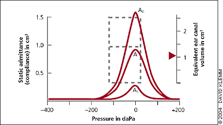
Figure 2A is a flattened, or type B tracing, with a low static admittance. The ear canal volume is normal. The most common cause of this pattern is decreased mobility of the tympanic membrane secondary to middle ear fluid (OME). Other causes are increased stiffness of the eardrum (from scarring), tympanosclerosis (the formation of dense connective tissue around the auditory ossicles), cholesteatoma, or middle ear tumor.26,31 When evaluating the efficacy and clinical usefulness of tympanometry, many studies consider only a type B tracing as definitely abnormal.13
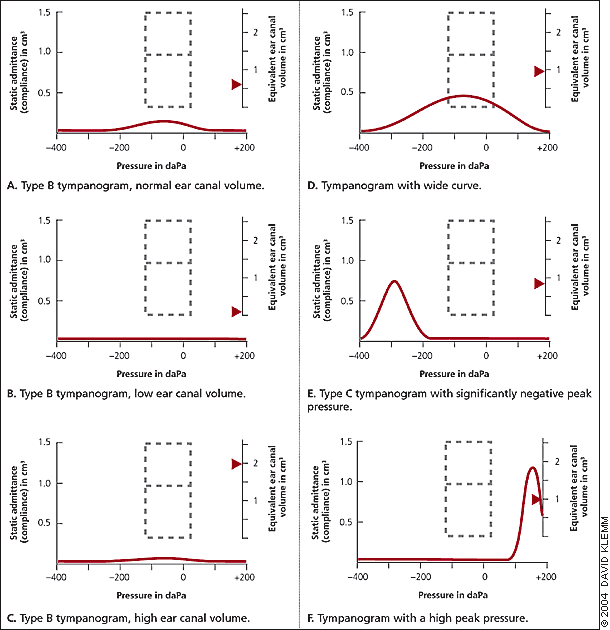
Figure 2B depicts a completely flat tracing with low ear canal volume, indicating partial ear canal occlusion with cerumen or improper placement of the probe.Figure 2C depicts a type B curve with a high measured volume. In the presence of a perforation of the tympanic membrane or a patent tympanostomy tube, acoustic energy also will be absorbed by air in the middle ear and possibly mastoid air cells, resulting in a higher than normal volume detected.32 Mastoidectomy also increases the measured volume.
Figure 2D is qualitatively somewhere between the preceding examples—the peak height falls within the normal range, but the tympanogram is too wide. Although this finding has been reported to be sensitive to middle ear disease when the static admittance is normal,33 most authorities do not consider its presence to be reliably diagnostic for middle ear pathology. It may occur with oncoming or resolving OME, or tympanosclerosis.
Figure 2E (or type C tracing) demonstrates a highly negative pressure in the middle ear, correlating to a retracted tympanic membrane. A viral upper respiratory infection may impair the ventilatory function of the eustachian tube. Negative middle ear pressure develops and nasopharyngeal contents are aspirated into the middle ear, resulting in AOM.4,34 This type of curve may indicate a transition between a normal ear and an ear that is full of fluid.35 The presence of a highly negative tympanic peak pressure observed during upper respiratory infection with no evidence of AOM may be a significant marker for increased risk for development of AOM.36 A type C curve may be clinically useful when correlated with other findings, but by itself is an imprecise estimate of middle ear pressure and does not have high sensitivity or specificity for middle ear disorders.17,37 Some authorities will subdivide C curves and distinguish C1 curves (moderately negative pressure) as normal and C2 curves (highly negative pressure) as abnormal or indefinite.22
Figure 2F indicates a highly positive peak pressure consistent with the bulging tympanic membrane that sometimes occurs with AOM.
The tympanometer may analyze the data and produce error messages. “BLOCKED” (very low equivalent ear canal volume) suggests the probe tip is not inserted properly, or the canal is occluded with cerumen. “OPEN” (very high equivalent ear canal volume) is caused by obtaining an inadequate seal with the probe tip, or it is sometimes secondary to a perforation of the tympanic membrane. “LEAK” indicates that the device was not able to produce the desired pressures in the ear canal secondary to an inadequate seal of the probe tip.
Final Comments
The tympanometer (handheld or desktop) is a useful, affordable diagnostic tool for the family physician (Table 3). AOM and OME require documentation of the presence of effusion in the middle ear, and both conditions frequently are misdiagnosed by history and physical examination (excluding pneumatic otoscopy) alone. Tympanometry may be helpful in diagnosing OME and AOM (a flat, type B curve with a normal ear canal volume), but it usually is not able to differentiate between the two. AOM also may produce a positive middle ear pressure (Figure 2F) or a high ear canal volume secondary to perforation of the tympanic membrane (Figure 2C). The AHRQ 2002 review of diagnostic methods concluded that pneumatic otoscopy performed by a skilled examiner (though level of skill was not clearly defined) has a more optimal combination of sensitivity and specificity than tympanometry. However, physical examination including pneumatic otoscopy and tympanometry may be the ideal method to document middle ear effusion in AOM and OME, though the clinical effectiveness of this approach has not been confirmed. Tympanometry also may provide useful information on positive or negative middle ear pressures that pneumatic otoscopy does not. There are no guidelines that recommend the performance of tympanometry on all patients with suspected AOM or OME.
| Model | Manufacturer | Style | Thermal printer | Graphic acoustic reflex display | Audiometry | Memory | Test time (seconds) | Weight (lb) | Other |
|---|---|---|---|---|---|---|---|---|---|
| GSI 37/38 series | Grason-Stadler, Inc. (888-647-0785) | Handheld/desktop | Yes | 37 no 38 yes | 37 no 38 yes | 1 patient, 8 tests | 2 to 12 | 6 | — |
| MTP 10 | Interacoustics (+45 6371 3555) | Handheld | Yes | Yes | Yes | 20 patients, full test | 2 to 12 | 1 | Optional pass/fail criteria box |
| MI 24/26 | Maico (888-941-4201) | Desktop | Yes | Yes | Yes | 1 patient, 2 tests | 2 to 12 | 10 | Bilingual (English/Spanish) |
| Race car and QT series | Maico | Desktop | Yes | Yes | Yes | 1 patient, 2 tests | 2 to 12 | 5 | Colorful overlay for children |
| Earscan | Micro Audiometrics (866-327-7226) | Desktop | Yes | Yes | Yes | 1 patient, 2 tests | 2 to 12 | 6 | — |
| MicroTymp2 | Welch Allyn (800-535-6663) | Handheld | Yes | No | No | 1 patient, 1 test | 2 | 0.61, handle 4, printer/charger | — |
| Tymp-Screen | Madsen (952-769-8100) | Handheld | Yes | Yes | No | 1 patient, 1 test | 8 | 1 | — |