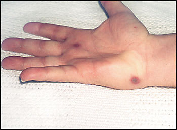
A 44-year-old man presents with pain in the muscles, right shoulder, and right wrist. Other symptoms include malaise, pharyngitis, fever, and chills. He denies intravenous drug use, contact with ill persons, or recent travel. Wrist examination shows tenosynovitis, and shoulder examination reveals pain, warmth, and erythema without effusion. Tender, hemorrhagic pustules ranging from 0.3 to 0.8 cm in diameter appear on his hands (see accompanying figure) and feet. His white blood cell count is 13,400 per mm3 with a neutrophil predominance, and his erythrocyte sedimentation rate is 52 mm per hour. Blood cultures showed no growth. Skin biopsy reveals leukocytoclastic vasculitis with intravascular thrombi and associated epidermal necrosis with perivascular inflammation. Gram stain is negative for bacterial organisms.
Question
Based on the above history, physical examination, and laboratory and biopsy results, which one of the following diagnoses best fits the etiology of this patient's condition?
A. Infective endocarditis.
B. Reactive arthritis.
C. Disseminated gonococcal infection.
D. Acute human immunodeficiency virus (HIV) infection.
E. Meningococcemia.
Discussion
The answer is C: Disseminated gonococcal infection. Neisseria gonorrhoeae infection typically produces urethral discharge and dysuria.1 However, 1 to 3 percent of patients with gonorrhea develop disseminated infection and present with one of two forms.2
The first form is an arthritis-dermatitis syndrome consisting of tenosynovitis, dermatitis, and polyarthralgias without purulent arthritis.2 Acute illness is characterized by fever, chills, and malaise. The tenosynovitis often involves multiple tendons. Skin lesions (three to 20 in number) include painful erythematous macules less than 1 cm in diameter that evolve to hemorrhagic pustules within 24 to 48 hours and are found near small joints of the hands and feet. The pustular vasculitis, intravascular thrombi, arthralgias, and tenosynovitis result from hematogenous dissemination of the gonococcus.3
The second form is a purulent arthritis without associated skin lesions. Most patients are afebrile and present with an asymmetric polyarthritis. These strikingly different clinical presentations may overlap, with the dermatitis arthritis syndrome progressing to purulent arthritis.2,4 In rare septicemic forms, disseminated infection may lead to hepatitis, meningitis, endocarditis, or other systemic involvement.5
Diagnosis is based on the characteristic clinical picture and clinical suspicion. Patients may not always admit to or know about their own or their partners' sexual exposures. For example, the patient in this case admitted only at follow-up to having sexual intercourse with prostitutes, rather than sharing this information at presentation. Disseminated gonococcal infection is three to four times more common in women than in men,2,4 and asymptomatic colonization of the oropharynx, urethra, anorectum, and endometrium is a primary predisposing factor. Other dissemination factors include menstruation, pregnancy, pelvic surgery, insertion of intrauterine devices, and congenital or acquired complement or immune deficiencies.1,6
Patients with disseminated gonorrhea are less likely to have positive blood cultures but more likely to have positive cultures of the urethra or cervix.7 Thus, this infection is best confirmed by culturing gonococci from the primary infected mucosal site.1 Cultures are positive in 75 to 90 percent of cases.8 It is imperative to obtain cultures from all possible mucosal surfaces to increase diagnostic yield. Selective medium (i.e., Thayer Martin) that contain antibiotics (i.e., vancomycin) may inhibit the growth of some gonococci and should be avoided.9 Cultures of joints, skin lesions, and blood are less likely (positive in 25 to 50 percent of cases) to grow organisms8 and should be cultured on antibiotic-free medium.9 Direct fluorescent antibody testing on skin biopsies is positive in more than 50 percent of cases.1 Synovial fluid analysis, culture, and nested polymerase chain reaction may help identify organisms and establish the diagnosis.8
Treatment consists of parenteral antibiotics for 24 to 48 hours and, after clinical improvement, oral therapy for a total of seven days. Initial therapy should include ceftriaxone, cefotaxime, or ceftizoxime. Oral treatments include cefixime, ciprofloxacin, ofloxacin, or levofloxacin. Penicillin-resistant strains of N. gonorrhoeae are widespread,10 and quinolone resistance is becoming more prevalent.11 Patients usually recover quickly and completely. Purulent arthritis, if present, may require joint drainage and a longer course of antibiotics. All patients should be treated presumptively for Chlamydia trachomatis.1,3,11
Patients with infective endocarditis may present with myalgias, arthralgias, fever, and chills. Hemorrhagic skin lesions are caused by septic emboli. However, most patients with infective endocarditis have cardiac abnormalities and positive blood cultures.12
Reactive arthritis (i.e., Reiter's syndrome or keratoderma blennorrhagicum) can present with the triad of urethritis, uveitis, and subacute arthritis involving the axial skeleton. Patients usually are afebrile. Hyperkeratotic skin lesions on the palms and soles are typical and circinate balanitis may be present.2,6
Acute HIV infection may present with a diffuse, painless, red, macular exanthem. Other symptoms include fever, joint and muscle pain, malaise, urticaria, and pharyngitis.4
Although meningococcal arthritis may be difficult to differentiate from disseminated gonorrhea, it usually is more severe. Patients may have clinical signs of sepsis or concurrent meningitis.
Selected Differential Diagnosis of Hemorrhagic Pustules, Tenosynovitis, and Arthritis
| Condition | Characteristics |
|---|---|
| Disseminated gonococcal infection | Erythematous macules evolving to tender hemorrhagic pustules or necrotic papules; usually near small joints of hands and feet; fever, asymmetric arthralgias, tenosynovitis, and arthritis are common. |
| Hypersensitivity vasculitis | Palpable purpura; may convert to hemorrhagic blisters, necrosis, ulcers in severe cases; found on lower legs, ankles, buttocks, arms; usually drug induced or post-infectious. |
| Infective endocarditis | Nonpainful, erythematous or hemorrhagic macules or nodules found on palms or soles (Janeway lesions); tender, purple, subcutaneous nodules in the pulp of fingers/toes (Osler's nodes); heart murmurusually present. |
| Korean hemorrhagic fever | Mucocutaneous petechiae, ecchymosis; fever, myalgia, headache, thrombocytopenia, renal involvement common; infectious etiology (Hantavirus). |
| Meningococcemia | Discrete pink macules/papules/petechiae; fulminant cases may include purpura, ecchymosis, necrosis, disseminated intravascular coagulation; highest incidence in children. |
| Reiter's syndrome | Red to brown papules, vesicles, or macules on soles or palms (keratoderma blennorrhagicum); lesions may become pustular, hyperkeratotic, crusted. Other findings include arthritis, uveitis, stomatitis, circinate balanitis. |
| Sweet's syndrome | Red to red-brown painful plaques and nodules; found on head, neck, upper extremities; fever, neutrophilia present; generally idiopathic or post-infectious; may indicate underlying malignancy. |
