A 48-year-old man presented with a flattening in the right pectoral region, absent musculature, and hypoplasia and brachysyndactyly of the right hand (Figures 1 and 2). No other pathologic findings were noted on physical examination, and there was no family history of these symptoms. Chest radiography showed diffuse hyperlucency on the right side of the thorax, presumably from a hypoplastic pectoral muscle.
Figure 1.

Flattening of the right pectoral region.
Figure 2.
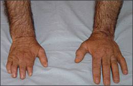
Hypoplasia of the right hand.
Question
Based on the patient’s physical examination and radiologic findings, which one of the following is the most likely diagnosis?
A. Apert’s syndrome.
B. Klippel-Feil syndrome.
C. Pectus excavatum.
D. Poland’s syndrome.
E. Spondylocostal dysostosis.
Discussion
The answer is D: Poland’s syndrome. In 1841, Alfred Poland described a case of unilateral absence of the pectoralis minor muscle and the sternal portion of the pectoralis major, and maldevelopment of the abdominal external oblique and serratus muscles.1 Poland’s syndrome involves unilateral absence of the sternal head of the pectoralis major; rib cage and upper extremity hypoplasia; breast and nipple hypoplasia; reduced fatty tissue, sweat glands, and hair in the affected area; and scoliosis.2 Thoracic computed tomography confirms the absence of the right pectoralis major and minor muscles (Figure 3). The syndrome more often affects the right side of the body and occurs in about one of every 30,000 live births with a male-to-female ratio of 3:1.1,3,4
Figure 3.
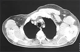
Computed tomography showing absence of the right pectoralis major and minor muscles.
The etiology of this syndrome is obscure, although two pathogenic mechanisms have been proposed. One is autosomal-dominant inheritance, but this is not universally accepted and no chromosomal alterations have been confirmed.1 Because this syndrome is rare, the literature provides only single studies or small samples with questionable genetic information. A familial component is rare and twin studies have failed to confirm transmission, making genetic and teratogenic etiologies less likely.3
A more accepted hypothesis centers on vascular pathogenesis. During the sixth to seventh week of pregnancy, a mesodermal defect may occur, leading to avascular thrombosis or embolus in the subclavian artery with resulting developmental alterations in the areas supplied. The side of the body affected and the degree of arterial obstruction determine the nature of the clinical symptoms. Maternal smoking may be a risk factor.5
Poland’s syndrome may coexist with other systemic malformations or derangements, such as thrombocytopenia, leukemia, lymphoma, Klippel-Feil syndrome, dextrocardia, renal agenesis, ureteral reflux, syndactyly, Adams-Oliver syndrome, Parry-Romberg syndrome, and Möbius’ syndrome.
Findings typical of Poland’s syndrome and other coexisting conditions should be assessed, and testing should include chest and upper limb radiography, echocardiography, abdominal ultrasonography, and complete blood count and urinalysis. Periodic lymph node and blood count examinations also should be considered.
Klippel-Feil syndrome is a rare congenital spinal disorder. Its main components are short neck, decreased range of motion in the cervical spine, and a low posterior hairline. Patients may have hearing loss and neurologic, cardiac, renal, and respiratory problems. Upper extremity abnormalities (i.e., syndactyly, hypoplastic thumb, supernumerary digits, and hypoplasia of the upper extremity) may be associated.6
Apert’s syndrome is an autosomal-dominant disorder with clinical findings of severe developmental disturbances of the craniofacial region, including bilateral coronal synostosis associated with midface hypoplasia, exophthalmia, hypertelorism, and symmetric syndactyly of the hands and feet.7 It also can be associated with cardiac and renal anomalies.
Pectus excavatum is the concave depression of the breast bone. In severe cases, it may cause cardiopulmonary insufficiency from compression of the right atrium and right ventricle and diminished pulmonary vital capacity.8
Spondylocostal dysostosis is a rare congenital condition in which development of the spinal column and rib cage is disrupted. It is characterized by severe rib and vertebral anomalies that cause respiratory problems, short stature, short trunk, and other abnormalities.9
Selected Differential Diagnosis of Upper Trunk Anomalies and Syndactyly
| Condition | Characteristics |
|---|---|
| Apert’s syndrome | Craniosynostosis, symmetric syndactyly, coronal suture synostosis, wide-set eyes, choanal atresia |
| Klippel-Feil syndrome | Short neck, decreased range of motion in the cervical spine, a low posterior hairline |
| Pectus excavatum | Concave sternal deformity |
| Poland’s syndrome | Absent pectoralis major; rib and upper extremity abnormalities; ipsilateral soft-tissue changes |
| Spondylocostal dysostosis | Severe rib and vertebral anomalies; short stature |
