Guideline source: American College of Cardiology, American Heart Association
Literature search described? Yes
Evidence rating system used? Yes
Published source: Circulation, September 20, 2005
Available at: http://circ.ahajournals.org/cgi/content/full/112/12/e154
As the number of treatments for heart failure has increased, uncertainty about the timing and sequence of initiating new medications and the appropriateness of combining them has made clinical decision making more complex. In addition, recognition of clinical heart failure in patients with a normal ejection fraction (EF) has highlighted the limitations of evidence-based therapy for these patients. These changes are reflected in the updated American College of Cardiology (ACC) and American Heart Association (AHA) guidelines for the diagnosis and management of chronic heart failure in adults.
Heart failure is a clinical syndrome characterized by specific symptoms (e.g., dyspnea, fatigue) and signs (e.g., edema, rales). The development of heart failure has been divided into four stages (Figure 1): A—high risk of heart failure but no structural disease or symptoms; B—structural heart disease but no signs or symptoms; C—structural heart disease with current or past symptoms; and D—refractory heart failure requiring specialized interventions. Possible structural or functional reasons for the development of heart failure include cardiomyopathy and left ventricular (LV) dysfunction. Patients with LV dysfunction or heart failure typically present with decreased exercise tolerance caused by dyspnea or fatigue, fluid retention in the legs or abdomen, or evidence of cardiac enlargement or dysfunction found during evaluation for another disorder. Diagnosis is largely clinical, based on a careful history and physical examination.
Figure 1. Recommended Therapy by Stage of Heart Failure Development
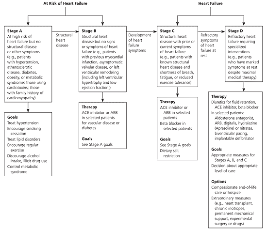
Recommended therapy for patients with heart failure, by disease stage. (ACE = angiotensin-converting enzyme; ARB = angiotensin receptor blocker)
Adapted with permission from Hunt SA, Abraham WT, Chin MH, Feldman AM, Francis GS, Ganiats TG, et al. ACC/AHA 2005 guideline update for the diagnosis and management of chronic heart failure in the adult—summary article: a report of the American College of Cardiology/American Heart Association Task Force on Practice Guidelines. Circulation 2005;112:1830.
Clinical Assessment
In the initial evaluation of patients with heart failure, physicians should locate structural and functional cardiac abnormalities and identify disorders and behaviors that could cause the disease or accelerate its progression. Physicians then should focus on clinical assessment to identify symptoms and determine their effects on function. An ongoing review of symptoms and function is critical to treatment selection.
IDENTIFICATION OF ABNORMALITIES
The history and physical examination may provide clues as to the nature of the underlying abnormality, but a specific diagnosis generally requires cardiac imaging. The most useful diagnostic tool for evaluating patients with heart failure is two-dimensional echocardiography with Doppler to assess left ventricular ejection fraction (LVEF), LV size, ventricular compliance, wall thickness, and valve function. This should be performed during the initial evaluation. Comprehensive evaluation is important because there often is more than one contributing cardiac abnormality, and the test also can be useful as a baseline for comparison.
Radionuclide ventriculography can be used to assess LVEF and volumes, and magnetic resonance imaging or computed tomography also may provide information in selected patients. Twelve-lead electrocardiography and chest radiography (postero-anterior and lateral) should be performed in all patients presenting with heart failure, but they should not be used as the primary basis for determining responsible abnormalities.
EVALUATION OF THE CAUSE
To evaluate the cause of heart failure, physicians should obtain a thorough history (Table 1), including current and past use of alcohol, illicit drugs, standard or alternative therapies, and chemotherapy drugs. A careful physical examination should be performed, including assessment of volume status, orthostatic blood pressure changes, weight and height, body mass index, and ability to perform routine and desired activities of daily living. Maximal exercise testing may be used to determine whether heart failure is the cause of exercise limitation; or, with measurement of respiratory gas exchange, to identify high-risk patients who may need advanced treatments such as cardiac transplantation.
TABLE 1 History-Taking in Patients with Heart Failure
| Patient history |
|---|
| Collagen vascular disease |
| Coronary or peripheral vascular disease |
| Current and past alcohol consumption |
| Diabetes |
| Dyslipidemia |
| Exposure to cardiotoxic agents |
| Exposure to sexually transmitted diseases |
| History or symptoms of sleep-disordered breathing |
| Hypertension |
| Mediastinal irradiation |
| Myopathy |
| Obesity |
| Pheochromocytoma |
| Rheumatic fever |
| Smoking |
| Thyroid disorder |
| Valvular heart disease |
| Family history |
| Cardiomyopathy (unexplained heart failure) |
| Conduction system disease (need for pacemaker) |
| Myopathy |
| Predisposition to atherosclerotic disease (e.g., history of myocardial infarction, stroke, peripheral arterial disease) |
| Skeletal myopathies |
| Sudden cardiac death |
| Tachyarrhythmias |
Adapted with permission from Hunt SA, Abraham WT, Chin MH, Feldman AM, Francis GS, Ganiats TG, et al. ACC/AHA 2005 guideline update for the diagnosis and management of chronic heart failure in the adult—summary article: a report of the American College of Cardiology/American Heart Association Task Force on Practice Guidelines. Circulation 2005;112:1832.
Laboratory tests should include complete blood count; urinalysis; lipid profile; liver function tests; and measurement of serum electrolyte levels (including calcium and magnesium), blood urea nitrogen, serum creatinine, fasting blood glucose levels, and thyroid-stimulating hormone levels. Elevated brain natriuretic peptide (BNP) levels may support a suspected diagnosis of heart failure or trigger its consideration, but this finding should not be used alone to confirm or exclude the diagnosis. BNP measurement may be useful in the emergency setting if heart failure is uncertain. Routine measurement of circulating neurohormone levels is not recommended.
Because patients with hemochromatosis may show improved LV function after treatment, screening for hemochromatosis is reasonable in selected patients. Screening for sleep-disturbed breathing also may be reasonable. Human immunodeficiency virus testing should be considered in those at high risk of infection, and diagnostic testing for rheumatologic disease, amyloidosis, or pheochromocytoma is reasonable if these conditions are suspected.
Coronary artery disease (CAD) often is an underlying cause of heart failure with low EF, and revascularization could play a role in treatment. Thus, determination of the presence and characteristics of CAD may be useful in selected patients. Coronary arteriography should be performed in patients who have angina or significant ischemia and are eligible for revascularization. It is a reasonable option in patients with chest pain who have not had coronary anatomy evaluation and have no contraindications to coronary revascularization, and in those with known or suspected CAD without angina and with no contraindications to revascularization of any kind. Noninvasive imaging to detect myocardial ischemia and viability is reasonable in patients eligible for revascularization who have known CAD without angina. Noninvasive imaging to define the likelihood of CAD may be considered in patients with LV dysfunction.
Although myocardial disorders often are responsible for cardiomyopathy, there typically is no identifiable causative factor. Endomyocardial biopsy may be useful to confirm a diagnosis that would influence therapy, but routine use is not recommended. Holter monitoring may be considered in certain patients. Signal-averaged electrocardiography is not recommended on a routine basis.
CLINICAL EVALUATION
At the initial and follow-up visits, physicians should assess patients' fluid or volume status, weight, blood pressure (sitting and standing), and ability to perform routine and desired activities of daily living. Evaluation of fluid or volume status is critical in determining whether diuretic therapy is needed and in detecting excesses or deficiencies of sodium, which could interfere with drug treatment. Physicians should determine the degree of jugular venous distension and its response to abdominal pressure, the presence and severity of organ congestion, and the scale of peripheral edema and ascites. A history of alcohol and drug use and diet (including sodium intake) also should be obtained.
Serum electrolyte levels (particularly potassium) and renal function should be monitored routinely because diuretic treatment may cause hypokalemia, which can lead to fatal arrhythmias and an increased risk of digitalis toxicity. Hyperkalemia can complicate therapy with certain drugs, and hyponatremia or anemia may indicate disease progression. Physicians should make every effort to prevent hypokalemia and hyperkalemia. Repeat measurement of EF and severity of structural remodeling may be useful if the clinical status has changed: improvement may reflect recovery from a previous condition or titration of therapies; deterioration may indicate disease progression or recurrent myocardial infarction.
Preventive Interventions
All patients at high risk of heart failure (stages A and B) or with heart failure symptoms (stage C) should be treated for risk factors such as hypertension, diabetes, lipid disorders, and thyroid disorders. Physicians should advise patients to avoid smoking, excessive alcohol consumption, use of illicit drugs, and other activities that may increase the risk of heart failure. Periodic evaluation of patients for signs and symptoms of heart failure is recommended. A noninvasive evaluation of LV function is recommended in patients who have a strong family history of cardiomyopathy and those receiving cardiotoxic interventions. Angiotensin-converting enzyme (ACE) inhibitors or angiotensin-II receptor blockers (ARBs) may be useful preventive therapies in patients at high risk of heart failure who have atherosclerotic valvular disease, diabetes, or hypertension with associated cardiovascular risk factors (Table 2).
TABLE 2 Indications for Medications Used in Treatment of Stages A, B, and C Heart Failure
| Drug | Stage A | Stage B | Stage C | |
|---|---|---|---|---|
| ACE inhibitors | ||||
| Benazepril (Lotensin) | H | — | — | |
| Captopril (Capoten) | H, DN | Post MI | HF | |
| Enalapril (Vasotec) | H, DN | HF | ||
| Fosinopril (Monopril) | H | — | HF | |
| Lisinopril (Zestril) | H, DN | Post MI | HF | |
| Moexipril (Univasc) | H | — | — | |
| Perindopril (Aceon) | H, CV risk | — | — | |
| Quinapril (Accupril) | H | — | HF | |
| Ramipril (Altace) | H, CV risk | Post MI | Post MI | |
| Trandolapril (Mavik) | H | Post MI | Post MI | |
| Aldosterone antagonists | ||||
| Eplerenone (Inspra) | H | Post MI | Post MI | |
| Spironolactone (Aldactone) | H | — | HF | |
| ARBs | ||||
| Candesartan (Atacand) | H | — | HF | |
| Eprosartan (Teveten) | H | — | — | |
| Irbesartan (Avapro) | H, DN | — | — | |
| Losartan (Cozaar) | H, DN | CV risk | — | |
| Olmesartan (Benicar) | H | — | — | |
| Telmisartan (Micardis) | H | — | — | |
| Valsartan (Diovan) | H, DN | Post MI | Post MI, HF | |
| Beta blockers | ||||
| Acebutolol (Sectral) | H | — | — | |
| Atenolol (Tenormin) | H | Post MI | — | |
| Betaxolol (Betoptic) | H | — | — | |
| Bisoprolol (Zebeta) | H | — | HF | |
| Carteolol (Cartrol) | H | — | — | |
| Carvedilol (Coreg) | H | Post MI | HF, Post MI | |
| Labetalol (Normodyne) | H | — | — | |
| Metoprolol succinate (Toprol XL) | H | — | HF | |
| Metoprolol tartrate (Lopressor) | H | Post MI | — | |
| Nadolol (Corgard) | H | — | — | |
| Penbutolol (Levatol) | H | — | — | |
| Pindolol (Visken) | H | — | — | |
| Propranolol (Inderal) | H | Post MI | — | |
| Timolol (Blocadren) | H | Post MI | — | |
| Digoxin | — | — | HF | |
note: Stage A = high risk of heart failure but no structural heart disease or heart failure symptoms; stage B = structural heart disease but no signs or symptoms of heart failure; stage C = structural heart disease with current or past symptoms of heart failure.
ACE = angiotensin-converting enzyme; H = hypertension; DN = diabetic nephropathy; post MI = reduction in heart failure or other cardiac events following myocardial infarction; HF = heart failure and asymptomatic left ventricular dysfunction; LVSD = left ventricular systolic dysfunction; CV risk = reduction in future cardiovascular events; ARB = angiotensin-II receptor blocker.
Adapted with permission from Hunt SA, Abraham WT, Chin MH, Feldman AM, Francis GS, Ganiats TG, et al. ACC/AHA 2005 guideline update for the diagnosis and management of chronic heart failure in the adult—summary article: a report of the American College of Cardiology/American Heart Association Task Force on Practice. Circulation 2005;112:1833.
ACE inhibitors are recommended in patients with a reduced EF but no heart failure symptoms. ACE inhibitors and beta blockers are recommended for all patients with a history of myocardial infarction regardless of EF or heart failure symptoms. Beta blockers also are indicated in patients with a reduced LVEF but no history of myocardial infarction or symptoms of heart failure. If patients who have had a myocardial infarction and have a low LVEF are intolerant of ACE inhibitors, ARBs should be administered. ACE inhibitors or ARBs may be beneficial in patients with hypertension and LV hypertrophy but no symptoms of heart failure.
Treatment of Heart Failure with Reduced LVEF
Most patients with heart failure and a reduced LVEF should receive a combination of a diuretic, an ACE inhibitor or ARB, and a beta blocker (Tables 2 through 5). Certain drugs can exacerbate heart failure syndrome and generally should be avoided or withdrawn in these patients, if possible. These include antiarrhythmic agents (except for amiodarone [Cordarone] and dofetilide [Tikosyn]), calcium channel blockers (except for those that are vasoselective), and nonsteroidal anti-inflammatory drugs.
TABLE 3 Oral Diuretics Recommended for Treatment of Fluid Retention in Chronic Heart Failure
| Drug | Initial daily dosage | Maximal daily dosage | Duration of action |
|---|---|---|---|
| Loop diuretics | |||
| Bumetanide (Bumex) | 0.5 to 1.0 mg one or two times | 10 mg | Four to six hours |
| Furosemide (Lasix) | 20 to 40 mg one or two times | 600 mg | Six to eight hours |
| Torsemide (Demadex) | 10 to 20 mg once | 200 mg | 12 to 16 hours |
| Potassium-sparing diuretics | |||
| Amiloride (Midamor) | 5 mg once | 20 mg | 24 hours |
| Spironolactone (Aldactone) | 12.5 to 25 mg once | 50 mg* | Two to three days |
| Triamterene (Dyrenium) | 50 to 75 mg two times | 200 mg | Seven to nine hours |
| Thiazide diuretics | |||
| Chlorothiazide (Diuril) | 250 to 500 mg one or two times | 1,000 mg | Six to 12 hours |
| Chlorthalidone (Hygroton) | 12.5 to 25 mg once | 100 mg | 24 to 72 hours |
| Hydrochlorothiazide (Esidrix) | 25 mg one or two times | 200 mg | Six to 12 hours |
| Indapamide (Lozol) | 2.5 mg once | 5 mg | 36 hours |
| Metolazone (Zaroxolyn) | 2.5 mg once | 20 mg | 12 to 24 hours |
*—Higher dosages may be used occasionally with close monitoring.
Adapted with permission from Hunt SA, Abraham WT, Chin MH, Feldman AM, Francis GS, Ganiats TG, et al. ACC/AHA 2005 guideline update for the diagnosis and management of chronic heart failure in the adult—summary article: a report of the American College of Cardiology/American Heart Association Task Force on Practice Guidelines. Circulation 2005;112:1836.
TABLE 4 Intravenous Diuretics Used in Treatment of Severe Heart Failure
| Drug | Initial dose | Maximal single dose |
|---|---|---|
| Loop diuretics | ||
| Bumetanide (Bumex) | 1.0 mg | 4 to 8 mg |
| Furosemide (Lasix) | 40 mg | 160 to 200 mg |
| Torsemide (Demadex) | 10 mg | 100 to 200 mg |
| Thiazide diuretics | ||
| Chlorothiazide (Diuril) | 500 mg | 1,000 mg |
| Sequential nephron blockade | ||
| Chlorothiazide | 500 to 1,000 mg one or two times plus loop diuretics once—multiple doses per day | |
| Metolazone (Zaroxolyn) | 2.5 to 5 mg orally one or two times per day with loop diuretic | |
| Intravenous infusions | ||
| Bumetanide | 1 mg load then 0.5 to 2 mg per hour | |
| Furosemide | 40 mg load then 10 to 40 mg per hour | |
| Torsemide | 20 mg load then 5 to 20 mg per hour | |
Adapted with permission from Hunt SA, Abraham WT, Chin MH, Feldman AM, Francis GS, Ganiats TG, et al. ACC/AHA 2005 guideline update for the diagnosis and management of chronic heart failure in the adult—summary article: a report of the American College of Cardiology/American Heart Association Task Force on Practice Guidelines. Circulation 2005;112:1836.
TABLE 5 Renin-Angiotensin-Aldosterone–System Inhibitors and Beta Blockers Commonly Used in the Treatment of Patients with Heart Failure and Low EF
| Drug | Initial daily dosage | Maximal daily dosage | ||
|---|---|---|---|---|
| ACE inhibitors | ||||
| Captopril (Capoten) | 6.25 mg three times | 50 mg three times | ||
| Enalapril (Vasotec) | 2.5 mg two times | 10 to 20 mg two times | ||
| Fosinopril (Monopril) | 5 to 10 mg once | 40 mg once | ||
| Lisinopril (Zestril) | 2.5 to 5 mg once | 20 to 40 mg once | ||
| Perindopril (Aceon) | 5 mg two times | 8 to 16 mg once | ||
| Quinapril (Accupril) | 5 mg two times | 20 mg two times | ||
| Ramipril (Altace) | 1.25 to 2.5 mg once | 10 mg once | ||
| Trandolapril (Mavik) | 1 mg once | 4 mg once | ||
| Aldosterone antagonists | ||||
| Eplerenone (Inspra) | 25 mg once | 50 mg once | ||
| Spironolactone (Aldactone) | 12.5 to 25 mg once | 25 mg one or two times | ||
| ARBs | ||||
| Candesartan (Atacand) | 4 to 8 mg once | 32 mg once | ||
| Losartan (Cozaar) | 25 to 50 mg once | 50 to 100 mg once | ||
| Valsartan (Diovan) | 20 to 40 mg two times | 160 mg two times | ||
| Beta blockers | ||||
| Bisoprolol (Zebeta) | 1.25 mg once | 10 mg once | ||
| Carvedilol (Coreg) | 3.125 mg two times | 25 mg two times (50 mg two times for patients greater than 85 kg) | ||
| Metoprolol succinate (Toprol XL) | 12.5 to 25 mg once | 200 mg once | ||
EF = ejection fraction; ACE = angiotensin-converting enzyme; ARB = angiotensin-II receptor blocker.
Adapted with permission from Hunt SA, Abraham WT, Chin MH, Feldman AM, Francis GS, Ganiats TG, et al. ACC/AHA 2005 guideline update for the diagnosis and management of chronic heart failure in the adult—summary article: a report of the American College of Cardiology/American Heart Association Task Force on Practice. Circulation 2005;112:1839.
Diuretics are recommended in patients with evidence of fluid retention. Loop diuretics generally are preferred, although thiazide diuretics may be preferred in hypertensive patients with mild fluid retention. Moderate sodium restriction and daily weight measurement is recommended to allow lower doses of diuretic drugs.
ACE inhibitors can alleviate symptoms, improve clinical status, enhance well-being, and reduce the risks of death and hospitalization in patients with heart failure. All patients with current or prior symptoms of heart failure and reduced LVEF should receive ACE inhibitors unless contraindicated. If target doses are poorly tolerated, intermediate doses should be used. Patients who are intolerant of ACE inhibitors can be given an ARB, but extreme caution should be used in patients who have had ACE inhibitor–associated angioedema.
ARBs are a reasonable alternative to ACE inhibitors as first-line therapy in patients with mild to moderate heart failure and reduced LVEF. An ARB in addition to conventional therapy may be considered in persistently symptomatic patients. Blood pressure, renal function, and potassium should be reassessed within one to two weeks after initiation of ARBs and monitored closely after changes in dosage.
Beta blockers (e.g., bisoprolol [Zebeta], carvedilol [Coreg], sustained-release metoprolol succinate [Toprol XL]) are recommended to reduce mortality rates in these patients. Long-term beta-blocker therapy can reduce heart failure symptoms, improve clinical status, and reduce the risks of death and hospitalization. Beta blockers should be initiated at very low dosages as soon as LV dysfunction is diagnosed, even when symptoms are mild or have responded to other treatments. They should be avoided or used with caution in patients with persistent symptoms of reactive airway disease or bradycardia. Institution of beta blockers should not be delayed because of a failure to reach target doses of ACE inhibitors and is reasonable in stable patients before the full target dosage of ARBs is reached.
The addition of digitalis may be beneficial to reduce the risk of hospitalization, reduce symptoms, control rhythm, and enhance exercise tolerance. Aldosterone antagonists may be added to therapy in select patients with moderately severe to severe symptoms who can be carefully monitored for hyperkalemia and renal dysfunction. However, routine combined use of ACE inhibitors, ARBs, and aldosterone antagonists is not recommended.
PREVENTION OF SUDDEN DEATH
Implantable cardioverter-defibrillator therapy is recommended for primary prevention of sudden cardiac death in patients who have nonischemic cardiomyopathy or ischemic heart disease with at least 40 days since their last myocardial infarction; an LVEF of 30 percent or less; New York Heart Association (NYHA) functional class II or III symptoms during long-term optimal medical therapy; and reasonable expectation of survival with good functional status for more than one year.
Placement of a cardioverter-defibrillator is recommended as secondary prevention of death from ventricular tachyarrhythmias to prolong survival in patients with a history of cardiac arrest, ventricular fibrillation, or hemodynamically destabilizing ventricular tachycardia. It is reasonable for patients with an LVEF of 30 to 35 percent of any origin who have NYHA functional class II or III symptoms, are taking long-term optimal medical therapy, and have reasonable expectation of survival with good functional status for at least one year.
INTERVENTIONS FOR CONSIDERATION
Adjunctive exercise training is beneficial in ambulatory patients. Nitrate therapy may decrease symptoms of dyspnea and improve exercise tolerance in patients with persistent limitations. The addition of hydralazine (Apresoline) and a nitrate is reasonable in patients who have persistent symptoms and are taking an ACE inhibitor and beta blocker for heart failure. This combination may be reasonable in patients who cannot take an ACE inhibitor or ARB because of drug intolerance, hypotension, or renal insufficiency.
Cardiac resynchronization therapy may improve quality of life when added to optimal medical therapy in persistently symptomatic patients. It is recommended in patients who have an LVEF of 35 percent or less, sinus rhythm, NYHA functional class III or ambulatory class IV symptoms despite recommended optimal therapy, and cardiac dyssynchrony.
Heart Failure with Normal LVEF
Heart failure with normal EF is most prevalent in older women, most of whom also have hypertension, diabetes, or both. CAD and atrial fibrillation also are common in these patients. Diagnosis generally is made with findings of typical signs and symptoms of heart failure in patients with a normal LVEF and no valvular abnormalities on echocardiography. Similarly presenting disorders should be ruled out (Table 6). A definitive diagnosis can be made when the rate of ventricular relaxation is slowed.
Management of patients with heart failure and normal LVEF is based on control of physiologic factors that are known to affect ventricular relaxation, such as blood pressure, blood volume, heart rate, and myocardial ischemia. Systolic and diastolic hypertension should be controlled, and ventricular rate should be controlled in those with atrial fibrillation. Diuretics are recommended to control pulmonary congestion and peripheral edema.
Physicians should treat diseases that are known to cause heart failure with normal LVEF, including CAD, hypertensive heart disease, and aortic stenosis. Coronary revascularization is reasonable in patients with CAD if myocardial ischemia may be adversely affecting cardiac function. Targeting of symptom reduction is reasonable. In patients with controlled hypertension, ACE inhibitors, ARBs, or calcium antagonists may be effective in minimizing heart failure symptoms. Restoration and maintenance of sinus rhythm may be useful to improve symptoms in patients with atrial fibrillation.
