Clavicle fractures are most common in children and young adults, typically occurring in persons younger than 25 years. Its superficial location, its thin midshaft, and the forces transmitted across it make the clavicle a common site for injury. The most common mechanism of injury is a forceful fall with the arm at the side, which commonly occurs during contact sports. Diagnosis can often be made by the history and physical examination, although appropriate radiography should be used to confirm the diagnosis and guide treatment options. Most clavicle fractures occur in the midshaft and can be treated nonoperatively. A prominent callus is common in children, and parents may require reassurance. If a child has no history of trauma, then malignancy, rickets, and physical abuse should be considered. Surgery is an option in fractures that have high potential for nonunion (e.g., displaced or communited fractures, fractures with more than 15 to 20 mm clavicle shortening). Distal fractures are classified based on the relationship to the coracoclavicular ligaments, which determines the likelihood of displacement. Most distal fractures can also be treated nonoperatively; however, certain factors must be considered in children.
Clavicle fractures constitute 5 to 10 percent of all fractures.1 Most occur in men younger than 25 years; however, they are also more common in men older than 55 and in women older than 75.2
The anatomic site of the fracture is typically described using the Allman classification, which divides the clavicle into thirds.3 Group I (midshaft) fractures occur on the middle third of the clavicle, group II fractures on the lateral (distal) third, and group III fractures on the medial (proximal) third.3 Midshaft fractures account for approximately 75 to 80 percent of all clavicle fractures and typically occur in younger persons. Distal third fractures represent about 15 to 25 percent of clavicle fractures. Medial third fractures are least common, accounting for less than 5 percent of clavicle fractures.1,4,5
SORT: KEY RECOMMENDATIONS FOR PRACTICE
| Clinical recommendation | Evidence rating | References |
|---|---|---|
| Nonoperative treatment is preferred for nearly all acute, nondisplaced midshaft clavicle fractures. | B | 12 |
| Treatment with an arm sling is preferred over a figure-of-eight dressing for acute midshaft clavicle fractures because it is better tolerated and leads to similar outcomes. | B | 10, 11 |
| Displaced midshaft clavicle fractures may be managed nonoperatively, but plate fixation should be considered. | B | 12, 13, 15 |
| Nonoperative treatment is preferred for distal clavicle fractures because outcomes are the same whether or not bony union is achieved. | B | 25, 26 |
A = consistent, good-quality patient-oriented evidence; B = inconsistent or limited-quality patient-oriented evidence; C = consensus, disease-oriented evidence, usual practice, expert opinion, or case series. For information about the SORT evidence rating system, see https://www.aafp.org/afpsort.xml.
Anatomy
The anatomy of the clavicle is shown in Figure 1. The clavicle is easily fractured because of its subcutaneous, relatively anterior location and frequent exposure to transmitted forces. The middle third, or midshaft, is the thinnest, least medullous area of the clavicle, and thus the most easily fractured; the lack of muscular and ligamentous support makes it vulnerable to injury. The acromioclavicular (AC) and sternoclavicular joints have robust ligamentous and capsular support. The sternal ossification center of the clavicle completes ossification and fuses with the shaft by age 30 years. This medial ossification center contributes to 80 percent of the longitudinal growth of the bone. Physeal injuries should be considered in adolescents with open growth plates.
Figure 1.
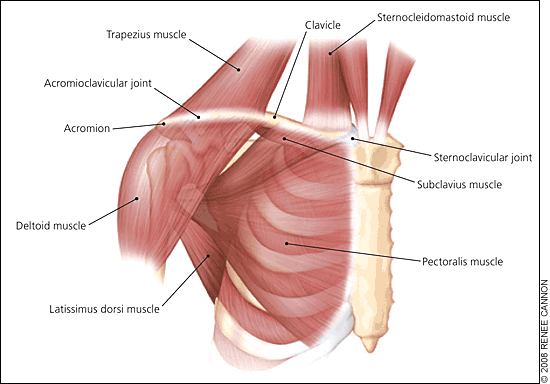
Anatomy of the clavicle.
The clavicle has many important functions: it connects the axial skeleton to the upper extremity; it contributes to the motion and stability of the upper extremity; and, with the subclavius muscle, it provides protection to the underlying neurovascular structures (i.e., subclavian vessels and brachial plexus).6,7 Malunion may impair these functions. In addition, callus formation or displacement can lead to thoracic outlet compression.
Evaluation
The usual mechanism of a clavicle fracture is a fall directly on the shoulder with the arm at the side. Rarely, clavicle fractures can occur from a direct blow or from a fall on an outstretched hand.8 In children and young adults, these injuries are typically related to sports participation, especially in contact sports such as football and rugby where participants are driven to the ground and land with the weight of their opponent on top of them.
Patients who have sustained clavicle fractures typically hold the affected arm adducted close to the body, often supporting the affected side with the opposite hand. This position is most comfortable because it limits the pull from the weight of the arm on the fractured bone. Physical examination may reveal ecchymosis, edema, focal tenderness, and crepitation on palpation over the clavicle. The defect in the bone may be seen by visual inspection or localized by palpation. Despite the low incidence of complications, it is important to perform a neurovascular and lung examination, because the subclavian vessels, brachial plexus, and lung apex can be injured in posteriorly displaced fractures.
Radiography should be performed on all patients with suspected clavicle fractures. Most fractures can be seen on a standard anteroposterior view of the clavicle; however, an anteroposterior view with 45-degree cephalic tilt minimizes the overlap of the ribs and scapula and allows for better assessment of displacement in the anterior and posterior plane9 (Figure 2). Advanced imaging such as computed tomography is indicated only in rare cases of distal or proximal fracture to assess the extent of intra-articular involvement.
Figure 2.
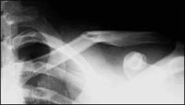
Anteroposterior radiograph of a nondisplaced midshaft clavicle fracture.
Midshaft Clavicle Fractures
Diagnosis of midshaft clavicle fractures by history, examination, and radiography is relatively straightforward (Figure 3). The medial segment is pulled superiorly by the sterno-cleidomastoid. The weight of the arm pulls the lateral segment inferiorly through the coracoclavicular ligaments, but is opposed by the trapezius. In addition, the pectoralis major and latissimus dorsi pull the lateral segment inferomedially with resultant shortening (Figure 4).
Figure 3.
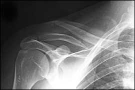
Radiograph of displaced midshaft clavicle fracture.
Figure 4.
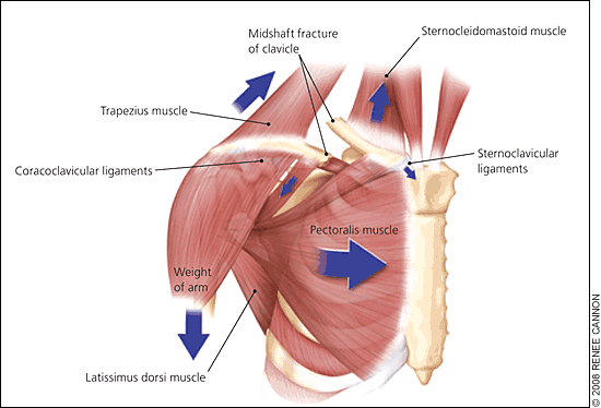
Deforming forces on displaced midshaft clavicle fracture.
The goals of treatment are to restore normal anatomy, limit pain, and promote a quick return to activity or play. Nonoperative management remains the most common approach to nondisplaced midshaft clavicle fractures. This consists of immobilization in a sling or figure-of-eight dressing. Although the figure-of-eight dressing is still widely used, several studies have demonstrated similar union rates and increased satisfaction in patients treated with a simple arm sling.10,11 The figure-of-eight dressing may cause discomfort and is often cumbersome to adjust. Immobilization is maintained for comfort and can be discontinued in one to two weeks or when the major pain subsides. Range-of-motion pendulum exercises can be started as soon as pain allows, with gradual progression to active range-of-motion and strengthening exercises over four to eight weeks.
Displaced midshaft clavicle fractures have higher rates of nonunion and a greater risk of long-term sequelae.12–14 Studies have shown that operative treatment results in a lower rate of fracture nonunion and improved patient-oriented outcomes compared with nonoperative treatment.14–16 Therefore, although nonoperative treatment is a viable option to treat displaced mid-shaft fractures, operative repair should be considered in patients with multiple risk factors for nonunion, especially significant fracture displacement or clavicle shortening (Table 1).13,14,16–18
Table 1 Risk Factors for Nonunion of Midshaft Clavicle Fractures
Options for operative management are open or closed reduction with plate fixation or, less commonly, intramedullary fixation. Intramedullary fixation uses smaller incisions and restores anatomy sufficiently. It also avoids the potential complications from plate pressure and the need for a second surgery. However, there is a small risk that the fixation device will migrate into an anatomically sensitive area. Consequently, a superior or anterior plate remains the preferred option for many surgeons.
Complications from midshaft clavicle fractures are rare, despite the proximity of neurovascular structures and lung apices, but include pneumothorax and neurovascular injury. Long-term sequelae include pain at rest or during activity, weakness, paresthesia, and cosmetic defects.19 Displacement of more than one bone width is the strongest radiographic risk factor for symptoms and sequelae.20
RETURN TO ACTIVITY CONSIDERATIONS
Return to activity recommendations for patients with midshaft clavicle fractures depend on patient age, level of contact, and presumed trauma risk. Before returning, an athlete should have full range of motion, normal shoulder strength, clinical and radiographic evidence of bony healing, and no tenderness to palpation.
Patients usually can return to noncontact sports and full daily activities six weeks after injury. Contact and collision sports should be delayed for two to four months until solid bony union occurs. If surgery is performed, some surgeons recommend removal of hardware before returning to sports.21 However, plate removal may delay return to sports and is not recommended by other surgeons.
MIDSHAFT CLAVICLE FRACTURES IN CHILDREN
For young adults, the most common cause of midshaft clavicle fractures is sports-related injuries. The mean age of patients with sports-related clavicle fractures is 21 years. The mean age of patients with non-neonatal clavicle fractures is eight years; 88 percent of these fractures are midshaft.4,22 Nearly all midshaft clavicle fractures in children heal well because of the great periosteal regenerative potential. Children often develop a significant callus formation, and it is important to educate parents about this normal progression of healing. Healing usually occurs within four to six weeks. If there is no history of trauma, then malignancy, rickets, osteogenesis imperfecta, and physical abuse must be considered.
Distal Clavicle Fractures
The primary restraints to vertical stability of the distal clavicle are the coracoclavicular ligaments. There are two distinct coracoclavicular ligaments: conoid and trapezoid. The classification and treatment of distal clavicle fractures depend on where the fracture occurs in relation to these ligaments.
The original classification by Neer in the 1960s described two types of distal clavicle fractures: type I, in which the coracoclavicular ligaments remain intact; and type II, in which the coracoclavicular ligaments are torn from the medial fragment and only the trapezoid ligament remains attached to the lateral fragment.23 The classification was later revised to include type III fractures, which involve extension into the AC joint; type IV fractures, which are seen in children and involve disruption of the periosteal sleeve rather than an actual bone fracture; and type V, which involve an avulsion of the ligaments with a small inferior cortical fragment.24
Types I and III fractures are inherently stable and do not displace; therefore, these types of fractures can be treated nonoperatively with a sling for comfort and early range-of-motion exercises as pain allows.25 Bony union occurs by six weeks, but return to contact sports should be delayed for two to four months until the bony union is solid.
Much of the controversy in the literature in treating distal clavicle fractures centers around how best to treat type II fractures, which have a tendency to displace. The lateral fracture fragment is held in place by the coracoclavicular ligaments, but pulled downward and medially by the weight of the arm and by the pectoralis and latissimus dorsi muscles, while the medial fragment is pulled superiorly by the trapezius and sternocleidomastoid muscles. Because of this displacement, there tends to be a high rate of nonunion.1,14 Despite this, outcome studies have demonstrated similar results in terms of function, strength, and pain in fractures united operatively, those united nonoperatively, and those that go on to nonunion.26,27 Therefore, even displaced fractures can be treated nonoperatively in the same way as types I and III fractures. However, the decision to treat a displaced type II fracture operatively in the acute setting may be made if the displacement is so severe that it is at risk of compromising skin integrity.
DISTAL CLAVICLE FRACTURES IN CHILDREN
Distal clavicle fractures are rare in children, and when they occur, the mechanism is often a fall on the point of the shoulder. Whereas this mechanism tends to cause AC joint disruption in older adolescents and adults, it much more commonly causes distal clavicle fractures in children.28
The fracture often occurs through the distal physis, with disruption of the thick periosteal sleeve surrounding the clavicle. The coracoclavicular ligaments remain attached to the periosteum while the clavicle herniates through the torn periosteum causing a “pseudodislocation.” These injuries are classified like adult AC joint injuries and treated similarly, with surgical treatment reserved for those with severe superior displacement, inferior displacement, or posterior displacement.
Proximal Clavicle Fractures
Fractures of the proximal third of the clavicle are uncommon. The proximal clavicle and sternoclavicular joint also have good ligamentous support, so when fractures do occur they typically do not displace. Nondisplaced fractures can be treated with a sling for comfort and gradual increase in range of motion, as pain allows.
Displaced fractures should be carefully evaluated for signs of neurovascular compromise; if present, this should be acutely reduced. In a setting capable of handling an airway or hemodynamic emergency, this reduction can be achieved acutely by grasping the clavicle with a towel clip and applying anterior traction. In the absence of neurovascular compromise computed tomography should be performed to fully visualize posteriorly displaced fragments; if necessary, reduction can be performed under anesthesia.
