Many patients in primary care present with ear pain (otalgia). When the ear is the source of the pain (primary otalgia), the ear examination is usually abnormal. When the ear is not the source of the pain (secondary otalgia), the ear examination is typically normal. The cause of primary otalgia is usually apparent on examination; the most common causes are otitis media and otitis externa. The cause of secondary otalgia is often difficult to determine because the innervation of the ear is complex and there are many potential sources of referred pain. The most common causes are temporomandibular joint syndrome, pharyngitis, dental disease, and cervical spine arthritis. If the diagnosis is not clear from the history and physical examination, options include a trial of symptomatic treatment without a clear diagnosis; imaging studies; and consultation with an otolaryngologist. Patients who smoke, drink alcohol, are older than 50 years, or have diabetes are at higher risk of a cause of ear pain that needs further evaluation. Patients whose history or physical examination increases suspicion for a serious occult cause of ear pain or whose symptoms persist after symptomatic treatment should be considered for further evaluation, such as magnetic resonance imaging, fiberoptic nasolaryngoscopy, or an erythrocyte sedimentation rate measurement.
Ear pain (otalgia) is a common symptom in primary care with many possible causes. When the cause arises from the ear (primary otalgia), the ear examination is usually abnormal and the diagnosis is typically apparent. In secondary or referred otalgia, the ear examination is usually normal, and the pain may be referred from a variety of sites.
SORT: KEY RECOMMENDATIONS FOR PRACTICE
| Clinical recommendation | Evidence rating | References |
|---|---|---|
| Magnetic resonance imaging and referral for nasolaryngoscopy should be considered for patients with otalgia who have a normal ear examination and who have signs, symptoms, or risk factors for tumor (e.g., tobacco or alcohol use, age older than 50 years). | C | 1, 5 |
| Young (i.e., younger than 40 years), otherwise healthy adults with otalgia and a normal ear examination can be treated symptomatically. Referral is appropriate if symptoms persist. | C | 1, 2 |
| Patients older than 50 years with unexplained otalgia and a normal ear examination should have an erythrocyte sedimentation rate measurement to help rule out temporal arteritis. | C | 25 |
A = consistent, good-quality patient-oriented evidence; B = inconsistent or limited quality patient-oriented evidence; C = consensus, disease-oriented evidence, usual practice, expert opinion, or case series. For information about the SORT evidence rating system, see page 579 or https://www.aafp.org/afpsort.xml.
The ear receives sensation fibers from cranial nerves V (trigeminal), VII (facial), IX (glossopharyngeal), and X (vagus), and cervical nerves C2 and C3. These nerves have long courses in the head, neck, and chest, which is why so many diseases can cause ear pain. The structures of the inner ear (i.e., cochlea and semicircular canals) are innervated by cranial nerve VIII (vestibulo-cochlear), which has no pain fibers. Therefore, most pathologic processes of the inner ear do not produce pain.1 However, inner ear diseases such as Meniere's disease can produce other sensations, such as pressure or fullness ( online Table A).1
It is often stated that 50 percent of pain in the ear is secondary otalgia,1 and that 50 percent of secondary otalgia results from dental causes2; however, these estimates are not based on published data. In a study of 500 patients visiting an ear, nose, and throat clinic, 58 presented with primary otalgia and 28 with secondary otalgia.3 In another study involving 615 patients, the most common causes of secondary otalgia were dental (38 percent), temporomandibular joint (TMJ) disorders (35 percent), cervical spine disorders (8 percent), and neuralgias (5 percent).4 The causes of otalgia in children are similar to those in adults, although middle ear disease (especially acute otitis media) is more common in children.5
Clinical Evaluation
HISTORY
Key points in the history include the patient's age, the location of pain (asking the patient to point with one finger), the radiation of pain, aggravating factors (e.g., chewing), associated symptoms (otologic and systemic), and risk factors for tumor (e.g., age older than 50 years, tobacco or alcohol use). Otologic symptoms that favor a primary cause include discharge, tinnitus, hearing loss, and vertigo. The severity of pain is not necessarily correlated with the seriousness of the cause. For example, the pain from tumors can be mild, whereas the pain from dental caries and otitis media can be severe.
PHYSICAL EXAMINATION
Key components of the physical examination include inspection of the auricle and periauricular region and a thorough otoscopic examination, which may require cerumen removal. Tenderness that occurs with traction on the auricle ( online Figure A) or pressure on the tragus ( online Figure B) indicates a condition of the external auditory canal, usually otitis externa.
When the ear examination is normal, the physician should palpate the TMJ for tenderness and crepitus as the patient opens and closes the mouth ( online Figure C).
In addition, the basic examination should include inspection of the nose and oropharynx, palpation of the head and neck, and examination of the cranial nerves. The gingiva should be inspected and palpated and the teeth inspected and percussed to assess tenderness. Fiberoptic nasolaryngoscopy is not usually necessary. Patients may need this procedure if they have risk factors for tumor or if conservative measures do not resolve symptoms.
DIAGNOSTIC TESTS
An assessment of hearing, by audiometry or simple testing (i.e., finger rub or whispered voice), is indicated in patients who notice hearing loss. An assessment of tympanic membrane mobility with pneumatic otoscopy or tympanometry can be helpful if there is suspicion of middle ear disease. When the physical examination is normal and the goal is to rule out tumor, the patient should have nasolaryngoscopy and magnetic resonance imaging (MRI) of the head and neck with gadolinium contrast.4 When the disease is evident on examination and the goal is to determine the extent of involvement, computed tomography (CT) with contrast media is generally indicated. For example, temporal bone trauma should be evaluated with CT scanning.
CLINICAL APPROACH TO DIAGNOSIS
Referring to a list of the causes of otalgia (Tables 1 through 4,1,4,6–39; online Table A) may be helpful, but in many patients these causes do not seem to fit. When the evaluation is unrevealing, a diagnosis of possible TMJ syndrome or eustachian tube dysfunction is often made. The physician must then decide whether to treat the patient symptomatically or to evaluate further with MRI or fiberoptic nasolaryngoscopy. Figure 1 provides one approach to this decision.1,4,6 In a patient at low risk of tumor or other serious illness, it is reasonable to offer symptomatic treatment (e.g., nonsteroidal anti-inflammatory drugs and a soft diet if TMJ syndrome is suspected). If conservative measures are not helpful, MRI or a more invasive examination should be considered.
Table 1 Common Causes of Ear Pain: Abnormal Ear Examination
| Cause | History | Physical findings | Comments |
|---|---|---|---|
| Otitis media7 | Recent upper respiratory infection Night restlessness in children | Red or cloudy tympanic membrane that is immobile on pneumatic otoscopy | Most common cause of primary ear pain More common in winter |
| Otitis externa8 | Recent swimming White discharge | Pain elicited by traction on auricle or pressure on tragus External auditory canal swollen and red with white debris1 | Findings can be subtle (consider empiric therapy) More common in summer Consider malignant (necrotizing) otitis externa in patients with diabetes or immunocompromise |
| Foreign body9 | Insects, small objects Commonly occurs in children | Foreign body visible in ear canal | May need sedation for removal |
| Barotrauma10 | Pain onset during descent of airplane or while scuba diving | Tympanic membrane hemorrhage Serous or hemorrhagic middle ear fluid | Otoscopic signs of barotrauma are present in 10 percent of adults and 22 percent of children after an airplane flight10 |
Table 2 Common Causes of Ear Pain: Normal Ear Examination
| Cause | History | Physical findings | Comments |
|---|---|---|---|
| TMJ syndrome11 | Pain or crepitus with talking or chewing | Tender TMJ Crepitus or clicking on motion of mandible May have restricted jaw movement | Risk factors include clenching and biting inside of lips and mouth |
| Dental causes (e.g., caries, periodontal abscess, impacted third molars, pulpitis)6 | May have dental complaints or history of dental disorders | Caries Abscess Gingivitis Facial swelling Teeth tender to percussion | Caries and abscess most common |
| Pharyngitis or tonsillitis4 | Often accompanied by sore throat | Pharyngeal or tonsillar erythema Swelling Exudate | Otalgia can be the primary symptom even if ear not involved |
| Cervical spine arthritis4,12 | Crepitus or pain with neck movement | Decreased neck range of motion Tender spinous processes or paraspinal muscles | Pain referred from C2, C3 cervical nerve roots |
| Idiopathic 4,6,13 | Variable | Normal | In practice, often labeled TMJ syndrome, neuropathic pain, or eustachian tube dysfunction |
TMJ = temporomandibular joint.
Table 3 Uncommon Causes of Ear Pain: Abnormal Ear Examination
| Cause | History | Physical findings | Comments |
|---|---|---|---|
| Malignant (necrotizing) otitis externa14 * | Suspect in refractory otitis externa in patients with diabetes, older patients, and those with immunocompromise Pain disproportionate to examination findings | Granulation tissue on floor of external auditory canal | Easy to miss, findings can be subtle Obtain technetium bone scan to determine extent of disease and gallium tagged white-cell scan as baseline to follow response to treatment |
| Ramsay Hunt syndrome (herpes zoster oticus)15,16 | Pain often precedes vesicles and is much worse than in Bell's palsy Patient may have vertigo, hearing loss, or tinnitus | Vesicular rash on auricle and external auditory canal Palsy of cranial nerve VII (facial) | Can involve other cranial nerves (e.g., V [trigeminal], IX [glossopharyngeal], X [vagus]) Pain can occur without significant vesicular eruption |
| Cellulitis/chondritis/perichondritis | Preceding insect bite, scratch, or piercing Rapid progression Perichondritis characterized by persistent redness, swelling, and pain | Earlobe usually involved with cellulitis | Perichondritis must be treated aggressively; sometimes requires parenteral antibiotics |
| Relapsing polychondritis17,18 | Recurrent swelling and redness of auricle Hearing loss frequent | Earlobe is spared because it has no cartilage | Noninfectious Can involve other cartilage such as trachea and bronchi |
| Trauma19 | Blunt or sharp trauma Frostbite Burns | Traumatic lesions of auricle, ear canal, or tympanic membrane | Most common injury is laceration of the auricle |
| Mastoiditis20 | Recent or concurrent otitis media Retroauricular pain | Protrusion of auricle Tender edematous mastoid | Prevalence increased in children with limited access to health care |
| Tumors or infected cysts in auricle or ear canal | Pain usually well localized to auricle or ear canal | May require meticulous examination of external auditory canal May need to remove cerumen | Diagnosis of ear canal tumors is often delayed because of misdiagnosis as chronic inflammation |
| Wegener's granulomatosis | Arthralgia Hearing loss Myalgias Oral or nasal ulcers Otorrhea Rhinorrhea | Often causes chronic otitis media or serous otitis | Consider testing for antineutrophil cytoplasmic antibodies |
| Viral myringitis21,22 | Presentation similar to acute otitis media | Tympanic membrane red, but not bulging; landmarks visible | Bullous myringitis is not pathognomonic of viral myringitis |
*— Rule out “worst-case scenario” diagnosis (see Table 5).
Table 4 Uncommon Causes of Ear Pain: Normal Ear Examination
| Cause | History | Physical findings | Comments |
|---|---|---|---|
| Tumors (e.g., parotid, hypopharynx, nasopharynx, base of tongue, tonsillar fossa, larynx, esophagus, intracranial, cervical spine)4 | Risk factors include smoking, alcohol use, age older than 50 years, hoarseness, dysphagia, radiation exposure, weight loss | May require fiberoptic nasolaryngoscopy | Consider referral for invasive examination and MRI |
| Neuralgias (e.g., trigeminal, glossopharyngeal, geniculate, sphenopalatine)1,4 | Pain usually brief (seconds), severe, lancing, jabbing, electric-shock–like, episodic | Usually none May have trigger point | Trigeminal neuralgia (tic douloureux) best defined |
| Bell's palsy 23,24 | Retroauricular pain, less severe than Ramsay Hunt syndrome; can precede or follow the palsy | Peripheral facial palsy (involvement of forehead) | Pain occurs in 25 to 50 percent of patients with Bell's palsy |
| Temporal arteritis25 * | Age older than 50 years Jaw claudication Diplopia | Temporal arteries may be tender, prominent, or beaded | Erythrocyte sedimentation rate usually greater than 50 mm per hour Biopsy and prompt treatment are indicated |
| Oral aphthous ulcers | Localized pain in mouth as well as ear | Shallow ulcers with gray, necrotic base | Often recurrent Etiology not well defined |
| Cervical adenopathy | May have recent upper respiratory infection or scalp lesion | Tender cervical or periauricular lymph nodes | Consider CT and fine needle aspiration for lymph nodes > 1.5 cm, lasting longer than six weeks |
| Myofascial pain, muscle spasm or inflammation of sternocleidomastoid or muscles of mastication26,27 | Pain aggravated by chewing or head movement | May have trigger point | Can be caused by clenching, bruxism, TMJ syndrome, and dental or oral disorders |
| Eagle's syndrome (elongation of styloid process)28 | Deep, unremitting pain exacerbated by swallowing, yawning, or chewing May have pain in neck, foreign body sensation in throat | Reproduce pain with tonsillar fossa palpation | Diagnosed with CT Most patients are 3 to 40 years of age and have had a tonsillectomy Styloid process longer than 1 inch (2.5 cm) |
| Sinusitis/sinogenic referred pain from allergy29 | Nasal congestion Pain in maxillary sinuses | Nasal congestion Tender over maxillary sinuses | Sinusitis is common but otalgia from sinusitis is unusual |
| Carotidynia30 | May have dysphagia and throat tenderness | Tender carotid artery | More common in women May have abnormal enhancement on MRI |
| Thyroiditis | May report pain in thyroid | Thyroid may be tender or enlarged | Referred pain from cranial nerve X (vagus) |
| Salivary gland disorders (e.g., stones, mumps) | Pain in preauricular area | Prominent, tender parotid glands | There have been recent epidemics of mumps in the United States |
| Cricoarytenoid arthritis31 | Ear pain and hoarseness Pain is worse with speaking, coughing, or swallowing | May have other features of inflammatory arthritis | Often caused by rheumatoid arthritis or systemic lupus erythematosus |
| Gastroesophageal reflux32,33 | Heartburn Acid reflux | Usually none | Pain caused by irritation of oropharynx (cranial nerves IX [glossopharyngeal] and X) or of eustachian tube orifice |
| Angina pectoris, myocardial infarction34 * | Cardiac risk factors | Usually none | If suspected, obtain electrocardiogram and serum troponin level |
| Thoracic aneurysms (e.g., innominate artery, thoracic aorta)* | More common in older men May have hypertension and other risk factors for atherosclerosis | May have chest or back pain | Obtain chest CT scan or magnetic resonance angiogram; plain chest radiography is insensitive |
| Psychogenic (e.g., depression, anxiety)35 | History of depression or anxiety | Blunted affect Depressed mood | Consider in patients with idiopathic otalgia |
| Other rare causes (e.g., subdural hematoma, lung cancer,36,37 * central line placement,38 pillow otalgia,39 carotid artery aneurysm) | Variable | Variable | Lung cancer is the best described of these rare causes |
MRI = magnetic resonance imaging; CT = computed tomography; TMJ = temporomandibular joint.
*— Rule out “worst-case scenario” diagnosis (see Table 5).
Figure 1. Management of Ear Pain
Algorithm for the management of ear pain. (TMJ = temporomandibular joint; ECG = electrocardiography; ESR = erythrocyte sedimentation rate; NSAIDs = nonsteroidal anti-inflammatory drugs.)
Information from references 1, 4, and 6.
RULE OUT WORST-CASE SCENARIO
As with any symptom, a “rule out worst-case scenario” strategy (in which certain diagnoses must be ruled out immediately) may help avoid serious diagnostic errors.40 In patients with otalgia, physicians should rule out several potential causes that can have serious consequences if the diagnosis is delayed; these are malignant (necrotizing) otitis externa, cholesteatoma, myocardial infarction, temporal arteritis, and malignant tumor. However, these diseases can often be ruled out on the basis of a nonworrisome history and physical examination rather than extensive testing. Risk factors that should prompt consideration of these diseases are outlined in Table 5.
Table 5 Risk Factors for “Worst-Case Scenario” Diagnoses in Patients with Ear Pain
| Risk factor | Possible diagnosis |
|---|---|
| Age older than 50 years, ESR greater than 50 mm per hour | Temporal arteritis |
| Coronary artery disease risk factors | Myocardial infarction |
| Diabetes or immunocompromise | Malignant (necrotizing) otitis externa |
| Tobacco and alcohol use, dysphagia, weight loss, age older than 50 years | Head or neck tumor |
| Superior tympanic membrane retraction pocket, otorrhea | Cholesteatoma |
| Unilateral hearing loss | Malignant otitis externa, cholesteatoma |
ESR = erythrocyte sedimentation rate.
Common Causes of Ear Pain
ABNORMAL EAR EXAMINATION
Acute otitis media is probably the most common cause of primary otalgia (online Figure D).1,7,41 The tympanic membrane is classically red and bulging, but it can also be white or pink, and the discoloration sometimes involves only part of the tympanic membrane.
Otitis externa (or swimmer's ear) generally leads to swelling and redness of the ear canal. There is often debris in the ear canal or covering the tympanic membrane.8 Subtle otitis externa can be difficult to identify on inspection, but it usually causes tenderness when the examiner pulls on the auricle or presses on the tragus (online Figures A and B).
Foreign bodies in the ear canal are most common in children. In one study, the most common objects removed were beads, paper, popcorn kernels, and insects.9 Most foreign bodies can be removed under direct visualization with a curette or alligator forceps. If this is not successful, the child should have removal of the foreign body under sedation and otomicroscopy.9 Although most foreign bodies in the ear canal can be managed nonurgently, hearing-aid batteries should be removed promptly to prevent alkali burns.
Barotrauma typically occurs while scuba diving or during an airplane flight with the onset of pain during descent.10 Eustachian tube dysfunction caused by an upper respiratory infection or allergic rhinitis increases the risk of barotrauma. The tympanic membrane is typically hemorrhagic, and there may be blood or serous fluid in the middle ear.
NORMAL EAR EXAMINATION
TMJ syndrome is characterized by pain and crepitus with talking or chewing, and tenderness or crepitus on palpation of the TMJ joint ( online Figure C).11 It causes ear pain, especially with chewing.11 However, TMJ crepitus is prevalent, and its presence should not prematurely halt further investigation into other causes of otalgia.1
Dental causes of otalgia generally involve the molar teeth. A variety of dental diseases can produce otalgia, but the most common are caries, periodontal abscesses, and impacted third molars. The physician should palpate the gingiva and tap on the teeth with a tongue blade to assess for tenderness.2
Pharyngitis and tonsillitis often cause referred pain to the ear. In some patients with pharyngitis, ear pain can be the primary complaint even when the ear is normal.
Idiopathic otalgia is common, but patients and physicians can be uncomfortable with this diagnosis.4,6,13 If a thorough evaluation is unrevealing and the physician suspects a benign cause, empiric treatment for TMJ syndrome with nonsteroidal anti-inflammatory drugs and a soft diet would be reasonable (Figure 11,4,6). If the physician suspects neuropathic pain, a trial of gabapentin (Neurontin) or amitriptyline is reasonable.
Uncommon Causes of Ear Pain
ABNORMAL EAR EXAMINATION
Malignant otitis externa is defined by osteitis of the skull base, typically caused by Pseudomonas infection, and it usually occurs in patients with diabetes or immunocompromise.1 It is characterized by severe, deep, unrelenting pain and by granulation tissue, which can be a subtle finding, on the inferior aspect of the external auditory canal at the bony-cartilaginous junction. Squamous cell carcinoma of the external auditory canal can mimic malignant otitis externa.
Ramsay Hunt syndrome (herpes zoster oticus) typically causes ear pain, facial paralysis, and vesicles in the external auditory canal. Other symptoms can include hearing loss, tinnitus, vertigo, taste disturbance, and decreased tearing.15 The syndrome is caused by herpes zoster involving the geniculate ganglion (cranial nerve VII), and it often involves cranial nerves V, IX, and X in addition to the facial nerve.
Relapsing polychondritis is a systemic disease that involves cartilage. It can affect many organs, including the eyes, nose, heart, kidneys, and nervous system, but the most commonly affected organ is the ear.17 Relapsing polychondritis often affects both ears, producing a red or violaceous auricle. Sparing of the earlobe, which lacks cartilage, helps distinguish auricular chondritis from cellulitis. It is diagnosed by its relapsing course and typical appearance.
Cholesteatomas are epidermal cysts composed of desquamating epithelium. They gradually enlarge and can erode the ossicular chain, inner ear, and bony facial nerve canal. Cholesteatomas generally do not cause severe pain, but may produce a sense of fullness. In patients with otorrhea or conductive hearing loss, it is important to visualize the most superior aspect of the tympanic membrane to exclude a superior retraction pocket leading to a cholesteatoma (Figure 2).
Figure 2.
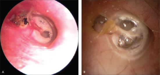
Two examples of cholesteatoma.
NORMAL EAR EXAMINATION
Tumors in the nose, nasopharynx, oral cavity, oropharynx, hypopharynx, infratemporal fossa, neck, or chest can cause ear pain. The most common sites are the base of the tongue, tonsillar fossa, and hypopharynx.4 Risk factors for head and neck tumors include tobacco or alcohol use, dysphagia, weight loss, radiation exposure, hoarseness, and age older than 50 years.24
Neuralgias can involve cranial nerves V and IX, the geniculate ganglion (cranial nerve VII), and the sphenopalatine ganglion (cranial nerves V and VII). The best known of these is trigeminal neuralgia (tic douloureux), which is characterized by paroxysmal, sharp, lancinating pain in the distribution of the maxillary and mandibular divisions. Glossopharyngeal neuralgia causes pain in the tonsillar area, pharynx, and, in some patients, the middle ear; this pain may be elicited by palpation of the tonsillar region.2 Sphenopalatine neuralgia results in pain around the eye and nose in addition to the ear and mastoid.2
Bell's palsy is characterized by the sudden onset of upper and lower facial paralysis. Postauricular pain occurs in about 25 percent of patients.23 Patients may also have hyperacusis, taste disturbances, and decreased tearing.
Temporal arteritis often causes temporal pain and tenderness that can involve the ear. Other symptoms include malaise, weight loss, fever, and anorexia. It is important to recognize temporal arteritis because it can cause permanent blindness, but this is usually preventable with prompt initiation of systemic corticosteroids. Only about 40 percent of patients have tenderness in the temporal arteries, but 65 percent have at least one temporal artery abnormality (e.g., tenderness, absent pulse, beading, prominence).25 Although temporal arteritis is unusual in patients younger than 50 years, it should be considered if there are multiple findings indicative of the disease.25 The disease is rare in patients with normal erythrocyte sedimentation rates and unusual if the erythrocyte sedimentation rate is less than 50 mm per hour.25
