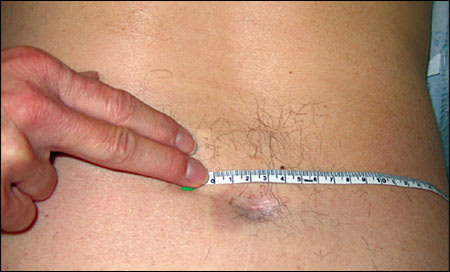
A 62-year-old man presented with a long history of intermittent low back pain and numbness. The pain was associated with lower extremity weakness and posterior leg numbness that occurred primarily with back stretching movements. He denied a history of trauma or a family history of major disease. The day of presentation, he had two episodes, each lasting a few minutes. The first occurred while he reached for a high object, and the second occurred as he got out of his car. The physical examination revealed a small dimple with hyperpigmented, hairy skin on the lower back (Figure 1). The neurologic examination was essentially unremarkable.
Figure 1.
Question
Based on the patient's history and physical examination, which one of the following is the most likely diagnosis?
A. Enterogenous cyst.
B. Herniated lumbar disc.
C. Spinal stenosis.
D. Syringomyelia.
E. Tethered spinal cord syndrome.
Discussion
The answer is E: tethered spinal cord syndrome. The patient's history and physical examination findings are classic for this syndrome. Magnetic resonance imaging (MRI) showing a low-lying tethered spinal cord ending at L3–4 with an associated neural tube defect (Figure 2) confirmed the diagnosis. Tethered spinal cord syndrome is a neurologic disorder caused by tissue attachments that stretch the spinal cord and limit its movement.1,2 It is associated with a number of congenital and acquired anomalies2,3 (Table 1). Other causes include dermal sinus tract, diastematomyelia (split spinal cord), trauma with scar tissue, and intradural dermoid cyst.3,4
Figure 2.
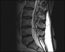
Magnetic resonance imaging showing low-lying tethered spinal cord ending at L3–4.
Table 1. Selected Conditions Associated with Tethered Spinal Cord Syndrome
| Condition | Characteristics |
|---|---|
| Diastematomyelia | Abnormal congenital division of the spinal cord by a bony spicule or fibrous band protruding from one or two vertebra; each half is surrounded by a dural sac |
| Fibrofatty terminal filum | Remnant of incomplete neural tube closure; caused by a failure of the surface ectoderm and dermal elements to separate from the neuroectoderm |
| Intradural dermoid cyst | Slow growing, congenital, benign tumors of the central nervous system |
| Low-lying conus medullaris | Failure of the conus medullaris to ascend may lead to tethered spinal cord syndrome; this ascension seems to occur during fetal development |
| Spina bifida | Incomplete closure of the embryonic neural tube leads to an incompletely formed spinal cord with unfused posterior vertebral structures |
| Meningocele is protrusion of the meninges through the gap in the spine; myelomeningocele is the protrusion of the spinal cord and the nerve roots from the gap in the spine; spina bifida occulta is the mildest form, and the spinal cord is often unaffected | |
| Spinal cord lipoma | Rare fatty-tissue tumors; mostly from a neural tube defect |
| Trauma | Trauma with scar tissue |
Symptoms may include low back pain, leg weakness, and numbness. In advanced disease, leg pain and loss of bladder and bowel control may occur. Examination may reveal hemangiomas, a tuft of hair, a dimple, fatty tissue, or a “tail.”5 Early surgery is recommended in children to prevent neurologic deterioration.1–5 In adults, surgery to detether the spinal cord can reduce complications. Other treatments are symptomatic and supportive, including pain control and physical therapy.6,7 MRI can confirm the diagnosis.
Enterogenous cysts of the central nervous system usually occur in the spinal canal, especially in the lower cervical and upper thoracic regions. They are benign epithelium-lined cysts. The cysts are often associated with developmental defects of the overlying skin or vertebral bodies and can cause spinal cord compression.8
Patients with a herniated disc may have sensory or motor dysfunction corresponding to the level of the nerve root compressed by the herniated disc. Diagnosis can be confirmed with MRI.9
Spinal stenosis is a narrowing of the spinal canal, lateral recess, or neural foramen with neurogenic claudication or pseudo-claudication.9
Syringomyelia includes cysts within the spinal cord that may cause pain, weakness, and stiffness in the back, shoulders, arms, or legs. MRI confirms the diagnosis.10
