Keloids and hypertrophic scars represent an exuberant healing response that poses a challenge for physicians. Patients at high risk of keloids are usually younger than 30 years and have darker skin. Sternal skin, shoulders and upper arms, earlobes, and cheeks are most susceptible to developing keloids and hypertrophic scars. High-risk trauma includes burns, ear piercing, and any factor that prolongs wound healing. Keloid formation often can be prevented if anticipated with immediate silicone elastomer sheeting, taping to reduce skin tension, or corticosteroid injections. Once established, however, keloids are difficult to treat, with a high recurrence rate regardless of therapy. Evidence supports silicone sheeting, pressure dressings, and corticosteroid injections as first-line treatments. Cryotherapy may be useful, but should be reserved for smaller lesions. Surgical removal of keloids poses a high recurrence risk unless combined with one or several of these standard therapies. Alternative postsurgical options for refractory scars include pulsed dye laser, radiation, and possibly imiquimod cream. Intralesional verapamil, fluorouracil, bleomycin, and interferon alfa-2b injections appear to be beneficial for treatment of established keloids. Despite the popularity of over-the-counter herb-based creams, the evidence for their use is mixed, and there is little evidence that vitamin E is helpful.
Keloids are elevated fibrous scars that extend beyond the borders of the original wound, do not regress, and usually recur after excision. The term is coined from the Greek word cheloides, meaning “crab's claw.”1 Hypertrophic scars are similar, but are confined to the wound borders and usually regress over time (Table 1).1,2 Scar hypertrophy usually appears within a month of injury, whereas keloids may take three months or even years to develop.3 Both represent abnormal responses to dermal injury, with exuberant deposition of collagen developing over three basic stages: (1) inflammation (first three to 10 days); (2) proliferation (next 10 to 14 days); and (3) maturation or remodeling (two weeks to years).1 The treatments for keloids and hypertrophic scars are similar, but hypertrophic scars have a better prognosis.
SORT: KEY RECOMMENDATIONS FOR PRACTICE
| Clinical recommendation | Evidence rating | References |
|---|---|---|
| Cryotherapy is useful for smaller lesions (e.g., acne keloids) and in combination with other techniques. | B | 8, 23 |
| Intralesional corticosteroid injections for prevention and treatment of keloids and hypertrophic scars are a practical first-line approach for the family physician. | B | 9, 22 |
| Silicone elastomer sheeting is a noninvasive, but time-intensive, first-line option for prevention and treatment of keloids and hypertrophic scars. | B | 8, 26, 31 |
| Pressure dressings or garments are effective for prevention of hypertrophic scars, especially in burns. | B | 10, 27, 31 |
| When first-line treatments for keloids and hypertrophic scars fail, combination therapy (surgery, silicone sheeting, and corticosteroid injections) is an effective second-line option. | B | 13, 14 |
| Intralesional verapamil, fluorouracil, bleomycin, and interferon alfa-2b injections, and topical imiquimod 5% cream (Aldara) are reasonable, but less studied, alternatives to corticosteroids for treatment and postoperative prevention of keloids. | B | 12, 13, 17–19, 28, 30–33 |
| Limited clinical trials have failed to demonstrate lasting improvement of established keloids and hypertrophic scars with onion extract topical gel (e.g., Mederma) or topical vitamin E. | B | 21, 34–37 |
A = consistent, good-quality patient-oriented evidence; B = inconsistent or limited-quality patient-oriented evidence; C = consensus, disease-oriented evidence, usual practice, expert opinion, or case series. For information about the SORT evidence rating system, go to https://www.aafp.org/afpsort.xml.
Table 1 Hypertrophic Scars vs. Keloids
| Hypertrophic scars | Keloids |
|---|---|
| Remain confined to border of original wound | Extend beyond border of original wound |
| Arise in any location; commonly occur on extensor surfaces of joints | Commonly occur on the sternal skin, shoulders and upper arms, earlobes, and cheeks |
| Regress with time | Grow for years |
| Fewer thick collagen fibers | Thick collagen |
| Scanty mucoid matrix | Mucoid matrix |
| Flatten spontaneously in time | Remain elevated more than 4 mm |
| Appear within one month | Appear at three months or later |
| Less association with skin pigmentation | More common in darker skin types |
Adapted with permission from Jackson IT, Bhageshpur R, DiNick V, Khan A, Bhaloo S. Investigation of recurrence rates among earlobe keloids utilizing various postoperative therapeutic modalities. Eur J Plast Surg. 2001;24(2):88, with additional information from reference 2.
Risk Factors and Etiology
The primary risk factor for keloids is darkly pigmented skin, which carries a 15- to 20-fold increased risk, perhaps because of melanocyte-stimulating hormone anomalies.4 Familial predisposition, with autosomal dominant and recessive genetic variants is recognized.5 Black, Hispanic, and Asian persons are far more likely to develop keloids than white persons.6,7 Hypertrophic scars, however, are less likely to be associated with skin pigmentation.
Keloids are more common in persons younger than 30 years, with risk peaking between 10 to 20 years of age, and in patients with elevated hormone levels (e.g., during puberty or pregnancy).8 Sternal skin, shoulders and upper arms, earlobes, and cheeks are most susceptible to developing keloids9 (Figure 1). Certain types of trauma and delayed healing (longer than three weeks) heighten keloid incidence even more, with burns carrying the highest risk. Acne, ear piercing, chickenpox, vaccinations (particularly bacille Calmette-Guérin vaccination), biopsy procedures, and lacerations may cause abnormal scarring (Figure 2). Acne keloids are particularly common. Keloids are more than just cosmetically unacceptable; many are also pruritic and painful. They often result in severe emotional distress.
Figure 1.
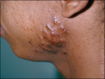
Cheeks are a common location for keloids, often secondary to acne.
Copyright © Logical Images, Inc.
Figure 2.
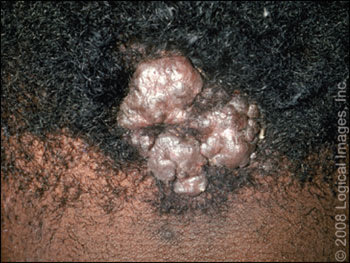
Mild trauma, often from shaving, can result in formation of a keloid, such as this one along the hairline.
Copyright © Logical Images, Inc.
Prevention
Before any surgical procedure, patients should be asked if they have had previous problems with scarring. Discuss the potential for keloids as part of informed consent, and discourage ear piercing and other elective procedures in persons with dark skin. If ears are pierced despite this advice, pressure earrings are commercially available for reducing keloid risk. If surgery cannot be avoided in a high-risk patient, immediate silicone elastomer sheeting or corticosteroid injections should be instituted. Anything that expedites wound healing and diminishes skin tension (e.g., postsurgical taping for 12 weeks) will diminish risk.10 The cosmetic outcome of wounds closed with standard suture techniques appears to be similar to that of those closed with 2-octyl cyanoacrylate dermal adhesive (Dermabond). One small study showed that hypertrophic scars occurred in five out of 24 repairs with Dermabond versus three out of 28 repairs with traditional suture.11
Treatment
Keloid and hypertrophic scar therapy is challenging and controversial (Table 2).1,7–9,12–21 Both conditions respond to the same therapies, but hypertrophic scars are easier to treat. The large number of treatment options is a reflection of the poor quality of research on this topic, with no single proven best treatment or combination of treatments. First-line options include silicone sheeting, pressure treatment, and corticosteroid injections, but all of these require exemplary adherence and follow-up. Cryotherapy is useful, but only for smaller lesions, such as those resulting from acne. Cryotherapy may cause hypopigmentation in patients with dark skin. Surgical removal of keloids, although temporarily gratifying, is almost invariably followed (50 to 100 percent) by even more aggressive regrowth of scar tissue.8 Therefore, all surgical options should be followed by corticosteroid injections, silicone sheeting, or these options combined with pulsed dye laser. A variety of other choices are emerging, but are less well studied.
Table 2 Prevention and Treatment Options for Keloids and Hypertrophic Scars
| Modality or treatment option | Response rate (%) | Recurrence rate (%) | Comments | Study design |
|---|---|---|---|---|
| Prevention | ||||
| Preventive silicone sheeting as postsurgery care | 0 to 75 | 25 to 36 | Multiple preparations available; tolerated by children | Review of multiple case studies8 |
| Expensive; should be avoided on open wounds; poor study designs | ||||
| Postsurgical intralesional corticosteroid injection (triamcinolone acetonide [Kenalog] 10 to 40 mg per mL at six-week intervals) | NA | 0 to 100 (mean 50) | Patient acceptance and safety | Review of multiple case studies9 |
| May cause hypopigmentation, skin atrophy, telangiectasia | ||||
| Postsurgical topical imiquimod 5% cream (Aldara) | NA | 28 | May cause hyperpigmentation, irritation | Case study12 |
| Postsurgical fluorouracil, triamcinolone acetonide, and pulsed dye laser (best outcomes) | 70 at 12 weeks | NA | Effective | Clinical trial13 |
| May cause hyperpigmentation, wound ulceration | ||||
| First-line treatment | ||||
| Cryotherapy | 50 to 76 | NA | Useful on small lesions; easy to perform | Review of multiple case studies9 |
| May cause hypopigmentation, pain | ||||
| Intralesional corticosteroid injection (triamcinolone acetonide 10 to 40 mg per mL at six-week intervals) | 50 to 100 | 9 to 50 | Inexpensive; available in family physician's office | Review of multiple case studies9 |
| Requires multiple injections | ||||
| May cause discomfort, skin atrophy, telangiectasia | ||||
| Silicone elastomer sheeting | 50 to 100 | NA | Multiple preparations available; tolerated by children | Review of multiple case studies8 |
| Expensive; poor study designs | ||||
| Pressure dressing (24 to 30 mm Hg) worn for six to 12 months | 90 to 100 | NA | Inexpensive | Review of multiple case studies9 |
| Difficult schedule; poor adherence | ||||
| Second-line and alternative treatment | ||||
| Surgical excision | NA | 50 to 100 | Z-plasty option for burns | Review of multiple case studies9 |
| Immediate postsurgical treatment needed to prevent regrowth | ||||
| Combined cryotherapy and intralesional corticosteroid injection | 84 | NA | See benefits of individual treatments | Case study1 |
| May cause hypopigmentation | ||||
| “Triple keloid therapy” (surgery, corticosteroids, and silicone sheeting) | 88 at 13 months | 12.5 at 13 months | Tedious; time intensive; expensive | Case study14 |
| Pulsed dye laser | NA | NA | Specialist referral needed; expensive; variable results depending on trial (controversial) | Case studies15,16 |
| Verapamil 2.5 mg per mL intralesional injection combined with perilesional excision and silicone sheeting | 54 at 18 months | NA | Repeated injections; limited experience | Clinical trial17 |
| May cause discomfort | ||||
| Fluorouracil 50 mg per mL intralesional injection two to three times per week | 88 | 0 | Effective | Review of multiple case studies7 |
| May cause hyperpigmentation, wound ulceration | ||||
| Bleomycin tattooing 1.5 IU per mL | 927 | NA | Effective | Review7 |
| 8819 | May cause pulmonary fibrosis, cutaneous reactions | Case study18 | ||
| Control trial19 | ||||
| Postsurgical interferon alfa-2b 1.5 million IU intralesional injection twice daily for four days | 30 to 50 | 8 to 19 | Expensive | Review of multiple case studies9 |
| May cause pruritus, altered pigmentation, pain | ||||
| Radiation therapy alone | 56 (mean) | NA | Local growth inhibition | Review of multiple case studies9 |
| May cause cancer, hyperpigmentation, paresthesias | ||||
| Postsurgical radiation therapy | 76 | NA | Local growth inhibition | Review of multiple case studies9,20 |
| May cause cancer | ||||
| Onion extract topical gels (e.g., Mederma) | NA | NA | Limited effect alone, better in combination with silicone sheeting | Prospective case study21 |
NA = not available.
Information from references 1, 7 through 9, and 12 through 21.
CORTICOSTEROID INJECTIONS
Corticosteroid injections for prevention and treatment of keloids and hypertrophic scars are perhaps the first-line option for family physicians. Corticosteroids suppress inflammation and mitosis while increasing vasoconstriction in the scar. Triamcinolone acetonide suspension (Kenalog) 10 to 40 mg per mL (depending on the site) is injected intralesionally, which, although painful, will eventually flatten 50 to 100 percent of keloids, with a 9 to 50 percent recurrence rate.9 Lidocaine (Xylocaine) may be combined with the corticosteroid to lessen pain, whereas using adjunctive cryotherapy immediately before injection may make the procedure easier by softening the scar (based on expert opinion).22 Combining cryotherapy and corticosteroid injections also improves outcomes more than either modality alone, although hypopigmentation is always a significant concern.23,24 Usually, two or three injections are given a month apart; however, therapy can continue for six months or longer.25 Newer keloids are more responsive to therapy than older, established lesions. Corticosteroid injections are more effective if combined with surgery; the sooner instituted, the greater the likelihood of success. Common adverse effects include atrophy, telangiectasias, and hypopigmentation.
SILICONE SHEETING
Silicone elastomer sheeting is a noninvasive and extensively studied approach to the prevention and treatment of keloids and hypertrophic scars. Silicone sheets are thought to work by increasing the temperature, hydration, and perhaps the oxygen tension of the occluded scar, causing it to soften and flatten.8 This technique should be avoided on open wounds, but can be applied as soon as the skin heals. More than 60 products have been marketed, including silicone sheets, strips, gels, sprays, and foams. Most are available over the counter, but can be expensive. To be effective, sheets must be worn over the scar for 12 to 24 hours per day for two to three months.8 The sheet and the scar should be washed daily with mild soap and water. The sheets can be reused until they start to disintegrate. Although most studies suggest silicone sheeting results in fewer scars in persons at risk, a recent Cochrane review concluded that most research in this area was of poor quality and highly susceptible to bias.26 Similar to silicone sheeting is the use of pressure dressings or garments, especially for the prevention of burn scars. However, pressure dressings (24 to 30 mm Hg) must be worn for six to 12 months, which is difficult and uncomfortable for most patients.27
COMBINATION THERAPY FOLLOWING SURGERY
If neither silicone nor corticosteroids are effective over 12 months, second-line surgical treatment followed by corticosteroids and possibly silicone sheeting should be considered. The use of corticosteroid injections following keloid surgery reduces the recurrence rate to lless than 50 percent.28 Scar excision may be complete, or a minute remnant of scar may be left on the wound margin, which may reduce recurrence (based on expert opinion). Immediate wound edge corticosteroid injection after the excision is followed by weekly injections for two to five weeks and monthly injections for three to six months.9 A “triple keloid therapy” combining surgery, corticosteroids, and silicone sheeting has been shown to be even more effective, with only a 12.5 percent recurrence rate after 13 months.14 However, this approach was described as tedious and time intensive by the author of the study and requires a motivated patient.
IMIQUIMOD
Imiquimod 5% cream (Aldara), an immune response modifier that enhances healing, has also been used to help prevent keloid recurrence after surgical excision. The cream is applied on alternate nights for eight weeks after surgery. Although the trials have been small, the postsurgical recurrence rate averaged only 28 percent over a six- to nine-month follow-up period, with best results (2.9 percent recurrence) in low skin tension areas such as earlobes.12 Adverse effects include irritation and hyperpigmentation.
PULSED DYE LASER
Treatment of keloids with short-pulsed, 585-nm pulsed dye laser has shown limited promise, with a 57 to 83 percent improvement rate.15 It is more vascular-specific than other laser therapies and appears to be most effective if used early and in conjunction with other techniques. Laser-treated portions of keloidal median sternotomy scars showed significant improvement in erythema, pruritus, and scar height compared with untreated portions of the same scars, and these improvements persisted for at least six months.16 The principal effect of a pulsed dye laser is on scar microvasculature, reducing erythema and pruritus and improving skin texture. The effectiveness of this therapy remains controversial, however, with other studies showing insignificant reduction in scar thickness.29 Disadvantages include significant expense and availability only through a specialist.
OTHER THERAPIES
Other therapies with limited studies include intralesional verapamil, fluorouracil, bleomycin, and interferon alfa-2b injections. Although all of these have results comparable or sometimes superior to corticosteroid injection and silicone sheeting, the optimal keloid therapy remains undefined. Combinations of therapies have proved superior to individual approaches.
Intralesional verapamil (2.5 mg per mL) in conjunction with silicone sheeting reduced keloid postsurgical recurrence by 90 percent at 18 months (54 percent of patients were keloid-free; 36 percent had partial success) compared with only 18 percent showing any improvement with silicone sheeting alone (no patients were keloid-free).17 Calcium antagonists appear to work by reducing collagen production and may be a reasonable and safe alternative to corticosteroid injection in the future.
Intralesional fluorouracil (50 mg per mL, two to three times per week) appears to shrink keloids safely while avoiding the tissue atrophy and telangiectasia that may occur with repeated corticosteroid injections.30 Combining fluorouracil with corticosteroid injections and pulsed dye laser produced superior results more rapidly than corticosteroid injections alone or corticosteroids with fluorouracil.13 Good to excellent responses at 12 weeks as rated by a blinded observer were 15 percent for triamcinolone acetonide, 40 percent for triamcinolone plus fluorouracil, and 70 percent for all three modalities (all significant). Combining corticosteroids and fluorouracil diminished the adverse effects of corticosteroids. Rare skin complications of fluorouracil may include hyperpigmentation and wound ulceration. No systemic adverse effects (e.g., anemia, leucopenia, thrombocytopenia) occurred in this study.
Bleomycin is another useful chemotherapeutic agent; a standard approach is bleomycin tattooing 0.1 mL (1.5 IU per mL) over two to six sessions, with a maximal dose of 6 mL.31 Results of one study showed a total regression of 84 percent.18 Multiple intralesional punctures are probably safe because it is likely that less than 5 percent of the dose ever reaches the bloodstream.18 Compared with triamcinolone injections combined with cryotherapy, bleomycin tattoo performed significantly better for keloids larger than 100 mm2 (P = .03).19 Systemically administered bleomycin is capable of causing pulmonary fibrosis (at doses greater than 400 U) and various cutaneous reactions (at doses of 200 to 300 U), including hair loss, hyperpigmentation, fibrosis, and vasospasm, any of which warrants cessation of treatment.32
Intralesional interferon alfa-2b (1.5 million IU twice daily for four days) reduced keloid size by 50 percent over nine days, proving superior to intralesional corticosteroids.31 Interferon alfa-2b was also more effective than corticosteroids for preventing keloid recurrence after excision. Injection pain and expense (about $100 per treatment) are the main concerns. A liposome-encapsulated interferon alfa-2b cream is also being investigated for scar reduction.33
Radiation, alone or (more commonly) after keloid excision, is a much more controversial option. It may pose a risk of local growth inhibition in children and possibly subsequent cancer. Commondosesrangebetween1,500to 2,000 rads over five to six sessions following surgery.28 The success rate for radiation alone is 56 percent (range of 10 to 94 percent), but this increases to 76 percent (range of 25 to 100 percent) if administered immediately after surgery.9 Another study showed a 67 percent success rate with radiation, increasing to 75 percent if delivered within 48 hours of surgery.20 Most physicians would reserve radiation as a last resort for keloids refractory to all other approaches.
OVER-THE-COUNTER TREATMENTS
Many patients use topical vitamin E (alpha-tocopherol) hoping its antioxidant properties will prevent scars. However, there is little evidence that it is helpful, and some patients develop a contact dermatitis that may delay healing.34 Used early on, vitamin E may also reduce the tensile strength of the scar, and its use should be discouraged.
Another over-the-counter option is onion extract topical gels (e.g., Mederma), but limited clinical trials have failed to demonstrate any clinical improvement in scar height, erythema, or pruritis.35,36 Contractubex gel (not available in the United States) contains onion extract with heparin, which is thought to promote scar maturity. Although one trial compared this product favorably with corticosteroids, another showed that it was ineffective in improving scar height and itching.21,37
Moist exposed burn ointment contains multiple herbs with betasitosterol, which provides hydration and possible benefits to wound healing.38 Another plant extract product contains Centella asiatica and Bulbine frutescens (Alpha Centella cream), which may increase wound strength if used in the first six to eight weeks.39 All of these commercially available products emphasize preventive use because they are unlikely to reverse well-established keloids.
