Background: Two ultrasound methods are used to diagnose suspected lower extremity deep venous thrombosis (DVT). The two-point strategy compresses the common femoral vein at the groin and the popliteal vein in the popliteal fossa. This method is easy to learn and apply, and is broadly available because it may be performed with almost any ultrasound scanner. However, if the test is initially normal, it may need to be repeated within one week.
The whole-leg ultrasound strategy examines the entire deep venous system and can better visualize small and calf veins. This method allows for one-test definitive diagnosis and has been assumed to be better because it can detect isolated calf DVT. Because this method requires specialized training and equipment, it may not be immediately available, potentially leading to delayed diagnosis. Bernardi and colleagues compared the effectiveness of the two diagnostic methods to exclude first episodes of suspected lower extremity DVT.
The Study: This multicenter, prospective, randomized controlled trial included 2,098 outpatients with symptoms of lower extremity DVT. Exclusion criteria included repeat DVT, current anticoagulation, pregnancy, and age younger than 18 years. Those randomized to the whole-leg protocol received calf-to-groin ultrasonography. If the evaluation was normal, the patients did not receive anticoagulation therapy and were not studied further. Those in the two-point arm who had normal ultrasonography then underwent d-dimer testing. If the d-dimer test was negative, no further testing was performed and the patient did not receive anticoagulation therapy. If the d-dimer result was abnormal, two-point ultrasonography was repeated within one week, and, if abnormal, the patients started anticoagulation therapy at that time (see accompanying figure). Participants with a normal evaluation were followed for up to three months by office visit or phone interview to assess the rates of symptomatic DVT in each study group.
Diagnosing Symptomatic Deep Venous Thrombosis
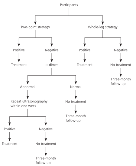
Figure. Algorithm for diagnosing suspected symptomatic deep venous thrombosis from a randomized controlled trial of two-point ultrasonography plus d-dimer versus whole-leg color-coded Doppler ultrasonography.
Results: Of the 1,045 patients randomized to the two-point strategy, 22.1 percent had abnormal findings on initial ultrasonography or on repeat testing after an abnormal d-dimer result. In the whole-leg strategy group, 26.4 percent of the 1,053 participants had abnormal ultrasound results, an absolute difference of 4.3 percent. Similarly low percentages were lost to follow-up in each group. There were few deaths in the two-point and whole-leg groups (1.1 and 0.9 percent, respectively), and none from pulmonary embolus. After three months, seven of 801 patients (0.9 percent) in the two-point group, and nine of 763 patients (1.2 percent) in the whole-leg group were diagnosed with venous thrombotic events; the difference (0.3 percent) fell within the equivalence limit.
Conclusion: Although the whole-leg method initially picks up more DVTs than the two-point strategy, the three-month outcome for those with negative test results is equivalent. This study shows that simpler, potentially more accessible two-point ultrasonography with or without a d-dimer method of diagnosing symptomatic lower extremity DVT is equivalent to more in-depth whole-leg ultrasonography.
