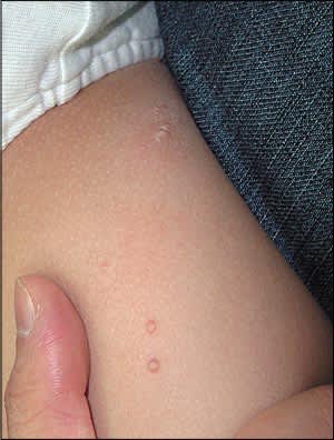
An otherwise healthy 18-month-old boy presented with asymptomatic papules on the inner aspect of the right thigh. The lesions appeared three months earlier and progressively increased in size. Physical examination revealed discrete, flesh-colored, dome-shaped papules (see accompanying figure) with central umbilication.
Question
Based on the patient's history and physical examination, which one of the following is the most likely diagnosis?
A. Common warts.
B. Molluscum contagiosum.
C. Papular acrodermatitis of childhood.
D. Papular urticaria.
E. Varicella.
Discussion
The answer is B: molluscum contagiosum. Discrete, smooth, pearly or flesh-colored, dome-shaped papules with central umbilication are characteristic of molluscum contagiosum. Although the lesions are usually cutaneous, they may appear in mucosal areas, such as the genital mucosa and conjunctiva.1 Lesions are typically 1 to 5 mm in diameter and number less than 20.2 Central umbilication may be difficult to observe in smaller lesions.1 In patients with immunodeficiency, the lesions can be extensive and larger in size.
Molluscum contagiosum is caused by a poxvirus of the Molluscipoxvirus genus. The virus is transmitted through close physical contact, autoinoculation, and fomites. The incubation period is two to seven weeks.3 Molluscum contagiosum occurs worldwide but is more common in warmer climates. The incidence is unknown. The condition usually occurs between two and 11 years of age,1 and boys are affected more often than girls. Although the condition occurs primarily in healthy persons, those with immunodeficiency, atopic disease, and Darier disease are at increased risk.1,2
Molluscum contagiosum lesions are asymptomatic and heal without scarring. Rare complications include secondary bacterial infection, conjunctivitis, and superficial punctate keratitis. Most lesions resolve spontaneously within six to nine months, but they may persist for years.
To decrease the risk of spreading the infection, patients should not share bath water or towels and should avoid swimming in public pools.1 Some suggest awaiting spontaneous resolution,2 but active treatment is usually recommended for cosmetic and epidemiologic reasons.1,4 Active treatment may be mechanical (e.g., curettage, cryotherapy with liquid nitrogen); chemical (e.g., tretinoin [Retin-A], cantharidin, podophyllotoxin); or immunologic (e.g., imiquimod [Aldara], cimetidine [Tagamet]).1,4
Common warts (verruca vulgaris) are dome-shaped lesions with an irregular surface. When the surface is pared away, characteristic punctate black dots become visible.
Papular acrodermatitis of childhood (Gianotti-Crosti syndrome) typically leads to symmetrical, erythematous, flat-topped papules. The papules appear in crops on the extremities.
Papular urticaria is intensively pruritic and is caused by hypersensitivity to insect bites. Lesions initially appear as wheals and progress to papules. Some lesions may have a central punctum.
The lesions of varicella appear in successive crops and consist of rose-colored macules, papules, vesicles, and pustules with crusting. The distribution of the lesions is typically central.
Summary Table
| Condition | Characteristics |
|---|---|
| Common warts (verruca vulgaris) | Dome-shaped lesions with irregular surface; punctate black dots visible when surface is pared away |
| Molluscum contagiosum | Discrete, smooth, pearly or flesh-colored, dome-shaped papules with central umbilication |
| Papular acrodermatitis of childhood (Gianotti-Crosti syndrome) | Symmetrical, erythematous, flat-topped papules appearing in crops on the extremities |
| Papular urticaria | Intensively pruritic lesions consisting initially of wheals and then papules; may have central punctum |
| Varicella | Lesions appear in successive crops; rose-colored macules, papules, vesicles, and pustules with crusting; distribution is typically central |
