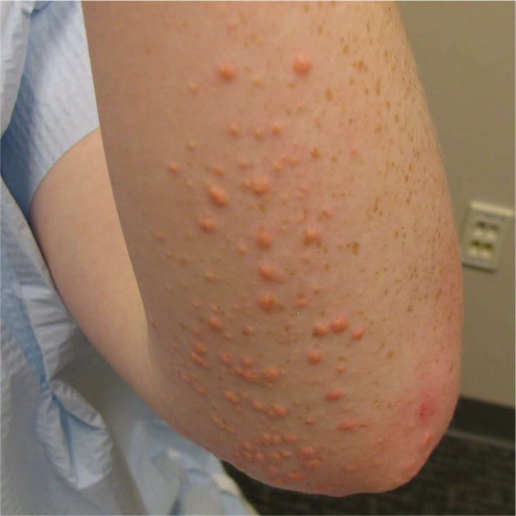
A 15-year-old boy presented with papules concentrated on his bilateral forearms, elbows, knees, and trunk that arose suddenly five weeks earlier. The patient reported that the lesions became pruritic after showering. He had no personal or family history of similar lesions and took no regular medications. He was obese and had a family history of hypercholesterolemia, but had no other notable medical history.
Physical examination revealed multiple erythematous-yellow, dome-shaped, non-follicular papules, some with an erythematous halo at the base (see accompanying figure). A few similar lesions were scattered on the abdomen and lower back. Some of the lesions were excoriated from scratching.
Question
Based on the patient's history and physical examination, which one of the following is the most likely diagnosis?
A. Eruptive xanthomas.
B. Juvenile xanthogranuloma.
C. Molluscum contagiosum.
D. Papular granuloma annulare.
Discussion
The answer is A: eruptive xanthomas. Eruptive xanthomas typically appear suddenly as multiple erythematous-yellow, dome-shaped papules on the extensor surfaces of the extremities, buttocks, and hands. The lesions are small (1 to 4 mm) and may have an inflamed base, or be tender or pruritic.1 Eruptive xanthomas sometimes koebnerize or appear following trauma.2
The condition may be the first presenting sign of an underlying lipid disorder and occurs almost exclusively in the setting of hypertriglyceridemia, with triglyceride levels usually in the thousands. Hypertriglyceridemia can be primary, as with familial hyperchylomicronemia or endogenous familial hypertriglyceridemia; or secondary, as with type 2 diabetes mellitus. The patient's lipid panel showed a triglyceride level of 553 mg per dL (6.25 mmol per L), low-density lipoprotein cholesterol level of 163 mg per dL (4.22 mmol per L), and high-density lipoprotein cholesterol level of 36 mg per dL (0.93 mmol per L). A1C and thyroid test results were normal.
Other factors associated with eruptive xanthomas include obesity, alcohol abuse, estrogen therapy, and retinoid therapy. These factors may also exacerbate primary hypertriglyceridemia.1 Biopsy is necessary to distinguish eruptive xanthomas from other causes of multiple papules. Biopsy of eruptive xanthomas demonstrates foamy macrophages and dermal extracellular lipids. Treatment is directed at the underlying cause. Dietary modifications and medications that lower serum lipid levels hasten the resolution of lesions.1
Juvenile xanthogranuloma, a non-Langerhans cell histiocytosis, typically appears in young children and infants. There are two clinical variants, a small nodular form and a large nodular form. The small nodular form consists of several pink or reddish-brown, dome-shaped papules that are 2 to 5 mm in diameter and distributed diffusely on the upper body. The large nodular form is associated with an isolated red-brown nodule on the head or neck. Juvenile xanthogranuloma is not associated with an underlying lipid disorder, however. Multiple lesions are associated with an increased risk of chronic myelogenous leukemia, although juvenile xanthogranuloma is usually self-limited with no sequelae.3
The molluscum contagiosum virus is part of the Poxviridae family. Molluscum contagiosum infection manifests as individual or grouped, pink-white, centrally umbilicated, dome-shaped papules that tend to occur in areas of friction (e.g., axillae, flank, genitals). They may become pustular or inflammatory and are highly contagious. The infection is spread through touching, autoinoculation, or trauma. Molluscum contagiosum infection resolves on its own, but is often treated to prevent spread or for cosmetic reasons using cryotherapy, imiquimod (Aldara), cantharidin, curettage, or topical retinoids.4
Papular granuloma annulare is a morphologic variant of granuloma annulare, which is a granulomatous dermatitis of unknown etiology. The condition occurs on the dorsal hands, fingers, and elbows as small, firm, pink-white papules. The lesions are typically isolated, but generalized outbreaks are possible. Papular granuloma annulare is usually asymptomatic and resolves on its own.5
Summary Table
| Condition | Characteristics |
|---|---|
| Eruptive xanthomas | Diffuse erythematous-yellow, dome-shaped papules with an inflamed base; appears on extensor surfaces of extremities, buttocks, and hands |
| Juvenile xanthogranuloma | Small nodular form: several pink or reddish-brown, domed-shaped papules distributed diffusely on the upper body |
| Large nodular form: isolated reddish-brown nodule on head or neck | |
| Molluscum contagiosum | Individual or grouped, pink-white, centrally umbilicated, dome-shaped papules; tends to occur on areas of friction, such as axillae, flank, and genitals |
| Papular granuloma annulare | Small, firm, pink-white, dome-shaped papules on dorsal hands, fingers, elbows; usually isolated |
