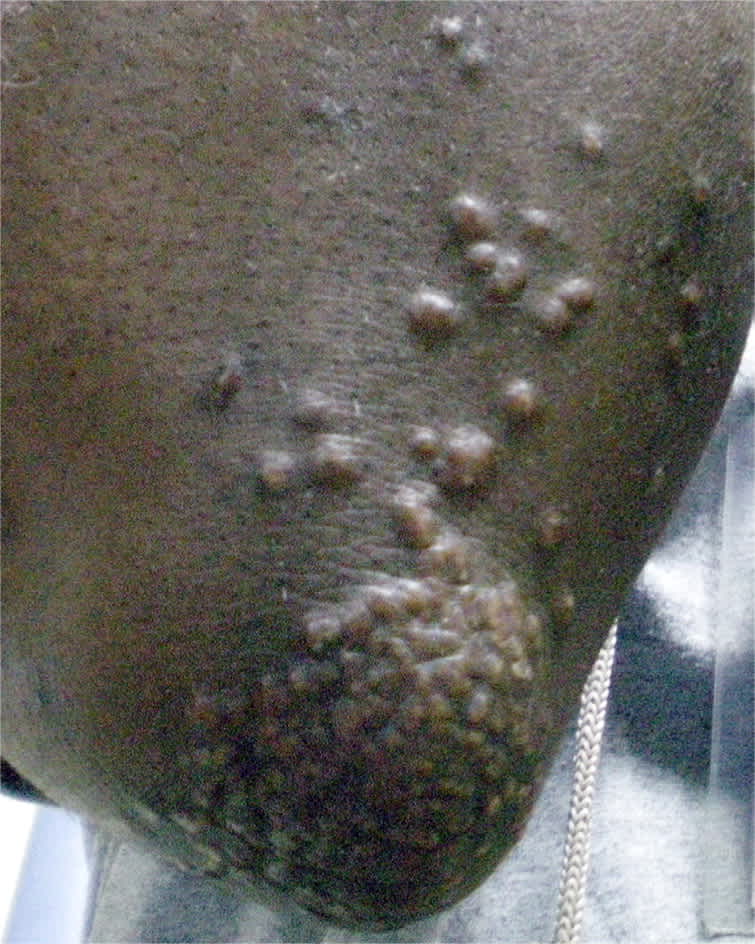
A 33-year-old man presented with a skin eruption that had appeared three months earlier. Examination revealed yellowish papules over the elbows, buttocks, knees, flank, and palmar creases (see accompanying figure). The lesions were 3 to 7 mm and firm to palpation. The patient had no significant medical history.
Question
Based on the patient's history and physical examination, which one of the following is the most likely diagnosis?
A. Cellulitis.
B. Eruptive xanthoma.
C. Psoriasis.
D. Smallpox.
E. Varicella.
Discussion
The answer is B: eruptive xanthoma. Xanthomas are localized lipid deposits in the skin, tendons, and subcutaneous tissue associated with a lipid abnormality. They are clinically classified as eruptive, planar, tuberous, or tendinous. Eruptive xanthomas are the most common type and appear as crops of discrete yellowish papules that may have erythematous bases. They often appear in areas subject to pressure or trauma, such as the buttocks, extensor surfaces, and flexural creases.1,2
Eruptive xanthomas are associated with hypertriglyceridemia and chylomicronemia due to a genetic disorder (primary hyperlipoproteinemia), or an underlying disease process (secondary hyperlipoproteinemia), such as diabetes mellitus, hypothyroidism, nephrotic syndrome, pancreatitis, or retinoid or estrogen therapy.3,4 Although eruptive xanthomas are usually associated with hyperlipoproteinemia, they can occur in normolipemic patients with local trauma.5–8 The condition may be associated with ophthalmologic and gastrointestinal findings, such as lipemia retinalis (salmon-colored retina with creamy white retinal vessels), abdominal pain, and hepatosplenomegaly.
Diagnosis of eruptive xanthoma includes measuring fasting blood glucose and lipid levels, including triglycerides, cholesterol, low-density lipoprotein, very-low-density lipoprotein, and high-density lipoprotein. The patient's biopsy result demonstrated a dermal infiltrate of foamy macrophages, with lymphocytes, and neutrophils, which is suggestive of eruptive xanthoma. Fasting cholesterol was greater than 1,000 mg per dL (25.90 mmol per L), and triglycerides were greater than 3,500 mg per dL (39.55 mmol per L). Treatment involves dietary restrictions and medications to control the hyperlipidemia.
Cellulitis is a bacterial infection. Fever is uncommon, but may be present. The rash appears as pustules and is red, warm to the touch, and can be tender to palpation.
Although psoriasis can occur over the extensor surfaces, it usually appears as erythematous, scaly plaques. Psoriasis is most common on the elbows and knees, but can also occur on the palms, soles, scalp, and back.
Smallpox may be similar to xanthomas in size and shape, but are vesicular and in the same stage of development. Smallpox lead to small red spots on the mucous membranes (enanthem).
Varicella usually causes vesicles on an erythematous base, which is the classic “dew drop on a rose petal” appearance. Patients have a viral prodrome and develop the rash over one week.
Summary Table
| Condition | Characteristics |
|---|---|
| Cellulitis | Bacterial infection; rash includes pustules and is red, warm to the touch, and can be tender |
| Eruptive xanthoma | Crops of discrete yellowish papules that may have erythematous bases; often appear in areas subject to pressure or trauma, such as the buttocks, extensor surfaces, and flexural creases |
| Psoriasis | Erythematous, scaly plaques, usually on the elbows and knees |
| Smallpox | Vesicles or pustules similar to eruptive xanthomas, but all in the same stage of development; small red spots on the mucous membranes (enanthem) are also present |
| Varicella | Vesicles on an erythematous base (“dew drop on a rose petal”); viral prodrome; rash develops over one week |
