
A more recent version of evaluation and treatment of nausea and vomiting is available.
This is a corrected version of the article that appeared in print.
Am Fam Physician. 2013;88(6):371-379
Author disclosure: No relevant financial affiliations.
In the absence of acute abdominal pain, significant headache, or recent initiation of certain medications, acute nausea and vomiting is usually the result of self-limited gastrointestinal infections. Nausea and vomiting is also a common adverse effect of radiation therapy, chemotherapy, and surgical anesthesia. Other potential diagnoses include endocrine conditions (including pregnancy), central nervous system disorders, psychiatric causes, toxin exposure, metabolic abnormalities, and obstructive or functional gastrointestinal causes. The likely cause of acute nausea and vomiting can usually be determined by history and physical examination. Alarm signs such as dehydration, acidosis caused by an underlying metabolic disorder, or an acute abdomen warrant additional evaluation. Based on the suspected diagnosis, basic laboratory testing may include urinalysis, urine pregnancy testing, complete blood count, comprehensive metabolic panel, amylase and lipase levels, thyroid-stimulating hormone level, and stool studies with cultures. Imaging studies include abdominal radiography, ultrasonography, and computed tomography. Computed tomography of the head should be performed if an acute intracranial process is suspected. Chronic nausea and vomiting is defined by symptoms that persist for at least one month. Patients with risk factors for gastric malignancies or alarm symptoms should be evaluated with esophagogastroduodenoscopy. If gastroparesis is suspected, a gastric emptying study is recommended. In addition to functional causes, it is also important to consider psychiatric causes when evaluating patients with chronic nausea and vomiting.
Nausea and vomiting is a common presenting symptom in primary care. Diagnostic and management strategies vary depending on the duration of symptoms. This article addresses acute and chronic nausea and vomiting, with illustrative cases.
| Clinical recommendation | Evidence rating | References |
|---|---|---|
| Findings from the history and physical examination should guide diagnostic testing in patients with nausea and vomiting. | C | 18 |
| Acute nausea and vomiting in the absence of alarm symptoms (e.g., altered mental status, abdominal pain, hematochezia, melena, focal neurologic deficit) may initially be treated supportively. | C | 31 |
| Ginger is effective at relieving nausea in pregnant women. | A | 36, 37 |
Acute Nausea and Vomiting
ILLUSTRATIVE CASE
A 49-year-old woman with type 2 diabetes mellitus presents with a three-day history of acute-onset nausea and intermittent vomiting. She rapidly becomes nauseous when eating solid food. She does not have fever, chills, abdominal pain, diarrhea, hematochezia, melena, or constipation. Over-the-counter antacids have been ineffective. Blood glucose measurements taken at home have been less than 180 mg per dL (10.0 mmol per L).
PRESENTATION
The duration of nausea and vomiting, associated symptoms, and alleviating and exacerbating factors can help determine the likely cause (Table 1).1–4 [ corrected] Physicians should ask about exposure to toxins; suspect food; sick contacts; and recent radiation therapy, surgery, or chemotherapy. The absence of significant abdominal pain, headaches, and other alarm signs or symptoms can narrow the differential diagnosis.
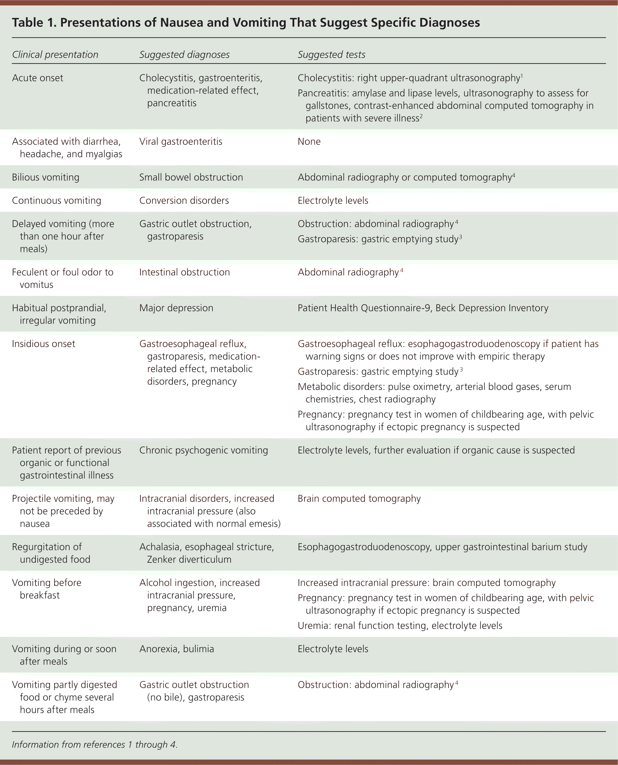
| Clinical presentation | Suggested diagnoses | Suggested tests |
|---|---|---|
| Acute onset | Cholecystitis, gastroenteritis, medication-related effect, pancreatitis | Cholecystitis: right upper-quadrant ultrasonography1 |
| Pancreatitis: amylase and lipase levels, ultrasonography to assess for gallstones, contrast-enhanced abdominal computed tomography in patients with severe illness2 | ||
| Associated with diarrhea, headache, and myalgias | Viral gastroenteritis | None |
| Bilious vomiting | Small bowel obstruction | Abdominal radiography or computed tomography4 |
| Continuous vomiting | Conversion disorders | Electrolyte levels |
| Delayed vomiting (more than one hour after meals) | Gastric outlet obstruction, gastroparesis | Obstruction: abdominal radiography4 |
| Gastroparesis: gastric emptying study3 | ||
| Feculent or foul odor to vomitus | Intestinal obstruction | Abdominal radiography4 |
| Habitual postprandial, irregular vomiting | Major depression | Patient Health Questionnaire-9, Beck Depression Inventory |
| Insidious onset | Gastroesophageal reflux, gastroparesis, medication-related effect, metabolic disorders, pregnancy | Gastroesophageal reflux: esophagogastroduodenoscopy if patient has warning signs or does not improve with empiric therapy |
| Gastroparesis: gastric emptying study3 | ||
| Metabolic disorders: pulse oximetry, arterial blood gases, serum chemistries, chest radiography | ||
| Pregnancy: pregnancy test in women of childbearing age, with pelvic ultrasonography if ectopic pregnancy is suspected | ||
| Patient report of previous organic or functional gastrointestinal illness | Chronic psychogenic vomiting | Electrolyte levels, further evaluation if organic cause is suspected |
| Projectile vomiting, may not be preceded by nausea | Intracranial disorders, increased intracranial pressure (also associated with normal emesis) | Brain computed tomography |
| Regurgitation of undigested food | Achalasia, esophageal stricture, Zenker diverticulum | Esophagogastroduodenoscopy, upper gastrointestinal barium study |
| Vomiting before breakfast | Alcohol ingestion, increased intracranial pressure, pregnancy, uremia | Increased intracranial pressure: brain computed tomography |
| Pregnancy: pregnancy test in women of childbearing age, with pelvic ultrasonography if ectopic pregnancy is suspected | ||
| Uremia: renal function testing, electrolyte levels | ||
| Vomiting during or soon after meals | Anorexia, bulimia | Electrolyte levels |
| Vomiting partly digested food or chyme several hours after meals | Gastric outlet obstruction (no bile), gastroparesis | Obstruction: abdominal radiography4 |
Assessment of the patient's hydration status and vital signs can help determine the severity of the illness and whether outpatient therapy is appropriate. The physical examination should evaluate for acute abdominal or intracranial processes and other causes (Table 2).5–16 [ corrected] If musculoskeletal pain is suspected, an increase in abdominal pain with tensing of abdominal muscles (i.e., Carnett sign) suggests that the abdominal wall is the source.17 The absence of severe abdominal pain and hematochezia excludes gastrointestinal malignancy (negative predictive value = 99%) and gastrointestinal ulcers (negative predictive value = 97%).8 Conversely, abdominal pain relieved by vomiting suggests bowel obstruction (specificity = 94%).15
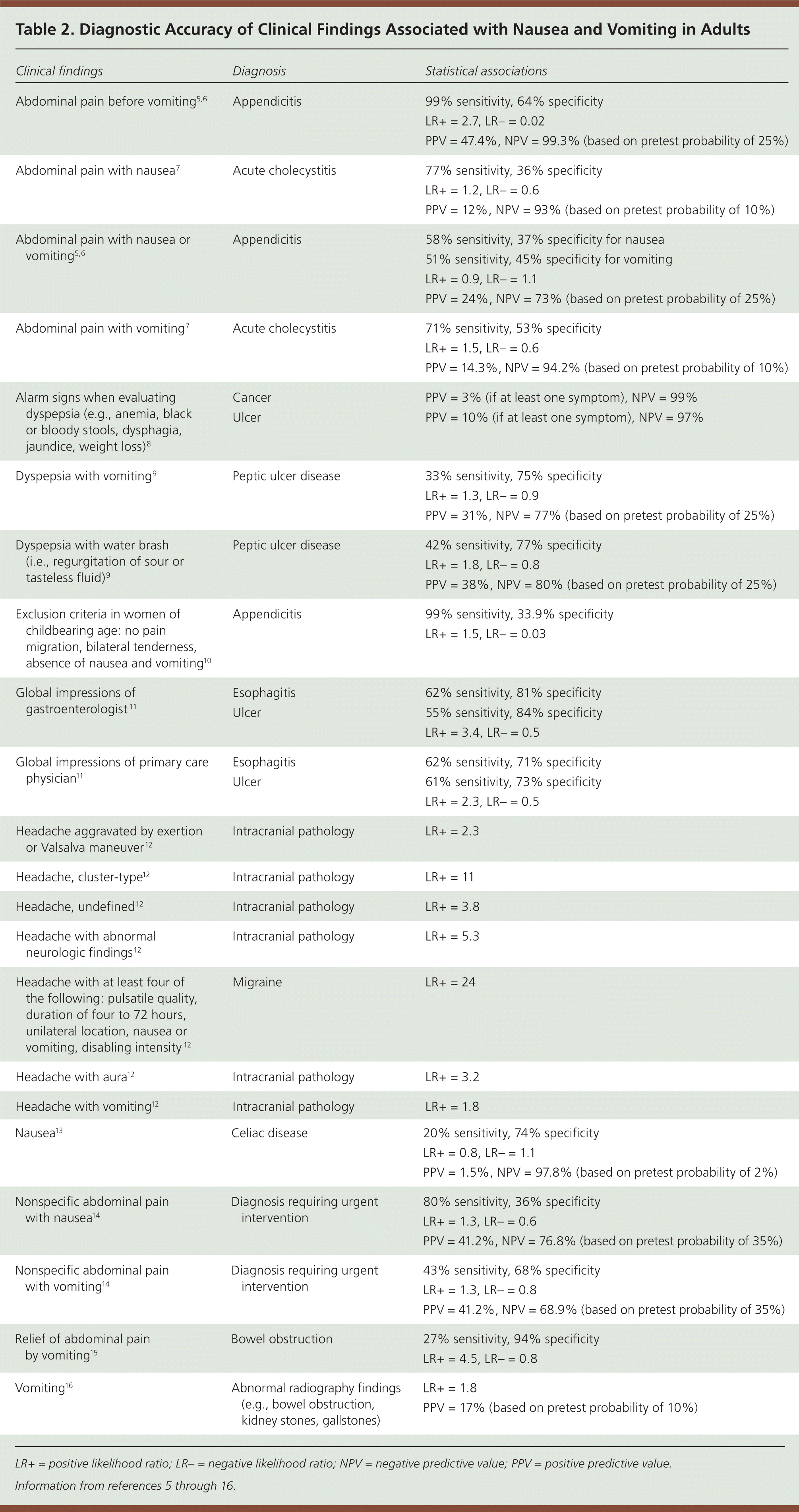
| Clinical findings | Diagnosis | Statistical associations | |
|---|---|---|---|
| Abdominal pain before vomiting5,6 | Appendicitis | 99% sensitivity, 64% specificity | |
| LR+ = 2.7, LR– = 0.02 | |||
| PPV = 47.4%, NPV = 99.3% (based on pretest probability of 25%) | |||
| Abdominal pain with nausea7 | Acute cholecystitis | 77% sensitivity, 36% specificity | |
| LR+ = 1.2, LR– = 0.6 | |||
| PPV = 12%, NPV = 93% (based on pretest probability of 10%) | |||
| Abdominal pain with nausea or vomiting5,6 | Appendicitis | 58% sensitivity, 37% specificity for nausea | |
| 51% sensitivity, 45% specificity for vomiting | |||
| LR+ = 0.9, LR– = 1.1 | |||
| PPV = 24%, NPV = 73% (based on pretest probability of 25%) | |||
| Abdominal pain with vomiting7 | Acute cholecystitis | 71% sensitivity, 53% specificity | |
| LR+ = 1.5, LR– = 0.6 | |||
| PPV = 14.3%, NPV = 94.2% (based on pretest probability of 10%) | |||
| Alarm signs when evaluating dyspepsia (e.g., anemia, black or bloody stools, dysphagia, jaundice, weight loss)8 | Cancer | PPV = 3% (if at least one symptom), NPV = 99% | |
| Ulcer | PPV = 10% (if at least one symptom), NPV = 97% | ||
| Dyspepsia with vomiting9 | Peptic ulcer disease | 33% sensitivity, 75% specificity | |
| LR+ = 1.3, LR– = 0.9 | |||
| PPV = 31%, NPV = 77% (based on pretest probability of 25%) | |||
| Dyspepsia with water brash (i.e., regurgitation of sour or tasteless fluid)9 | Peptic ulcer disease | 42% sensitivity, 77% specificity | |
| LR+ = 1.8, LR– = 0.8 | |||
| PPV = 38%, NPV = 80% (based on pretest probability of 25%) | |||
| Exclusion criteria in women of childbearing age: no pain migration, bilateral tenderness, absence of nausea and vomiting10 | Appendicitis | 99% sensitivity, 33.9% specificity | |
| LR+ = 1.5, LR– = 0.03 | |||
| Global impressions of gastroenterologist11 | Esophagitis | 62% sensitivity, 81% specificity | |
| Ulcer | 55% sensitivity, 84% specificity | ||
| LR+ = 3.4, LR– = 0.5 | |||
| Global impressions of primary care physician11 | Esophagitis | 62% sensitivity, 71% specificity | |
| Ulcer | 61% sensitivity, 73% specificity | ||
| LR+ = 2.3, LR– = 0.5 | |||
| Headache aggravated by exertion or Valsalva maneuver12 | Intracranial pathology | LR+ = 2.3 | |
| Headache, cluster-type12 | Intracranial pathology | LR+ = 11 | |
| Headache, undefined12 | Intracranial pathology | LR+ = 3.8 | |
| Headache with abnormal neurologic findings12 | Intracranial pathology | LR+ = 5.3 | |
| Headache with at least four of the following: pulsatile quality, duration of four to 72 hours, unilateral location, nausea or vomiting, disabling intensity12 | Migraine | LR+ = 24 | |
| Headache with aura12 | Intracranial pathology | LR+ = 3.2 | |
| Headache with vomiting12 | Intracranial pathology | LR+ = 1.8 | |
| Nausea13 | Celiac disease | 20% sensitivity, 74% specificity | |
| LR+ = 0.8, LR– = 1.1 | |||
| PPV = 1.5%, NPV = 97.8% (based on pretest probability of 2%) | |||
| Nonspecific abdominal pain with nausea14 | Diagnosis requiring urgent intervention | 80% sensitivity, 36% specificity | |
| LR+ = 1.3, LR– = 0.6 | |||
| PPV = 41.2%, NPV = 76.8% (based on pretest probability of 35%) | |||
| Nonspecific abdominal pain with vomiting14 | Diagnosis requiring urgent intervention | 43% sensitivity, 68% specificity | |
| LR+ = 1.3, LR– = 0.8 | |||
| PPV = 41.2%, NPV = 68.9% (based on pretest probability of 35%) | |||
| Relief of abdominal pain by vomiting15 | Bowel obstruction | 27% sensitivity, 94% specificity | |
| LR+ = 4.5, LR– = 0.8 | |||
| Vomiting16 | Abnormal radiography findings (e.g., bowel obstruction, kidney stones, gallstones) | LR+ = 1.8 | |
| PPV = 17% (based on pretest probability of 10%) | |||
Table 3 lists common and uncommon causes of acute nausea and vomiting.18 Self-limited viral gastroenteritis is the most common cause.19 Approximately 179 million episodes of acute gastroenteritis occur each year in the United States and result in roughly 600,000 hospitalizations. Although this illness typically resolves in three to five days, it results in significant time lost from work and accounts for an estimated $1 billion per year in direct and indirect costs.20 Only 20% of acute gastroenteritis cases are attributed to a specific etiology.21–23 Viruses are the most common cause; norovirus is the most common in adults.
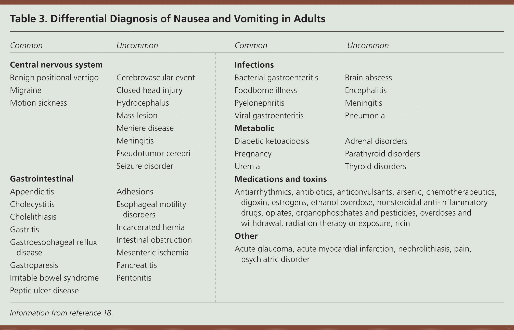
| Central nervous system |
| Common |
| Benign positional vertigo |
| Migraine |
| Motion sickness |
| Uncommon |
| Cerebrovascular event |
| Closed head injury |
| Hydrocephalus |
| Mass lesion |
| Meniere disease |
| Meningitis |
| Pseudotumor cerebri |
| Seizure disorder |
| Gastrointestinal |
| Common |
| Appendicitis |
| Cholecystitis |
| Cholelithiasis |
| Gastritis |
| Gastroesophageal reflux disease |
| Gastroparesis |
| Irritable bowel syndrome |
| Peptic ulcer disease |
| Uncommon |
| Adhesions |
| Esophageal motility disorders |
| Incarcerated hernia |
| Intestinal obstruction |
| Mesenteric ischemia |
| Pancreatitis |
| Peritonitis |
| Infections |
| Common |
| Bacterial gastroenteritis |
| Foodborne illness |
| Pyelonephritis |
| Viral gastroenteritis |
| Uncommon |
| Brain abscess |
| Encephalitis |
| Meningitis |
| Pneumonia |
| Metabolic |
| Common |
| Diabetic ketoacidosis |
| Pregnancy |
| Uremia |
| Uncommon |
| Adrenal disorders |
| Parathyroid disorders |
| Thyroid disorders |
| Medications and toxins |
| Antiarrhythmics, antibiotics, anticonvulsants, arsenic, chemotherapeutics, digoxin, estrogens, ethanol overdose, nonsteroidal anti-inflammatory drugs, opiates, organophosphates and pesticides, overdoses and withdrawal, radiation therapy or exposure, ricin |
| Other |
| Acute glaucoma, acute myocardial infarction, nephrolithiasis, pain, psychiatric disorder |
DIAGNOSTIC STRATEGY
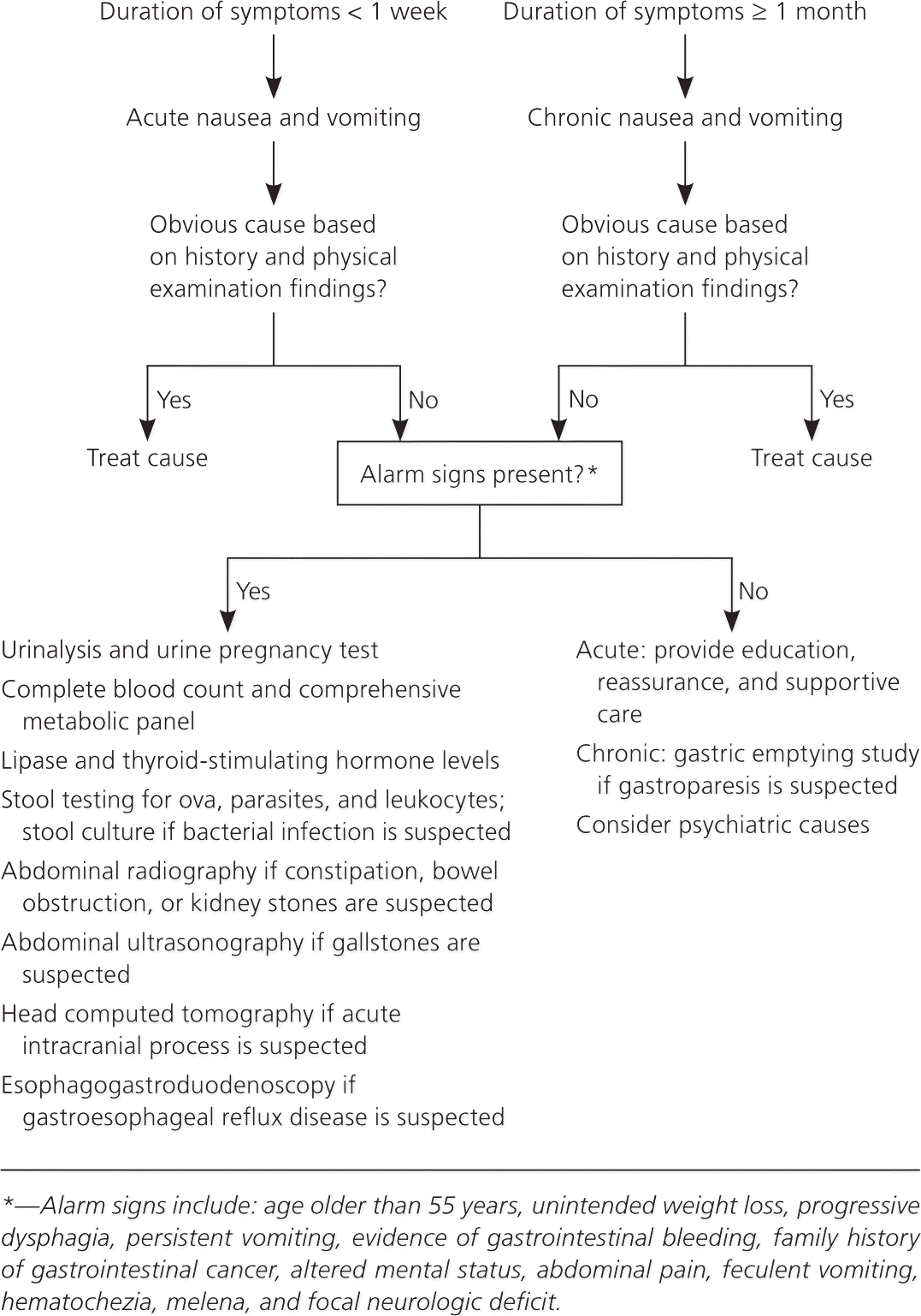
Figure 1 outlines a suggested approach to the evaluation of nausea and vomiting in adults. Most conditions can be diagnosed by findings from the history and physical examination. Diagnostic testing may be warranted in patients with signs of significant dehydration (e.g., decreased urine output, skin tenting, dry mucous membranes), signs of acidosis caused by diabetic ketoacidosis or another underlying disorder (e.g., markedly increased respiratory rate, fruity odor to breath, altered mental status), severe abdominal pain or distension, hematochezia, jaundice, melena, severe headache, urinary tract infection symptoms, abdominal pain relieved by vomiting, hematemesis, or feculent vomiting.18 If any of these signs or symptoms are present, initial evaluation and testing can be guided by the suggested diagnoses in Table 1.1–4
IMAGING
Abdominal radiography is helpful in detecting bowel obstructions and kidney stones.4 Computed tomography of the abdomen is useful for detecting infections (e.g., appendicitis, cholecystitis) and for additional testing for bowel obstruction and kidney stones that are not detected on radiography.24 In adults, abdominal radiography combined with clinical examination and laboratory analysis (complete blood count and basic metabolic panel) is useful for predicting the need for urgent intervention in the first 24 hours of illness (sensitivity = 56%; specificity = 81%).4 Table 4 lists the accuracy of diagnostic tests in adults with nausea and vomiting.1,2,25–29 [ corrected]
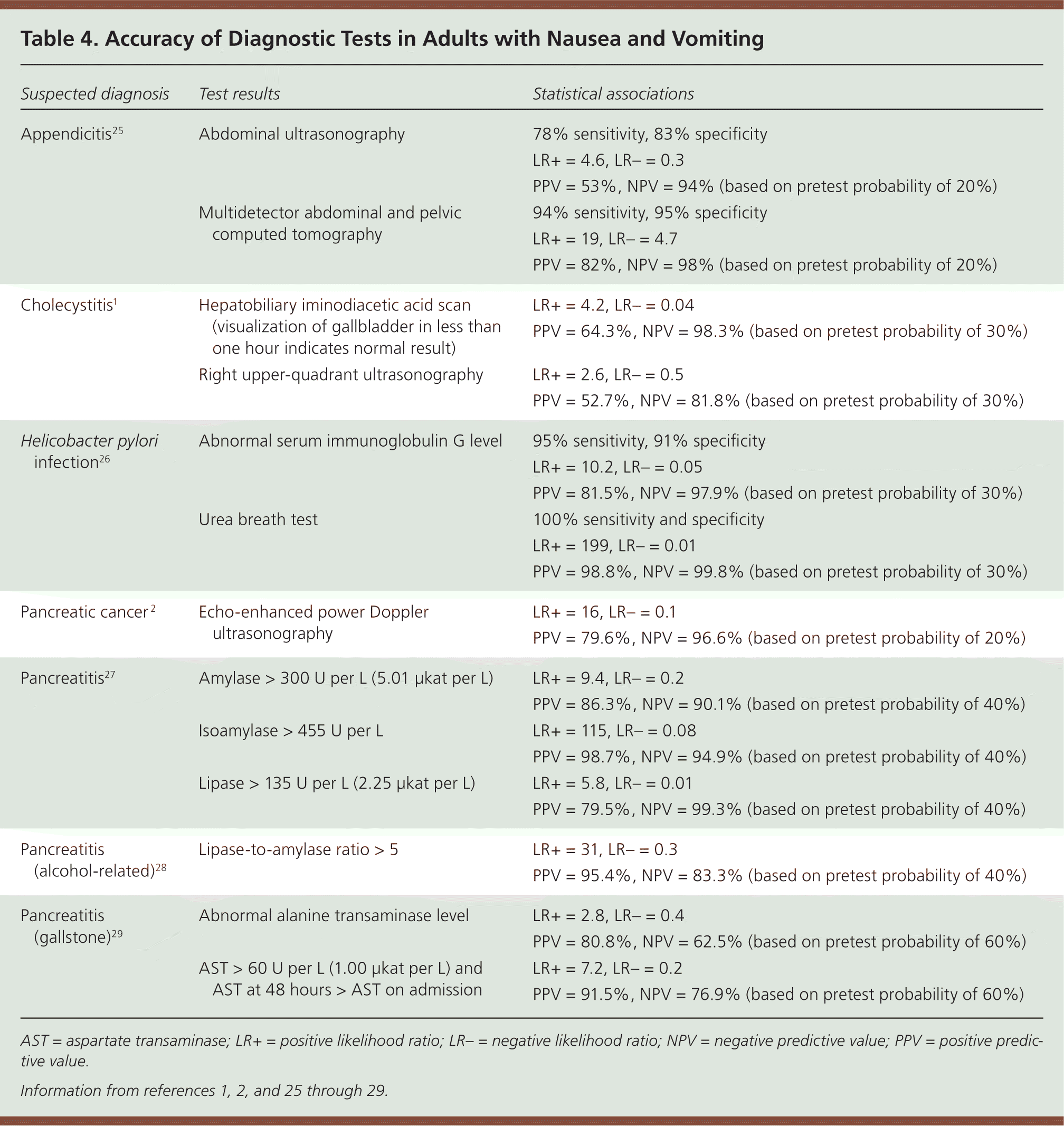
| Suspected diagnosis | Test results | Statistical associations |
|---|---|---|
| Appendicitis25 | Abdominal ultrasonography | 78% sensitivity, 83% specificity |
| LR+ =4.6, LR– = 0.3 | ||
| PPV = 53%, NPV = 94% (based on pretest probability of 20%) | ||
| Multidetector abdominal and pelvic computed tomography | 94% sensitivity, 95% specificity | |
| LR+ = 19, LR– = 4.7 | ||
| PPV = 82%, NPV = 98% (based on pretest probability of 20%) | ||
| Cholecystitis1 | Hepatobiliary iminodiacetic acid scan (visualization of gallbladder in less than one hour indicates normal result) | LR+ = 4.2, LR– = 0.04 |
| PPV = 64.3%, NPV = 98.3% (based on pretest probability of 30%) | ||
| Right upper-quadrant ultrasonography | LR+ = 2.6, LR– = 0.5 | |
| PPV = 52.7%, NPV = 81.8% (based on pretest probability of 30%) | ||
| Helicobacter pylori infection26 | Abnormal serum immunoglobulin G level | 95% sensitivity, 91% specificity |
| LR+ = 10.2, LR– = 0.05 | ||
| PPV = 81.5%, NPV = 97.9% (based on pretest probability of 30%) | ||
| Urea breath test | 100% sensitivity and specificity | |
| LR+ = 199, LR– = 0.01 | ||
| PPV = 98.8%, NPV = 99.8% (based on pretest probability of 30%) | ||
| Pancreatic cancer2 | Echo-enhanced power Doppler ultrasonography | LR+ = 16, LR– = 0.1 |
| PPV = 79.6%, NPV = 96.6% (based on pretest probability of 20%) | ||
| Pancreatitis27 | Amylase > 300 U per L (5.01 μkat per L) | LR+ = 9.4, LR– = 0.2 |
| PPV = 86.3%, NPV = 90.1% (based on pretest probability of 40%) | ||
| Isoamylase > 455 U per L | LR+ = 115, LR– = 0.08 | |
| PPV = 98.7%, NPV = 94.9% (based on pretest probability of 40%) | ||
| Lipase > 135 U per L (2.25 μkat per L) | LR+ = 5.8, LR– = 0.01 | |
| PPV = 79.5%, NPV = 99.3% (based on pretest probability of 40%) | ||
| Pancreatitis (alcohol-related)28 | Lipase-to-amylase ratio > 5 | LR+ = 31, LR– = 0.3 |
| PPV = 95.4%, NPV = 83.3% (based on pretest probability of 40%) | ||
| Pancreatitis (gallstone)29 | Abnormal alanine transaminase level | LR+ = 2.8, LR– = 0.4 |
| PPV = 80.8%, NPV = 62.5% (based on pretest probability of 60%) | ||
| AST > 60 U per L (1.00 μkat per L) and AST at 48 hours > AST on admission | LR+ = 7.2, LR– = 0.2 | |
| PPV = 91.5%, NPV = 76.9% (based on pretest probability of 60%) |
Right upper-quadrant ultrasonography is used to evaluate for gallstones. Hepatobiliary iminodiacetic acid scans can determine whether delayed gallbladder emptying is the cause of nausea and abdominal pain when initial ultrasonography is negative.
Migraine should be diagnosed in patients who have headaches with at least four of the following characteristics: pulsatile quality, duration of four to 72 hours, unilateral location, nausea or vomiting, and disabling intensity (likelihood ratio = 24).12 Cluster-type headaches, headaches with abnormal neurologic findings, undefined headaches, and headaches aggravated by exertion or the Valsalva maneuver are more likely to have associated intracranial pathology (likelihood ratio = 11, 5, 4, and 2, respectively).12 Computed tomography or magnetic resonance imaging of the brain should be ordered for patients with these symptoms, and in those with other abnormal neurologic signs or symptoms.
MANAGEMENT
For the patient described previously, the focus should be on determining the severity of illness from the history and physical examination findings. Treatment should be supportive, including limiting foods likely to trigger vomiting and providing antiemetics with an appropriate hydration strategy. Patients with mild to moderate dehydration may benefit from oral rehydration therapy (i.e., a combination of electrolytes, glucose, and water) or sports drinks.30
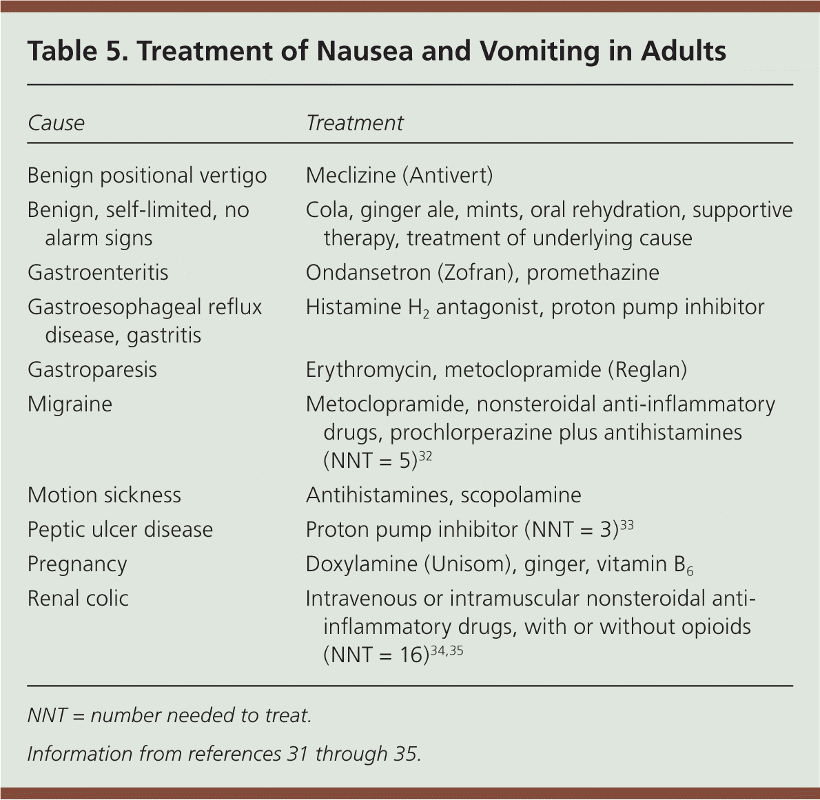
| Cause | Treatment |
|---|---|
| Benign positional vertigo | Meclizine (Antivert) |
| Benign, self-limited, no alarm signs | Cola, ginger ale, mints, oral rehydration, supportive therapy, treatment of underlying cause |
| Gastroenteritis | Ondansetron (Zofran), promethazine |
| Gastroesophageal reflux disease, gastritis | Histamine H2 antagonist, proton pump inhibitor |
| Gastroparesis | Erythromycin, metoclopramide (Reglan) |
| Migraine | Metoclopramide, nonsteroidal anti-inflammatory drugs, prochlorperazine plus antihistamines (NNT = 5)32 |
| Motion sickness | Antihistamines, scopolamine |
| Peptic ulcer disease | Proton pump inhibitor (NNT = 3)33 |
| Pregnancy | Doxylamine (Unisom), ginger, vitamin B6 |
| Renal colic | Intravenous or intramuscular nonsteroidal anti-inflammatory drugs, with or without opioids (NNT = 16)34,35 |
Chronic Nausea and Vomiting
ILLUSTRATIVE CASE
The patient described previously returns two months later with intermittent nausea and vomiting that is much less severe than on initial presentation. Her current symptoms have lasted two days, and she has had six bouts of emesis. She has no fever, chills, or headache, but has intermittent epigastric discomfort associated with nausea and vomiting. Her symptoms are not relieved by antacids, and she has no melena or blood in her stool. She says she feels full quickly when eating and often feels bloated. She has not had contact with any sick persons or toxins, does not drink alcohol, and appears well hydrated.
BACKGROUND
Chronic nausea and vomiting is defined by symptoms that persist for at least one month. A history and physical examination can help determine the most likely cause. A food and nausea diary may help determine patterns of symptoms and triggers. Diagnostic testing, including imaging and laboratory evaluation, may be indicated based on history and physical examination findings. Esophagogastroduodenoscopy may be necessary depending on the course and the results of diagnostic testing.
DIAGNOSTIC STRATEGY
Diabetes-associated gastroparesis, gastroesophageal reflux disease, gastritis, and gastric ulcer are common causes of chronic nausea and vomiting. Initial testing should be guided by findings from the history and physical examination. Esophagogastroduodenoscopy is recommended in patients with risk factors or alarm signs (e.g., age older than 55 years, unintended weight loss, progressive dysphagia, persistent vomiting, evidence of gastrointestinal bleeding, family history of gastrointestinal cancer).38
For this patient, the chronic nature of her symptoms and the waxing and waning course suggest that the most likely cause is diabetes-associated gastroparesis. In this patient and in others with suspected gastroparesis, a gastric emptying study should be performed.3
In patients with recurrent or chronic nausea and vomiting who also have abdominal pain with no definable physiologic cause, physicians should consider major depressive disorders, schizophrenia, somatoform and somatization disorders, hypochondriasis, factitious disorder, pain disorder, and generalized anxiety disorder. The Carnett sign can also support or exclude a diagnosis of psychogenic abdominal pain (positive likelihood ratio = 2.91 [95% confidence interval, 2.71 to 3.13]; negative likelihood ratio = 0.19 [95% confidence interval, 0.11 to 0.34]).39
MANAGEMENT
A gastric emptying study can detect delayed solid and liquid emptying, which may be present in up to 55% of patients with diabetes. Enhancing glycemic control in these patients can improve symptoms. In addition, a low-fat and low-residue diet that includes small, frequent meals may reduce symptoms of gastroparesis. If possible, medications that delay gastric emptying (e.g., opioids, calcium channel blockers, anticholinergics) should be discontinued.40 Alcohol, cannabis, and nicotine also delay gastric emptying.
Impaired gastric emptying may be treated with metoclopramide or erythromycin, which result in up to 60% improvement in symptoms.3 However, symptom relief must be balanced against the risk of Parkinson-like symptoms or tardive dyskinesia with the use of metoclopramide and the risk of tachyphylaxis with the use of erythromycin, which limits its long-term effectiveness. Patients with gastroparesis and significant weight loss, recurrent dehydration, or electrolyte disturbances should be referred to a gastroenterologist for possible endoscopic therapies and nutritional supplementation.40
Gastritis is suggested by the presence of dyspepsia symptoms, including postprandial fullness, early satiety, epigastric pain, or burning. Helicobacter pylori testing should be performed,41 and, if negative, a gastric emptying study and esophagogastroduodenoscopy can be conducted, particularly if nausea and vomiting persists. Patients with dyspepsia who are not taking nonsteroidal anti-inflammatory drugs and who have negative H. pylori serology are unlikely to have a gastric ulcer.42
Data Sources: A PubMed search was completed in Clinical Queries using the key terms nausea and vomiting, diagnosis, and treatment. Also searched were Essential Evidence Plus and the Cochrane database. The search included meta-analyses, randomized controlled trials, and reviews. Search dates: March 10, 2012, and August 20, 2012.
