
Am Fam Physician. 2014;89(7):581-582
Author disclosure: No relevant financial affiliations.
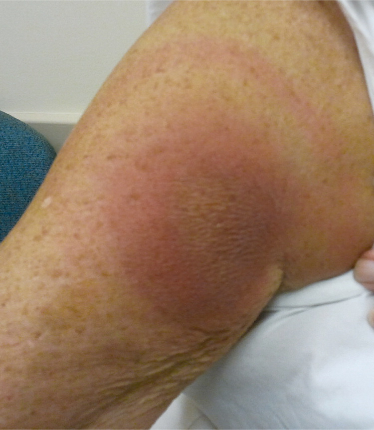
A 72-year-old woman presented with an insect bite on her right upper extremity. She first noticed the bite approximately five days before presentation. Over the subsequent five days, the rash enlarged and became more red, swollen, and painful. The center of the rash was warm and tender. She had nausea that began two days after the bite. She did not have fever, sore throat, joint aches, or body aches. She had a history of hypertension, gastroesophageal reflux disease, hyperlipidemia, and osteoarthritis. She had no history of atopic dermatitis.
Examination revealed a solitary, 9-cm, indurated, violaceous, mildly tender plaque on the right anteromedial portion of the deltoid. A 14-cm red ring surrounded the central tender area of the rash (see accompanying figure). She did not have lesions on her palms, soles, feet, or elbows.
Question
Discussion
The correct answer is C: Lyme disease. Lyme disease can be contracted from a deer tick (Ixodes) bite containing Borrelia species of bacteria. Erythema migrans is the pathognomonic skin finding associated with Lyme disease and is often described as a bull's-eye or target rash. It typically is an annular, erythematous plaque with central clearing. Lyme disease can be associated with myalgias and arthralgias, fever, anorexia and nausea, fatigue, and regional lymphadenopathy, but the rash may be the only finding at presentation.
Lesions are usually 5 to 68 cm, although they can vary in size.1 They appear three to 30 days after the tick bite, but most commonly within seven to 14 days. The classic lesion occurs in approximately 80% of cases.2 Lyme disease has been reported in all 50 states but is endemic in the Northeast and in parts of Minnesota, Wisconsin, and northern California.3 Most cases occur from May to September when the Ixodes tick is in the nymph stage.
There are three distinct clinical stages of Lyme disease. The early localized stage (three to 30 days after tick exposure) includes the influenza-like symptoms of fever, fatigue, arthralgias, and myalgias. The early disseminated stage (days to weeks after tick exposure) includes multiple lesions, neurologic symptoms (palsies, radiculopathy, or peripheral neuropathy), or cardiac symptoms (myocarditis and varying degrees of atrioventricular block). The late stage (months to years after tick exposure) includes arthritis, primarily affecting the knee, and possible cognitive disturbances. Treatment should be initiated promptly to avoid progression to late stages of the disease.
Granuloma annulare is a benign, self-limited, annular eruption that does not require treatment. It presents as skin-colored plaques or papules on distal portions of the extremities, specifically the hands, wrists, and feet. It is idiopathic and occurs in adults and children, although it is most common between 40 and 50 years of age. It is more common in women than in men.6
Nummular eczema is an idiopathic papulovesicular dermatitis commonly associated with asthma and atopic dermatitis. It typically manifests as coin-shaped lesions and is most prominent in cold or dry months.
Rheumatic fever is an inflammatory disease that can develop after group A streptococcal infection and is associated with erythema marginatum. This annular rash is typically slightly elevated, mildly erythematous, and nonpruritic, and is primarily found on extensor surfaces of extremities, sparing the face.
| Condition | Cause | Characteristics |
|---|---|---|
| Erythema multiforme | Hypersensitivity reaction to medication use or an infection, such as herpes simplex virus infection | Papules or plaques with erythematous borders; usually located on the palms, soles, elbows, or knees |
| Granuloma annulare | Idiopathic and benign | Skin-colored plaques or papules on distal portions of the extremities; typically occurs between 40 and 50 years of age |
| Lyme disease | Borrelia species of bacteria transmitted by a deer tick bite | Annular, erythematous plaque with central clearing; described as a bull's-eye or target rash |
| Nummular eczema | Idiopathic but associated with asthma and atopic dermatitis | Papulovesicular, coin-shaped lesions |
| Rheumatic fever | Complication of group A streptococcal infection | Annular rash; slightly elevated, mildly erythematous, and nonpruritic; primarily appears on extensor surfaces of extremities; spares the face |
