
Am Fam Physician. 2014;89(10):793-794
Author disclosure: No relevant financial affiliations.
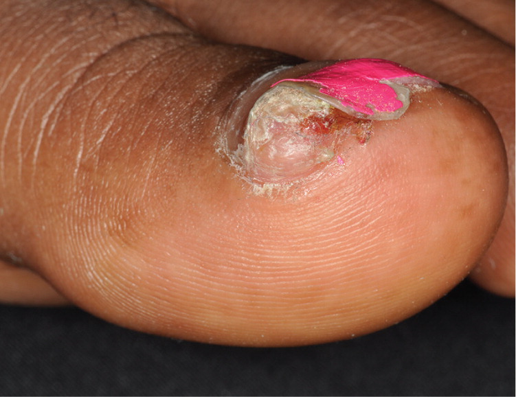
A 19-year-old woman presented with a slowly enlarging, painful nodule under her left great toenail that appeared two years earlier. She had attempted to remove part of the nodule on her own, but it grew back. She did not have a history of trauma to the area, but she noted increased pain when she wore tight or high-heeled shoes. The patient was a track athlete, and the pain of the nodule caused her to miss a season of competition.
The physical examination revealed a tender 5-mm × 5-mm nodule under the medial side of the distal nail plate (Figure 1). The nodule was firm and displaced the nail plate upward. There was distal onycholysis, but no destruction of the overlying nail plate. No other toes were affected.
Question
Discussion
The answer is D: subungual exostosis. Subungual exostosis is a tender, epithelialized, slowly growing papule or nodule most common in children and young adults. The benign osteocartilaginous tumor occurs on the distal phalanx of a digit, usually on the great toe, under or adjacent to the nail plate.1 The nail plate is displaced but not altered. Subungual exostosis is thought to be caused by repetitive trauma or chronic infection.1
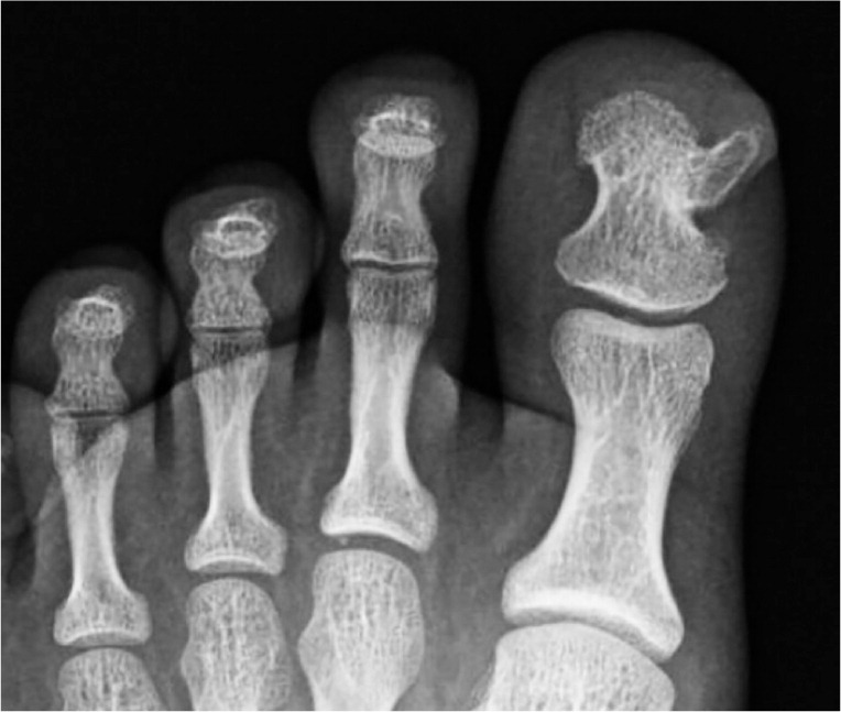
Because of the similar appearance to other conditions, subungual exostosis is often misdiagnosed. To narrow the differential diagnosis of a suspicious subungual mass, fine-detail radiography is recommended before biopsy. In this case, the radiograph was diagnostic of exostosis, which was continuous with the underlying cortex and medullary space (Figure 2). Recurrence is uncommon with proper surgical debridement of the base.2
Although acral-lentiginous melanomas are uncommon, they account for most melanomas in persons with darker skin.3 They present as irregularly shaped, pigmented macules or papules on the palm, sole, or nail bed. Amelanotic melanomas are unpigmented papules but can also present as nail dystrophy.3 Subungual pigment that extends proximal or lateral to the nail fold is called Hutchinson sign and should be biopsied to rule out a melanoma.4
Patients with a glomus tumor often present with pain in the digit. Glomus tumors are erythematous papules or nodules that grow from the glomus body, a specialized smooth muscle that links arterioles and venules.3 These lesions occur subungually, altering the distal nail plate and causing pain in response to cold and pressure. A vertical red band (erythronychia) often presents from the proximal to distal nail.
Verrucae are hyperkeratotic, rough papules with an irregular surface that occur in skin infected with human papillomavirus. The papules alter the normal skin lines and often contain characteristic red or brown dots (thrombosed capillary loops).3 Periungual verrucae can be especially resistant to treatment.4
| Condition | Characteristics |
|---|---|
| Acral-lentiginous melanoma | Irregularly shaped, pigmented macules or papules on the palm, sole, or nail bed; pigment often extends beyond the proximal nail fold |
| Glomus tumor | Painful, erythematous subungual papule or nodule; affects the distal nail plate; a vertical red band (erythronychia) often presents from the proximal to distal nail; sensitive to cold and pressure |
| Pyogenic granuloma | Tender vascular papules or nodules that bleed or erode easily; sudden appearance and rapid growth |
| Subungual exostosis | Tender, epithelialized, slowly growing papule or nodule; nail plate is displaced but not altered |
| Verruca vulgaris (common wart) | Hyperkeratotic, rough papule with irregular surface; alters normal skin lines; red or brown dots (thrombosed capillary loops) |
