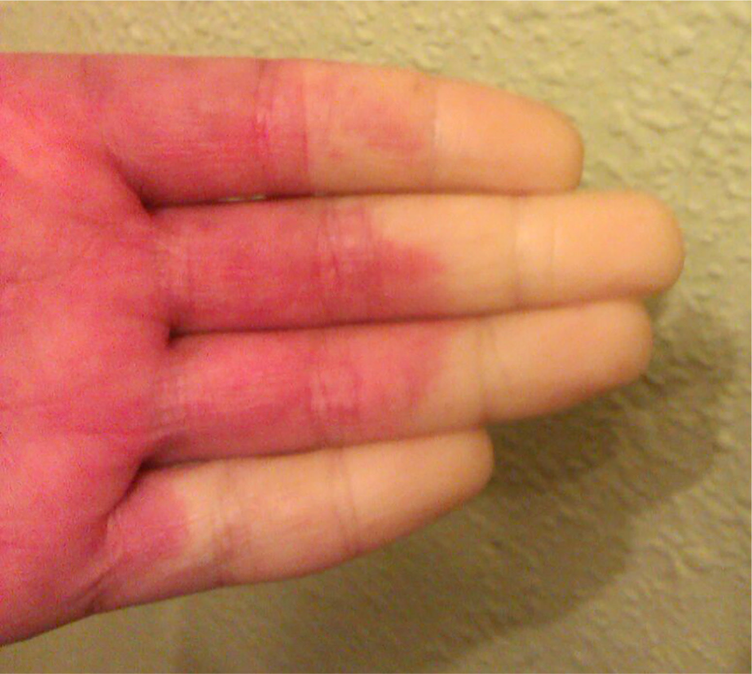
A 42-year-old woman had pain in her hands and discoloration on the tips of her fingers and toes when exposed to cold (Figure 1). She had chronic systemic lupus erythematosus that was diagnosed seven years earlier.
Her urinalysis findings were abnormal, including the presence of proteinuria and hematuria. Her creatinine level was normal, and her antinuclear antibody titer was 1:2,560. Renal biopsy, which was performed because of the proteinuria, revealed stage V membranous lupus nephritis.
Figure 1.
Question
Based on the patient's history and physical examination findings, which one of the following is the most likely diagnosis?
A. Acrocyanosis.
B. Acute peripheral arterial occlusion.
C. Frostbite.
D. Raynaud phenomenon.
Discussion
The correct answer is D: Raynaud phenomenon. Raynaud phenomenon is a discoloration of the fingertips and toes after exposure to cold or emotional stress. First, the skin turns white or pale because of exaggerated vasoconstriction, then blue when the oxygen supply is depleted and red as the blood flow returns. Raynaud phenomenon may be primary or idiopathic (not associated with other conditions) or secondary (associated with other conditions). If secondary, Raynaud phenomenon can present before the diagnosis or during the natural history of the associated condition.1,2
In this patient, Raynaud phenomenon presented with a decline in kidney function. Secondary Raynaud phenomenon is often associated with autoimmune rheumatic diseases such as systemic lupus erythematosus, scleroderma, mixed connective tissue disease, Sjögren syndrome, dermatomyositis, and polymyositis. Nail fold capillaroscopy can be used to distinguish between primary and secondary Raynaud phenomenon. An abnormal result can suggest an underlying autoimmune rheumatic disease.3,4
Treatment of Raynaud phenomenon is based on lifestyle changes (e.g., keeping the hands warm, quitting smoking, exercising regularly to avoid further attacks) and on medical treatments (e.g., calcium channel blockers, topical nitroglycerin, phosphodiesterase-5 inhibitors). Local injections of onabotulinumtoxinA (Botox) or parenteral prostaglandins can also be used.2,5
Acrocyanosis is a condition characterized by a symmetric, persistent, blue or cyanotic discoloration of the hands, feet, or face. It is caused by vasospasm of the small vessels of the skin and often occurs in newborns. It is painless and considered benign.
Acute peripheral arterial occlusion because of an embolism causes an acute stoppage of blood flow. It is often associated with cardiac conditions, such as atrial fibrillation. The patient presents with acute pain, the skin turns pale and cold, and there can be paralysis of the affected area. There is no pulse to the extremity. If blood flow is not restored promptly, tissue in the area will die.
Frostbite is caused by exposure of the skin to freezing temperatures, which damages the skin and underlying tissues. Initially, the skin becomes very cold and red. If the exposure persists, the skin becomes numb, hard, and pale, then blisters and ulcers appear.
Summary Table
| Condition | Characteristics | Examination findings |
|---|---|---|
| Acrocyanosis | Benign condition caused by mild vasospasm | Symmetric, persistent, blue or cyanotic discoloration of the hands, feet, or face |
| Acute peripheral arterial occlusion | Caused by an acute stoppage of blood flow as a result of an embolism | Acute pain, skin turns pale and cold, no pulse, possible paralysis of the affected area |
| Frostbite | Caused by freezing of the skin and underlying tissues | Skin becomes very cold and red; if the exposure persists, the skin becomes numb, hard, and pale, and blisters and ulcers appear |
| Raynaud phenomenon | Occurs with exposure to cold or emotional stress; caused by an exaggerated vasoconstriction | Skin turns white or pale because of vasoconstriction, then blue when the oxygen supply is depleted and red as the blood flow returns |
