
Am Fam Physician. 2016;94(4):314-316
Author disclosure: No relevant financial affiliations.
A female infant was born at 35 weeks' gestation by spontaneous vaginal delivery, following induction of labor for premature rupture of membranes. The pregnancy was otherwise uncomplicated. The newborn required three minutes of positive pressure ventilation, but transitioned well on room air over the next hour and did not require further treatment in the neonatal intensive care unit.
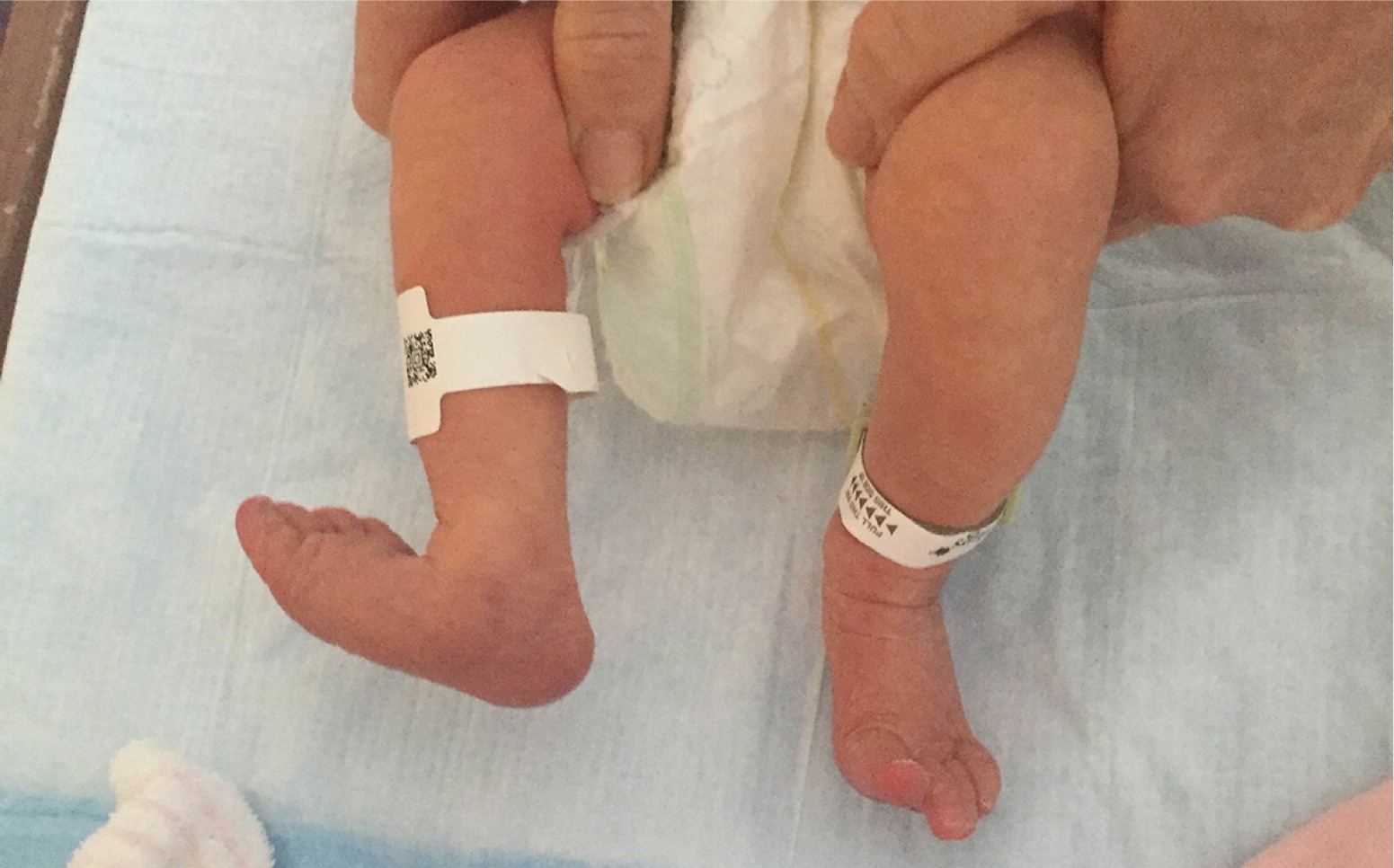
At the time of birth, physical examination showed that the newborn's right foot was grossly externally rotated (Figure 1). There was no crepitus on palpation of the foot, ankle, or leg, and a bilateral hip examination was unremarkable. The newborn spontaneously dorsiflexed and plantar-flexed the foot. The foot could be easily moved into normal alignment with gentle traction but returned when released. Her legs were equal in length. There were no dysmorphic features, no evidence of sacral dimple, and no signs of spina bifida. The remainder of the physical examination, including musculoskeletal and neurologic findings, was normal.
Question
Discussion
The answer is D: talipes calcaneovalgus. Talipes calcaneovalgus, also known as positional calcaneovalgus foot deformity, is relatively common and associated with intrauterine positioning. Although studies are limited, one study estimated an incidence of seven or eight per 1,000 births.1 On physical examination, the affected foot will be in a slightly abducted and valgus position. There may be tibial torsion. The infant is able to spontaneously move the affected limb, the foot is easily reduced into a normal or near-normal position, and the legs are of equal length.
Typically, the abnormality resolves spontaneously over a few months; however, stretching or splinting is sometimes needed for full resolution.2 Because talipes calcaneovalgus is associated with congenital hip dislocation, particularly from breech delivery, hip instability should be ruled out.3 Many other conditions in the differential diagnosis do not resolve spontaneously and should be ruled out.
Congenital vertical talus, or rocker-bottom foot, has a similar presentation. However, unlike talipes calcaneovalgus, the foot is rigidly fixed in valgus rotation because of structural deformity. The deformity includes a dorsal dislocation of the medial column of the foot at the talonavicular joint or dislocation of the entire midfoot on the hindfoot.2 It cannot be passively reduced by the examiner and will not resolve spontaneously, requiring prompt orthopedic referral for surgery or serial casting. This condition is often associated with neuromuscular disease or genetic syndromes, such as trisomies 13 and 18.3
A paralytic calcaneus foot deformity presents as valgus rotation and dorsiflexion. There is partial to full paralysis of plantar flexion, and the patient is unable to move it back into alignment spontaneously; it can be reduced by the examiner. It may occur with a number of disorders, including meningocele,4 trisomies, and polio.2,3
Posteromedial bowing of the tibia is a more proximal deformity that can cause the foot to be abducted with valgus rotation. It is most commonly associated with leg-length discrepancy. The length discrepancy and the foot deformity often improve during skeletal growth, but nearly all patients need some treatment ranging from orthotics (e.g., shoe lift) to surgical management, including epiphysiodesis or osteotomy.2
Talipes equinovarus, commonly known as club foot, is a relatively common diagnosis occurring in approximately one per 1,000 births, and affects boys twice as often as girls.5,6 The foot is excessively plantar-flexed, and the forefoot is medially rotated in the varus position so that the sole of the foot points medially and sometimes superiorly. This deformity does not resolve spontaneously. Treatment includes stretching and splinting, with minor surgical procedures to more extensive reconstructive surgery.6
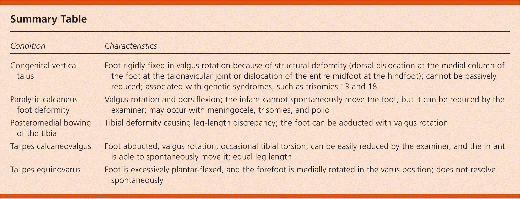
| Condition | Characteristics |
|---|---|
| Congenital vertical talus | Foot rigidly fixed in valgus rotation because of structural deformity (dorsal dislocation at the medial column of the foot at the talonavicular joint or dislocation of the entire midfoot at the hindfoot); cannot be passively reduced; associated with genetic syndromes, such as trisomies 13 and 18 |
| Paralytic calcaneus foot deformity | Valgus rotation and dorsiflexion; the infant cannot spontaneously move the foot, but it can be reduced by the examiner; may occur with meningocele, trisomies, and polio |
| Posteromedial bowing of the tibia | Tibial deformity causing leg-length discrepancy; the foot can be abducted with valgus rotation |
| Talipes calcaneovalgus | Foot abducted, valgus rotation, occasional tibial torsion; can be easily reduced by the examiner, and the infant is able to spontaneously move it; equal leg length |
| Talipes equinovarus | Foot is excessively plantar-flexed, and the forefoot is medially rotated in the varus position; does not resolve spontaneously |
The opinions and assertions contained herein are the private views of the authors and are not to be construed as official or as reflecting the views of the U.S. Army Medical Department or the U.S. Army Service at large.
