
Am Fam Physician. 2017;95(4):255-256
Author disclosure: No relevant financial affiliations.
A 25-year-old Hispanic woman delivered at 40 weeks' gestation by spontaneous vaginal delivery. Prenatal care was started late at 28 weeks' gestation, and routine laboratory screening showed no abnormalities. Prenatal ultrasonography at 29 weeks' gestation showed a horseshoe, pelvic kidney in the mother. She had no history of infections or recent travel and was not taking any medications other than prenatal supplements. Her two prior deliveries were unremarkable.
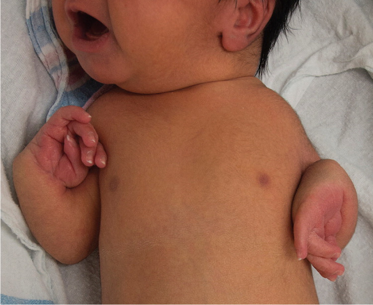
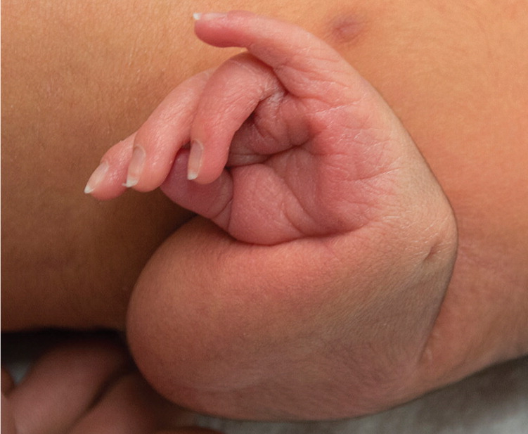
At birth, the newborn appeared healthy, and had a lusty cry and no visible craniofacial or cutaneous abnormalities. Cardiovascular and pulmonary assessments were normal. Examination revealed bilateral wrist flexion and shortened forearms (Figures 1 and 2). The humerus was palpable in the upper arm bilaterally. The newborn had normal thumb development and handgrip bilaterally. He had all 10 fingers, with webbing noted on both hands. Skeletal and joint examination was otherwise unremarkable, including normal hip rotation and lower extremity length.
Birth weight was 6 lb, 9 oz (2,975 g). Apgar score was 9 at one and five minutes. Pulse and respiratory rate were normal. Initial laboratory results showed a leukocyte count of 24.8 per μL (0.02 × 10⁹ per L), normal range: 9,000 to 30,000 per μL (9 to 30 × 109 per L); hematocrit of 42.8%, normal range: 42.0% to 60.0%; and platelet count of 146 × 103 per μL (146 × 109 per L), normal range: 150 to 400 × 103 per μL (150 to 400 × 109 per L).
Question
Discussion
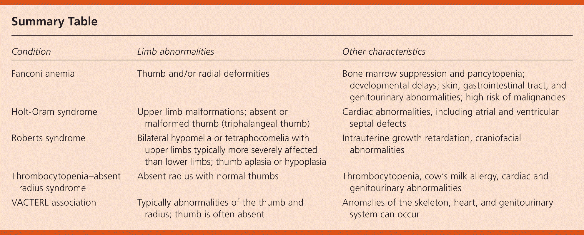
The answer is D: thrombocytopenia–absent radius (TAR) syndrome. This syndrome is a rare autosomal recessive disorder characterized by absent radius and thrombocytopenia.1 Fewer than one out of 100,000 newborns are affected each year.1 The thrombocytopenia associated with TAR syndrome can develop at birth or within the first few weeks or months of life. Cow's milk allergy is common and can be associated with exacerbation of thrombocytopenia. Anomalies of the skeleton (e.g., upper and lower limbs, ribs, and vertebrae), heart, and genitourinary system (e.g., renal anomalies and agenesis of the uterus, cervix, and the upper part of the vagina) can occur, but the thumbs are generally unaffected.2
The diagnosis of TAR syndrome is primarily clinical. The absent radius, which is noticed immediately, combined with thrombocytopenia and normal thumbs, suggests the diagnosis. Treatment consists of platelet transfusion for thrombocytopenia if needed. Orthopedic interventions may be needed to maximize limb function. Cow's milk should be avoided to reduce the severity of gastroenteritis and exacerbations of thrombocytopenia.
Fanconi anemia is a rare autosomal recessive disorder caused by a defect in DNA repair. It is characterized by bone marrow suppression with pancytopenia and thumb and/or radial deformities.3 Patients with Fanconi anemia may have developmental delays and skin, gastrointestinal tract, and genitourinary abnormalities. They are also at high risk of malignancies such as squamous cell carcinoma and bone marrow cancers. The diagnosis is a challenge because of the heterogeneous nature of the condition, but chromosome breakage tests, mutation analyses, and bone marrow chromosome analyses can be used.
Holt-Oram syndrome is an autosomal dominant disorder characterized by upper limb malformations with cardiac abnormalities (e.g., atrial and ventricular septal defects). The thumb is often absent or malformed (triphalangeal thumb). Most individuals with Holt-Oram syndrome have an atrial septal defect or ventricular septal defect, rarely with conduction defects and the tetralogy of Fallot.4 Upper limb abnormalities can involve the entire extremity; they are often bilateral but may not be symmetrical.
Roberts syndrome is an autosomal recessive disorder characterized by intrauterine growth retardation, craniofacial abnormalities (e.g., microcephaly, cleft lip or palate) and limb malformations (e.g., bilateral symmetric tetraphocomelia or hypomelia, thumb aplasia or hypoplasia).5 The upper limbs are more severely affected than the lower limbs. Mortality is high in those with severe disease, but those with mild disease may survive into adulthood. The diagnosis relies on cytogenetic testing.
The VACTERL association is defined by the presence of at least three of the following congenital malformations: vertebral defects, anal atresia, cardiac defects, tracheoesophageal fistula, renal anomalies, limb abnormalities.6 The most common limb finding involves the thumb and radius, but the thumb is usually absent. Diagnosis of VACTERL association is made clinically, based on the presence of these congenital malformations.
| Condition | Limb abnormalities | Other characteristics |
|---|---|---|
| Fanconi anemia | Thumb and/or radial deformities | Bone marrow suppression and pancytopenia; developmental delays; skin, gastrointestinal tract, and genitourinary abnormalities; high risk of malignancies |
| Holt-Oram syndrome | Upper limb malformations; absent or malformed thumb (triphalangeal thumb) | Cardiac abnormalities, including atrial and ventricular septal defects |
| Roberts syndrome | Bilateral hypomelia or tetraphocomelia with upper limbs typically more severely affected than lower limbs; thumb aplasia or hypoplasia | Intrauterine growth retardation, craniofacial abnormalities |
| Thrombocytopenia–absent radius syndrome | Absent radius with normal thumbs | Thrombocytopenia, cow's milk allergy, cardiac and genitourinary abnormalities |
| VACTERL association | Typically abnormalities of the thumb and radius; thumb is often absent | Anomalies of the skeleton, heart, and genitourinary system can occur |
