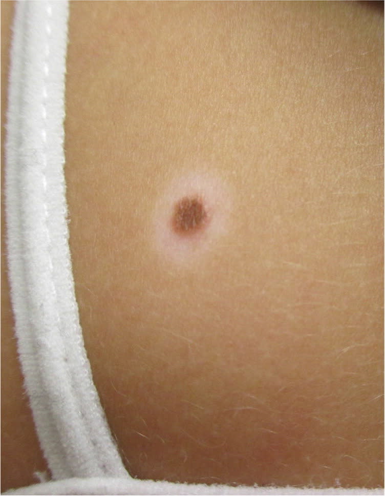
A 15-year-old girl presented with an asymptomatic pigmented mole that had been present for about 10 years. The mole had developed a white halo around it two years earlier. Over the past year, the halo enlarged, while the size and pigmentation of the mole decreased. She was in good health and was not taking any medications. There was no personal or family history of autoimmune disorders or melanoma.
Physical examination revealed a 4-mm, melanocytic nevus surrounded by a rim of depigmentation on the right scapula (Figure 1). No other cutaneous or systemic abnormalities were noted.
Figure 1.
Question
Based on the patient's history and physical examination findings, which one of the following is the most likely diagnosis?
A. Blue nevus.
B. Halo nevus.
C. Melanoma.
D. Nevus spilus.
E. Spitz nevus.
Discussion
The answer is B: halo nevus. This is a benign lesion characterized by the appearance of a depigmented halo around a central melanocytic nevus. It is common in children and young adults, with a mean age at onset of 15 years, and it most often appears on the upper back.1 Over time, the central nevus may remain brown in color—the classic form of halo nevus (stage I)—or the pigment can disappear, leading to a pink-colored papule (stage II).2 The central nevus may eventually disappear, leading to a circular area of depigmentation (stage III).2 Finally, the depigmented area may repigment, leaving no evidence of its existence (stage IV). Not all halo nevi evolve through each stage.3
A blue nevus typically presents as a solitary, smooth, well-demarcated, dome-shaped papule or nodule that is less than 1 cm in diameter. The lesion is homogeneously blue to blue-gray or blue-black and is asymptomatic. Common sites include the face, and dorsal surfaces of the hands and feet. The condition is usually first noted in adults younger than 40 years.
Melanoma is a malignancy that develops from melanocytes. It is more common in whites, and the mean age at onset is 55 years. The diagnosis should be suspected if a pigmented skin lesion displays the ABCDE characteristics: asymmetrical (one-half of the lesion does not match the other in terms of shape and color); irregular or indistinct border; color variegation (the color is mottled with different shades); diameter greater than 6 mm; and evolution over time with progressive enlargement or changes in color or symptoms. A halo phenomenon may occasionally occur with a regressing melanoma.
Classically, nevus spilus presents as a light brown, circumscribed pigmentation that is stippled with dark brown punctate macules or papules. Although nevus spilus can be congenital, most develop in the first year of life, with some developing later in childhood or adolescence. Common sites include the abdomen and back.
A Spitz nevus is a distinct subtype of melanocytic nevus that occurs in childhood. Although they are most often acquired, approximately 7% of the lesions are congenital.4 Typically, Spitz nevi are solitary, well circumscribed, smooth, dome shaped, firm, and hairless. The color varies from pink to tan or dark brown. The size varies from 2 mm to a few centimeters. Common sites include the head and neck. When Spitz nevi are examined through a glass slide compressing the surface of the lesion, brown pigmentation is often seen, which confirms the melanocytic nature.
Summary Table
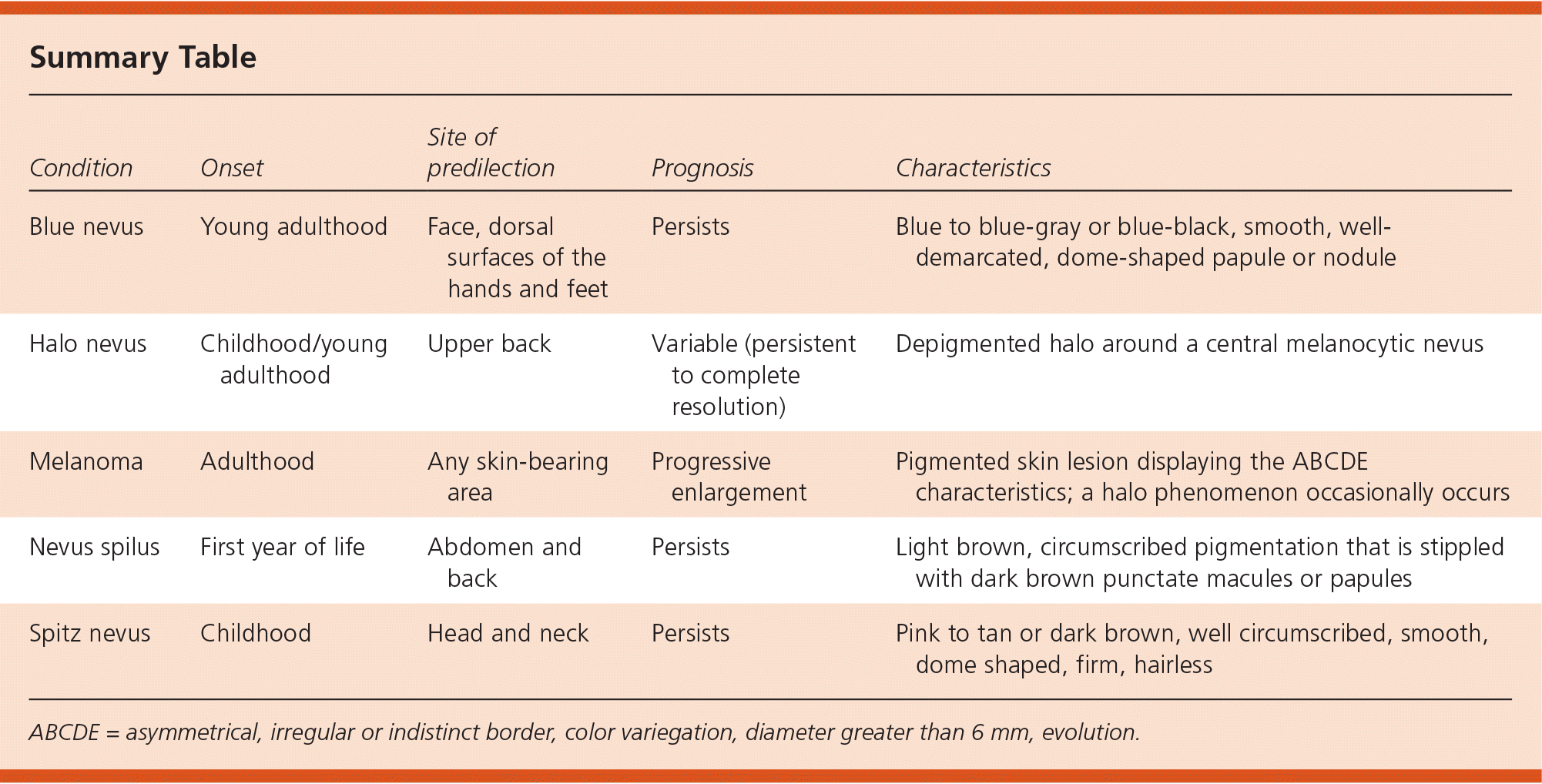
| Condition | Onset | Site of predilection | Prognosis | Characteristics |
|---|---|---|---|---|
| Blue nevus | Young adulthood | Face, dorsal surfaces of the hands and feet | Persists | Blue to blue-gray or blue-black, smooth, well-demarcated, dome-shaped papule or nodule |
| Halo nevus | Childhood/young adulthood | Upper back | Variable (persistent to complete resolution) | Depigmented halo around a central melanocytic nevus |
| Melanoma | Adulthood | Any skin-bearing area | Progressive enlargement | Pigmented skin lesion displaying the ABCDE characteristics; a halo phenomenon occasionally occurs |
| Nevus spilus | First year of life | Abdomen and back | Persists | Light brown, circumscribed pigmentation that is stippled with dark brown punctate macules or papules |
| Spitz nevus | Childhood | Head and neck | Persists | Pink to tan or dark brown, well circumscribed, smooth, dome shaped, firm, hairless |
ABCDE = asymmetrical, irregular or indistinct border, color variegation, diameter greater than 6 mm, evolution.
