
Am Fam Physician. 2002;65(10):2069-2073
A more recent article on Paget Disease of Bone is available.
Paget's disease of bone (also known as osteitis deformans) is a nonmalignant disease involving accelerated bone resorption followed by deposition of dense, chaotic, and ineffectively mineralized bone matrix. The origin of the disease is unknown, and it is frequently asymptomatic; however, the patient may present with symptoms depending on the bones involved. The most common symptom is pain in the affected bone; neurologic, hearing, vision, cardiac, and oncologic complications are possible. Diagnosis is primarily made by radiographs. Bisphosphonates are the most common treatment.
A side from osteoporosis, Paget's disease is the most common bone disorder.1 Paget's disease is equally prevalent in men and women, with increased incidence in persons older than 50 years. It affects approximately 3 percent of persons in the United States, and as many as 10 percent of persons older than 80 years.2,3
Paget's disease occurs in three phases.1,4 The initial phase consists of intense osteoclastic activity and bone resorption, with bone turnover as high as 20 times the normal rate.5 This phase is followed by an osteolytic-osteoblastic phase during which osteoblasts begin to produce an abundance of woven bone, but mineralization is ineffective. In the final phase, dense cortical and trabecular bone deposition dominates, but it is sclerotic, disorganized, and weaker than normal bone.4
| Bones | Percentage |
|---|---|
| Pelvis | 72 |
| Lumbar spine | 58 |
| Femur | 55 |
| Thoracic spine | 45 |
| Skull | 42 |
| Tibia | 35 |
| Humerus | 31 |
| Cervical spine | 14 |
Etiology
Although the etiology of Paget's disease is unknown, studies have provided some support for both viral and hereditary causes. Viral antigens have been detected in affected osteoclasts by numerous methods and research teams.5 In the United States, the measles virus antigen is most commonly detected in patients with Paget's disease.6–8
Diagnosis
| Bone pain from microfractures or osteoarthritis; if the jaw is involved, teeth may become loose |
| Headaches and loss of hearing or vision from pressure on nerves, brain, or spinal cord and reduced blood flow |
| Pain or neuropathy from pressure on nerves |
| Increased head size, bowing of a limb, or curvature of the spine |
| Hip pain |
| Damage to cartilage of joints, which may lead to osteoarthritis |
| Heart failure (only in severe cases, especially in patients with heart disease) |
| Kidney stones (more common in patients with Paget's disease) |
| Sarcoma, in less than 1 percent of patients with Paget's disease |
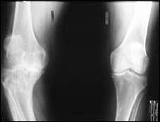
Patients with bone pain caused by Paget's disease usually describe the pain as continuous. Unlike osteoarthritis, pagetic bone pain usually increases with rest, on weight bearing, when the limbs are warmed, and at night.1,4 Paget's disease can cause osteoarthritis if the affected section of bone is near a joint.1
Neurologic symptoms arise from the compression of nerves, which is caused by osseous growth. Resulting sequelae include cranial nerve, brain stem and cerebellar deficits, and deficits caused by spinal stenosis. Although cardiovascular involvement is uncommon, patients with widespread Paget's disease and extensive hypervascularization of the bone marrow can present with an arteriovenous shunt leading to high output cardiac failure.
The incidence of malignant degeneration of pagetic bone ranges from less than 1 percent to 10 percent, depending on disease severity.1 Usually, these tumors are highly malignant osteosarcomas, fibrosarcomas, or undifferentiated spindle cell sarcomas.13,14 Radiologic characteristics suggestive of malignant transformation include cortical breakthrough and soft tissue masses.15
Patients with Paget's disease may also develop several types of pseudomalignancy, including pseudosarcoma and pseudo giant cell tumors that are responsive to corticosteroids. Paget's pseudosarcoma typically presents as a slow-growing, localized, periosteal mass on pagetic bone. It has a predilection for long bones, especially the femur, and differs from a true sarcoma in that it does not destroy cortical bone or invade surrounding soft tissue.16 Patients may present with metabolic abnormalities, including hypercalcemia, hypercalcuria, and hyperuricemia.1
Diagnosis of Paget's disease may be suspected based on the symptoms, but radiographs are the most specific diagnostic test. Asymptomatic patients with a first-degree relative with Paget's disease should be screened with a serum alkaline phosphatase test every two to three years. If the serum alkaline phosphatase level is elevated, a bone scan can be performed to determine the extent and activity of Paget's disease. Radiographs should be taken to confirm the diagnosis in a patient with bone scans suggestive of Paget's disease.
BIOCHEMICAL MARKERS
There are many biochemical markers for Paget's disease, but the two most important are total serum alkaline phosphatase and urinary pyridinoline.17 These markers may be normal in patients with the monostotic form of Paget's disease (15 percent of patients) therefore, serum bone-specific alkaline phosphatase measurements may be useful. Urinary hydroxyproline is no longer considered an accurate marker of activity or extent of the disease.18–20
RADIOGRAPHY
Radiographs include both lytic (early) and sclerotic findings (Table 3). Many patients are diagnosed incidentally in the asymptomatic phase by plain radiographs that show localized enlargement of bone. These radiographs often have a high specificity because of their classic nature, but a low sensitivity. Bone scans can be used to increase the sensitivity in patients suspected of having Paget's disease; however, the bone scan is less specific and should be interpreted cautiously.21 Once a diagnosis of Paget's disease is confirmed, repeat radiographs are required only to monitor degeneration around weight-bearing joints. Computed tomography and magnetic resonance imaging are not necessary.
| Radiographic |
| Osteoporosis circumscripta in skull |
| Flame-shaped lesions in long bones |
| Osteolytic lesions near thickened lesions |
| Sclerotic bone |
| Bowed limbs |
| Fractures, including “banana” or “chalk” transverse fractures |
| Bone scintigraphy |
| Areas of increased uptake of technetium-99m |
| “Mouse face” pattern on scan of affected vertebra |
If the results of the biochemical markers and radiography are inconclusive, a biopsy of the affected bone may be indicated in rare cases.
Treatment
Treatment of Paget's disease does not cure the disease but can provide prolonged periods of remission. Bisphosphonates, which decrease bone resorption by inhibiting osteoclast resorption, are the treatment of choice. Disease activity stays low for months or years after cessation of these medications. Alendronate (Fosamax) and pamidronate (Aredia) are used most frequently and will cause about a 70 percent decrease in biochemical markers in one half of patients.2 Newer-generation bisphosphonates that are still in development are thousands of times more potent than first-generation agents. Calcitonin (Calcimar) also inhibits osteoclastic bone resorption. Calcitonin-salmon is available in injectable and nasal-spray forms, but only the injectable form is approved for treatment of Paget's disease by the U.S. Food and Drug Administration22,23 (Table 412). Compared with the bisphosphonates, calcitonin is not as powerful and does not suppress the disease activity for as long after cessation. Patients with Paget's disease should receive adequate doses of calcium (1,000 to 1,500 mg per day) and vitamin D (400 IU per day). Exercise is recommended to maintain skeletal health in patients with Paget's disease; however, exercise programs should be individualized to prevent stress on affected bones.
| Medications | Dosage | |
|---|---|---|
| Bisphosphonates | Bisphosphonate tablets should be taken with 6 to 8 oz of tap water on an empty stomach. Do not eat or lie down for 30 minutes after taking medication. Bisphosphonates should be avoided in patients with kidney disease. | |
| Alendronate (Fosamax) | 40 mg orally once a day for six months. May reinstate treatment after six months, if necessary. | |
| Pamidronate (Aredia) | 30 mg intravenously over a four-hour period on three consecutive days. Reinstate treatment at intervals, as necessary. More commonly used regimen is 60 mg over a two- to four-hour period for two or more consecutive or nonconsecutive days.15 | |
| Tiludronate (Skelid) | 400 mg daily for three months. May reinstate treatment after three months, if necessary. | |
| Risedronate (Actonel) | 30 mg daily for two months. May reinstate treatment after two months, if necessary. | |
| Etidronate (Didronel) | 5 mg per kg per day (if ineffective, 11 to 20 mg per kg per day for a maximum of six months). May reinstate treatment after three months, if necessary. | |
| Calcitonin (Miacalcin injection) | ||
| Calcitonin-salmon (Calcimar) | 200 U per mL; 100 U subcutaneously or intramuscularly once daily for six to 18 months. | |
SURGICAL
Rarely, surgical treatment with elective joint replacements or osteotomy may be required in patients with the following conditions: progressive bowing of the tibia or femur; delayed union of fractures; unstable fractures; arthritis refractory to medical treatment; or focal nerve compression of the spine or cranium.1,23
INITIATION OF TREATMENT
Although no established standard for initiating therapy exists, the literature generally supports treating the following patients: all symptomatic patients; asymptomatic patients whose biochemical markers suggest an increase in bone remodeling; and patients with pagetic lesions located at weight-bearing regions or adjacent to joints.1,3,4 Many sources2,21 recommend initiating treatment when the serum alkaline phosphatase level rises to 125 to 150 percent of normal values. Recommendations for follow-up serum alkaline phosphatase monitoring range from every three months to annually. Patients should be followed indefinitely because of the increased risk of malignant transformation in patients with longstanding Paget's disease. Treatment goals should also include aggressive pain control.