
Am Fam Physician. 2014;89(8):663-664
Author disclosure: No relevant financial affiliations.
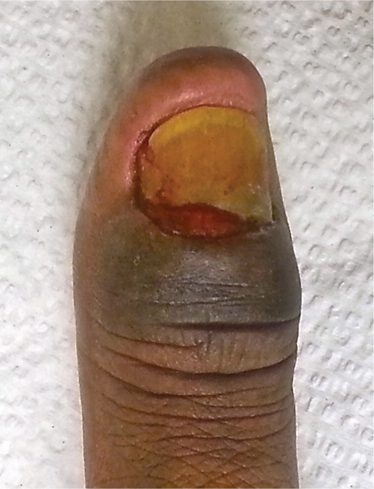
A 78-year-old man with a history of diabetes mellitus and hypertension presented with a four-month history of progressively worsening, painless swelling at the base of his left thumbnail. The swelling was accompanied by a serosanguineous discharge at the cuticle. There was no history of trauma to the area. Treatment with levofloxacin (Levaquin), 500 mg, for 10 days was ineffective.
Physical examination revealed hyperpigmented skin just below the cuticle of the left thumb with swelling (see accompanying figure). The base of the nail appeared fleshy with loss of the cuticle and dystrophy of the nail plate.
Question
Discussion
The answer is A: chronic paronychia. Paronychia is inflammation of the nail fold secondary to a bacterial infection.1,2 Retraction of the proximal nail fold, nail disfigurement, and loss of the cuticle are common. Chronic paronychia lasts longer than acute paronychia, often more than six weeks, and is usually nonsuppurative. Predisposing factors include exposure of hands to a moist environment (e.g., bartending, dish washing, swimming), immunocompromise, diabetes, and repeated minor trauma (e.g., nail biting, thumb sucking).
Treatment of chronic paronychia involves keeping the hands dry, wearing protective gloves if working in a moist environment, and keeping hands away from irritants. Medium- to high-potency topical steroids are the first-line treatment because chronic paronychia is thought to be primarily an inflammatory, eczematous process.3 Because colonization with Candida albicans is common, a topical antifungal cream can be added if there is no resolution. Surgery with proximal nail fold and nail plate excision may be indicated in refractory cases. Resolution of chronic paronychia can take several weeks to months.4
A felon is an acute subcutaneous pyogenic infection of the pulp space of the thumb and index finger commonly caused by Staphylococcus or Streptococcus species. Infection usually begins with minor trauma to the dermis overlying the finger pad. Felons that are untreated or incorrectly treated, or that have a prolonged course may lead to osteomyelitis.
Herpetic whitlow is caused by herpes simplex virus infection. Herpes simplex virus 1 infection is usually the cause in children, whereas herpes simplex virus 2 infection is more common in adults. Patients with herpetic whitlow have a tingling or burning sensation and fluid-filled vesicles, usually without nail plate involvement. A Tzanck test can confirm the diagnosis.5
Onychomycosis is a fungal infection that may involve any portion of the nail unit. Discoloration and thickening of the nail plate are characteristic. Subungual debris may separate the nail plate from the nail bed.6
Subungual hematomas result from trauma to the nail plate that causes bleeding and pain. The nail may separate from the nail plate, although puncturing the nail plate with a handheld cautery device or heated needles relieves the pressure. Subungual hematomas usually cause a bluish or violaceous discoloration of the nail plate that remains until the nail plate grows out.
| Condition | Characteristics |
|---|---|
| Chronic paronychia | Inflammation of the nail fold with retraction of the proximal nail fold, nail disfigurement, and loss of the cuticle |
| Felon | Acute subcutaneous pyogenic infection of the pulp space of the thumb and index finger, associated with minor trauma to the dermis overlying the finger pad |
| Herpetic whitlow | Tingling or burning sensation and fluid-filled vesicles, usually occurs without nail plate involvement |
| Onychomycosis | Discoloration and thickening of the nail plate, subungual debris separating the nail plate from the nail bed |
| Subungual hematoma | History of trauma; bluish or violaceous discoloration under the nail plate, pain and separation of the nail from the nail plate may occur |
