Am Fam Physician. 2019;99(3):online
Author disclosure: No relevant financial affiliations.

Narrative: Dyspnea is a common acute symptom in patients presenting to the emergency department and who are ultimately diagnosed with acute heart failure syndrome (AHFS).1 However, in patients with undifferentiated dyspnea, an accurate diagnosis of AHFS may be difficult with the standard initial evaluation that includes patient history, physical examination, electrocardiography (ECG), chest radiography, and natriuretic peptide testing. This systematic review and meta-analysis comprehensively evaluated the diagnostic accuracy of the clinical assessment and index tests that physicians may use to distinguish AHFS from other clinical conditions in patients presenting to the emergency department with dyspnea.2
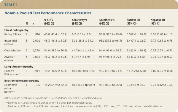
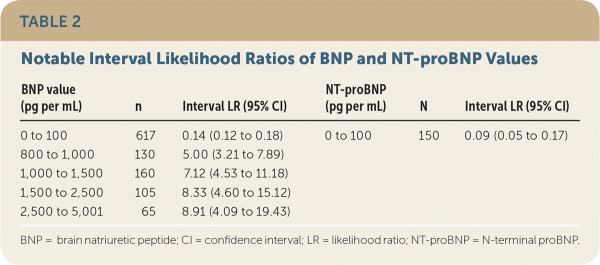
This review included 57 mostly prospective or cross-sectional studies, 52 unique cohorts, and a total of 17,893 patients.2 There was no single historical variable, symptom, or physical examination finding that could significantly reduce the likelihood of AHFS. An S3 gallop marginally increased the likelihood of AHFS (positive likelihood ratio [LR+] = 4.0; 95% confidence interval [CI], 2.7 to 5.9) but was an insensitive finding. None of the abnormal ECG findings substantially increased or decreased the probability of AHFS. The presence of radiographic findings that represented edema moderately increased the likelihood of AHFS (LR+ = 4.8 to 6.5; Table 1), but a negative chest radiograph was unhelpful. Serum B-type natriuretic peptide (BNP) testing increased the probability of AHFS (LR+ > 5.0) but only at markedly elevated concentrations (more than 800 pg per mL [800 ng per L]; Table 2); both BNP and serum N-terminal proB-type natriuretic peptide (NT-proBNP) testing were useful at excluding AHFS at very low concentrations (less than 100 pg per mL [100 ng per L]). Bedside lung ultrasonography of three or more B-line artifacts in two bilateral lung zones had the greatest discriminatory value among index tests (LR+ = 7.4; 95% CI, 4.2 to 12.8; negative likelihood ratio [LR–] = 0.16; 95% CI, 0.05 to 0.51). Bedside echocardiography of visually estimated reduced ejection fraction was somewhat helpful (LR+ = 4.1; 95% CI, 2.4 to 7.2), and the finding of restrictive mitral pattern was highly predictive of AHFS (LR+ = 8.3; 95% CI, 4.0 to 16.9).

| N | n | % AHFS (95% CI) | Sensitivity % (95% CI) | Specificity % (95% CI) | Positive LR (95% CI) | Negative LR (95% CI) | |
|---|---|---|---|---|---|---|---|
| Chest radiography | |||||||
| Kerley B lines | 2 | 814 | 46.8 (43.4 to 50.2) | 9.2 (6.5 to 12.5) | 98.8 (97.3 to 99.6) | 6.5 (2.6 to 16.2) | 0.88 (0.69 to 1.13) |
| Interstitial edema | 3 | 2,001 | 48.3 (46.2 to 50.5) | 31.1 (28.2 to 34.2) | 95.1 (93.6 to 96.3) | 6.4 (3.4 to 12.2) | 0.73 (0.68 to 0.78) |
| Cephalization | 5 | 1,338 | 54.0 (51.3 to 56.6) | 44.7 (41.1 to 48.4) | 94.6 (92.6 to 96.3) | 5.6 (2.9 to 10.4) | 0.53 (0.39 to 0.72) |
| Alveolar edema | 3 | 2,001 | 48.3 (46.2 to 50.5) | 5.7 (4.7 to 6.9) | 98.9 (98.4 to 99.3) | 5.3 (3.3 to 8.5) | 0.95 (0.94 to 0.97) |
| Lung ultrasonography | |||||||
| Positive B-line scan* | 8 | 1,914 | 48.2 (46.0 to 50.5) | 85.3 (82.8 to 87.5) | 92.7 (90.9 to 94.3) | 7.4 (4.2 to 12.8) | 0.16 (0.05 to 0.51) |
| Bedside echocardiography | |||||||
| Restrictive mitral pattern† | 1 | 125 | 43.2 (34.9 to 52.0) | 81.5 (68.6 tp 90.7) | 90.1 (80.7 to 95.9) | 8.3 (4.0 to 16.9) | 0.21 (0.12 to 0.36) |

| BNP value (pg per mL) | n | Interval LR (95% CI) | NT-proBNP (pg per mL) | N | Interval LR (95% CI) |
|---|---|---|---|---|---|
| 0 to 100 | 617 | 0.14 (0.12 to 0.18) | 0 to 100 | 150 | 0.09 (0.05 to 0.17) |
| 800 to 1,000 | 130 | 5.00 (3.21 to 7.89) | |||
| 1,000 to 1,500 | 160 | 7.12 (4.53 to 11.18) | |||
| 1,500 to 2,500 | 105 | 8.33 (4.60 to 15.12) | |||
| 2,500 to 5,001 | 65 | 8.91 (4.09 to 19.43) |
Caveats: Patients presenting to the emergency department with dyspnea are different than patients presenting to the primary care clinic with more subacute, less severe dyspnea. Although this possible spectrum bias may potentially influence study validity, the review analyzed all emergency department patients who presented with dyspnea and did not discriminate between time courses of presenting illness.2 Therefore, index test operative characteristics should maintain applicability, but considering the likely lower AHFS prevalence (and lower pretest probability) in the clinic population, relatively more tests may be required to confirm AHFS in the primary care clinic setting.
The estimation of each index test's diagnostic accuracy was limited by the lack of a true, objective criterion or “gold” standard against which the test was measured. The criterion standard for AHFS was typically a subjective assessment by two or more physicians with data from the clinical record. The review did not evaluate the diagnostic accuracy of historical elements, symptoms, or examination findings in combination, also known as clinical gestalt.2 Clinical gestalt likely outperforms these diagnostic elements in isolation and plays an important role in determining the pretest probability of AHFS.
The studies that were included in the review varied in quality.2 Differences in inclusion and exclusion criteria among the included studies put them at varying degrees of risk for spectrum bias, and the consideration of pooled results should factor in the moderate to high statistical heterogeneity among included studies. With regards to the natriuretic peptide analyses, this review did not consider age as a variable that may alter BNP and NT-proBNP values. Age-stratified NT-proBNP cut points have since been demonstrated to be moderately useful in the diagnosis of AHFS.3
The accuracy of lung ultrasonography will be provider-dependent, but emergency physician sonographers in most included studies were workshop-trained only. Furthermore, trainees may rapidly achieve procedural competency,4 and lung ultrasonography has been shown to have fair predictive value for pulmonary edema from AHFS when performed by novices and experts.5 Lastly, the performance of lung ultrasonography is likely time-sensitive because B-line artifacts appear to decrease coincident with treatment for lung edema caused by AHFS6 or other etiology.7
Conclusion: The individual components of the clinical history and physical examination, ECG, and chest radiography are not useful independently for confirming or excluding the diagnosis of AHFS in patients presenting to the emergency department. BNP concentrations above 800 pg per mL are helpful for establishing the diagnosis. BNP and NT-proBNP results are also valuable in ruling out AHFS when concentrations are below the threshold of 100 pg per mL. Lung ultra-sonography, although provider-dependent, may be learned quickly and appears to have the best combination of test characteristics to confirm or exclude the diagnosis of AHFS.
