
Am Fam Physician. 1998;58(5):1126-1130
Stress test parameters indicating the presence and extent of coronary artery disease have traditionally included such variables as exercise duration, and the blood pressure and ST-segment responses to exercise. The three-minute systolic blood pressure ratio, another important indicator of significant coronary artery disease, is a useful and readily obtainable measure that can be applied in all patients who are undergoing stress testing for the evaluation of known or suspected ischemic heart disease. The ratio is calculated by dividing the systolic blood pressure three minutes into the recovery phase of a treadmill exercise test by the systolic blood pressure at peak exercise. A three-minute systolic blood pressure ratio greater than 0.90 is considered abnormal and has a diagnostic accuracy of approximately 75 percent for the detection of coronary artery disease (i.e., an accuracy comparable to that of ST-segment depression). Higher values for the ratio are associated with more extensive coronary artery disease, as well as an adverse prognosis after myocardial infarction. Thus, the three-minute systolic blood pressure ratio provides information that is complementary to the traditional exercise test parameters for identifying high-risk ischemic heart disease.
Over the past decade, a number of studies1–9 have established an association between delayed recovery of the systolic blood pressure to the preexercise baseline value and the presence and extent of coronary artery disease. Yet, even though the postexercise systolic blood pressure response is a readily obtainable parameter, it is often overlooked in the interpretation of exercise test results.
This review focuses on the clinical application of the postexercise systolic blood pressure response in the assessment of ischemic heart disease. The mechanism of this response and its diagnostic and prognostic implications are also discussed.
Background
During treadmill exercise in normal patients, systolic blood pressure is expected to rise 5 to 10 mm Hg per metabolic equivalent of effort (MET), with 1 MET being roughly equivalent to the energy expended in the resting state. Thus, for a middle-aged patient with an average exertional tolerance, the systolic blood pressure can be expected to rise at least 40 to 50 mm Hg during treadmill exercise testing.
Systolic blood pressure generally reaches a plateau at peak exercise and remains at or near this value through the first minute of the recovery period following exercise. In normal patients, this plateau is followed by a rapid and uniform decline of the systolic blood pressure throughout the recovery phase. The systolic blood pressure generally reaches the baseline (preexercise) value within five minutes.8 A delay in the return of the systolic blood pressure to normal represents an abnormal response.
The postexercise systolic blood pressure ratio (SBPR) is an expression of the rate of decline of the postexercise blood pressure relative to the peak exercise value. This ratio is calculated as the systolic blood pressure at a specified time in recovery divided by the peak exercise systolic blood pressure value. For example, if a patient's systolic blood pressure is 200 mm Hg at peak exercise and falls to 180 mm Hg at three minutes into recovery, the SBPR is 180/200, or 0.90.
Several studies1,3,8 indicate that using the three-minute recovery blood pressure to calculate the SBPR provides the best discrimination between normal and abnormal patients. Higher values for the three-minute SBPR (i.e., greater than 0.90) reflect abnormal delay in the postexercise recovery of systolic blood pressure.
Hemodynamic Correlates of the Three-Minute SBPR
Studies evaluating intracardiac hemodynamics and their relationship to the SBPR have been helpful in determining the mechanism of the abnormal three-minute SBPR.2,4 In these studies, higher values for the calculated SBPR were associated with a significantly higher pulmonary capillary wedge pressure and a lower cardiac index at peak exercise.4
By three minutes into the recovery phase, pulmonary capillary wedge pressure and cardiac index were similar in patients with either high or low SBPR values.2,4 However, the patients with higher SBPR values had significantly greater systemic vascular resistance, elevated catecholamine levels and a pronounced delay in the return of stroke index to baseline.
Study findings suggest that an abnormal SBPR is related to the following: (1) recovery from exercise-induced ischemia with ischemic left ventricular dysfunction, and (2) the effects of catecholamine-related enhanced peripheral vasomotor tone during recovery from exercise.
Diagnostic Implications of the SBPR
The SBPR was first used to describe the delay in the fall of the systolic blood pressure that occurred immediately after exercise in unselected patients with coronary artery disease.8 Systolic blood pressure measurements taken during treadmill exercise testing and again during seated postexercise recovery showed that patients with angiographically proven coronary artery disease had a significantly higher three-minute SBPR than patients without coronary artery disease (0.93 ± 0.13 versus 0.73 ± 0.06, respectively; P < 0.001). The suggestion that an increased SBPR was superior to exercise-induced ST-segment depression in diagnosing cardiac disease was controversial.7,9,10
Subsequently, symptom-limited treadmill exercise testing was studied in 181 patients who had not taken any medications.3 Cardiac catheterization was performed in all patients, and coronary artery disease was identified in 106 patients. The patients with coronary artery disease had a significantly higher three-minute SBPR value than the patients without coronary artery disease (0.99 ± 0.16 versus 0.84 ± 0.13, respectively). Furthermore, the SBPR value was found to be related to the severity of the coronary artery disease as measured by the number of diseased coronary arteries.
When an empirically derived cutoff value of 0.91 or greater was used to define an abnormal three-minute SBPR, the ratio had a 74.4 percent sensitivity and a 77.1 percent specificity for the identification of coronary artery disease, comparable to the 81.7 percent sensitivity and 66.7 percent specificity of a 1-mm or greater depression of the ST segment. Thus, an increased SBPR was related to the presence and extent of coronary artery disease.
Recently, we reported the results of a study on the thallium-201 scintigraphic correlates of the three-minute SBPR.1 The goal was to further define the relationship of this ratio to the extent of coronary artery disease. The study included 133 patients (81 men and 52 women with a mean age of 56 ± 12 years) who had been referred to the University of Virginia Health Sciences Center for the evaluation of known or suspected coronary artery disease using combined exercise testing and thallium-201 myocardial perfusion scintigraphy.
The patients in the study underwent upright treadmill exercise testing while on medications.1 Postexercise blood pressures were recorded with the patients in a standing position. Significant associations between exercise test variables and higher values for the three-minute SBPR included a blunted exercise blood pressure response, a submaximal heart rate response to symptom-limited exercise, poor maximal exercise capacity and the presence of exercise-induced ST-segment depression. The results of thallium-201 scintigraphy yielded significantly higher three-minute postexercise SBPR values in association with redistribution defects, persistent defects and left ventricular dilation.
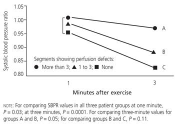
In this study,1 higher values for the three-minute SBPR were related not only to the presence of perfusion defects but also to the extent of the defects. In an 11-segment model with three planar views (anterior, 45-degree left anterior oblique and 75-degree left anterior oblique), patients showing redistribution or persistent defects in more than three scan segments had significantly higher three-minute postexercise SBPR values than patients with three or fewer abnormal segments (Figure 1).1
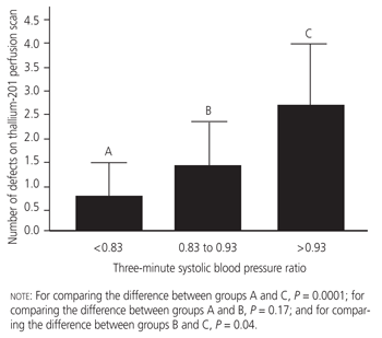
The three-minute SBPR could be used to divide the patients into groups for which the thallium-201 scintigraphic findings were anticipated to be low risk (SBPR less than 0.83), intermediate risk (SBPR between 0.83 and 0.93) or high risk (SBPR greater than 0.93). The study results are presented in Figure 2 and Table 1.1 A three-minute SBPR value of 0.93 or greater carried a 3.85 relative risk for extensive hypoperfusion (defined as more than three abnormal scan segments).
Based on the available data, it is possible to define the relative roles of ST-segment analysis and the three-minute SBPR in the assessment of coronary artery disease. The three-minute SBPR and ST-segment depression have comparable accuracy in diagnosing coronary artery disease. However, the three-minute SBPR may be particularly useful when baseline ST-segment abnormalities are present and the accuracy of the electrocardiographic response to exercise is therefore of limited value.11 In evaluations of the extent of coronary artery disease, the available angiographic and scintigraphic data indicate that the three-minute SBPR is related to the number of diseased coronary vessels and the extent of myocardial perfusion abnormalities. In contrast, the degree of ST-segment depression is generally of limited value in this assessment.
Prognostic Implications
An abnormal SBPR may also be useful in determining the prognosis for patients with coronary artery disease. One Japanese study5 reported the association between an abnormal SBPR (defined as a value of 0.90 or greater) and the prognosis in 271 survivors of myocardial infarction. After a mean follow-up period of four years, the SBPR was found to have a 62 percent sensitivity and a 68 percent specificity for cardiac events (i.e., cardiac death and myocardial infarction).
In the Japanese study,5 univariate associations with abnormal SBPR values included anterior myocardial infarction, congestive heart failure, short exercise duration, ST-segment depression, angina pectoris, reduced ejection fraction and multivessel coronary artery disease. Among these factors, many of which are associated with an adverse cardiovascular prognosis, an abnormal SBPR value was the most important risk factor for cardiac mortality, with a relative risk of 15.4.
When the results of all available studies are considered together, the possible prognostic importance of the three-minute SBPR is consistent with the association of this ratio with extensive abnormalities in myocardial perfusion.
Clinical Implications
The three-minute postexercise SBPR is an easily and readily obtainable measurement that is applicable to all patients (including those with resting ST-segment abnormalities and hypertension) who are undergoing treadmill testing for the evaluation of ischemic heart disease. In patients who are undergoing upright symptom-limited treadmill exercise testing with either supine or upright postexercise blood pressure measurements, a three-minute SBPR value of 0.91 or greater is considered abnormal.
Values of 0.91 or higher have been associated with the presence of coronary artery disease (reflecting both myocardial ischemia and scarring) and abnormal hemodynamics during exercise. In addition, higher values for the three-minute post-exercise SBPR indicate an increased likelihood of more extensive coronary artery disease and carry an adverse prognosis after myocardial infarction (Table 2).
| Lower exercise cardiac index, higher pulmonary capillary wedge pressure and higher systemic vascular resistance |
| Increased likelihood of angiographic coronary artery disease |
| Increased extent of angiographic coronary artery disease |
| More extensive thallium-201 perfusion defects |
| Worse prognosis following acute myocardial infarction |
The three-minute postexercise SBPR should be added to the list of traditional high-risk exercise test indicators in the assessment of ischemic heart disease.