Adolescent idiopathic scoliosis is present in 2 to 4 percent of children between 10 and 16 years of age. It is defined as a lateral curvature of the spine greater than 10 degrees accompanied by vertebral rotation. It is thought to be a multigene dominant condition with variable phenotypic expression. Scoliosis can be identified by the Adam's forward bend test during physical examination. Severe pain, a left thoracic curve or an abnormal neurologic examination are red flags that point to a secondary cause for spinal deformity. Specialty consultation and magnetic resonance imaging are needed if red flags are present. Of adolescents diagnosed with scoliosis, only 10 percent have curves that progress and require medical intervention. The main risk factors for curve progression are a large curve magnitude, skeletal immaturity and female gender. The likelihood of curve progression can be estimated by measuring the curve magnitude using the Cobb method on radiographs and by assessing skeletal growth potential using Tanner staging and Risser grading.
Recent research has led to a better understanding of the natural history of scoliosis. However, the optimal strategy for screening, diagnosing and treating this common spinal deformity remains controversial. Of adolescents diagnosed with scoliosis, only 10 percent have curve progression requiring medical intervention.1 The ability to estimate which curves require therapy has led to more appropriate treatment with observation, bracing or surgery.
Family physicians need to differentiate patients with stable or minimally progressive scoliosis who can be observed from patients with scoliosis that is at high risk for progression. They need to determine the patients they can follow and those who need referral to an orthopedic surgeon. Unnecessary referrals of adolescents with minimal scoliosis who are at low risk for progression can cause marked anxiety and lost time from school and work, and lead to unnecessary radiation exposure.2
Delayed referrals of patients with high-risk curves can lead to increased morbidity. In either situation, the psychologic and social effects of this disease can be profound.2 This article describes an approach to diagnosing and treating scoliosis that allows physicians to lessen the adverse psychologic, medical and economic effects of over-referral or delayed referral of adolescents to orthopedic subspecialists.
Classification
The Scoliosis Research Society has defined scoliosis as a lateral curvature of the spine greater than 10 degrees as measured using the Cobb method on a standing radiograph.3 Idiopathic scoliosis is a structural curve with no clear underlying cause. Secondary causes for scoliosis can usually be identified by radiography and clinical examination (Table 1).
Idiopathic scoliosis is classified based on the age of the patient when it is first identified. Infantile scoliosis has an onset before three years of age. The infantile form accounts for fewer than 1 percent of all cases. Juvenile scoliosis is first detected between three and 10 years of age. The juvenile form occurs in 12 to 21 percent of all patients with idiopathic scoliosis.4 Adolescent idiopathic scoliosis is found between age 10 and skeletal maturity.4 The adolescent form accounts for the majority of cases of idiopathic scoliosis.
Prevalence
Scoliosis is present in 2 to 4 percent of children between 10and 16yearsofage.5 The ratio of girls to boys with small curves of 10 degrees is equal but increases to a ratio of 10 girls for every one boy with curves greater than 30 degrees.5 Scoliosis in girls tends to progress more often and, therefore, girls more commonly need treatment than boys.5 The prevalence of curves greater than 30 degrees is approximately 0.2 percent, and the prevalence for curves greater than 40 degrees is approximately 0.1 percent.1 Improved understanding of the natural history and prognosis of this disease can help the physician predict the patients with scoliosis who need treatment.
TABLE 1 Secondary Causes of Scoliosis
| Inherited disorders of connective tissue | Neurologic disorders | Musculoskeletal |
|---|---|---|
| Ehlers-Danlos Syndrome Marfan syndrome Homocystinuria | Tethered cord syndrome* Syringomyelia Spinal tumor Neurofibromatosis Muscular dystrophy Cerebral palsy Poliomyelitis Friedreich's ataxia Familial dysautonomia (Riley-Day syndrome) Werdnig-Hoffmann disease | Leg length discrepancy Developmental dysplasia of the hip Osteogenesis imper-fecta Klippel-Feil syndrome |
*—A cord unable to change position in the spinal canal because of growth related to scarring, diastematomyelia or other etiology.
Natural History/Prognosis
Once a diagnosis of scoliosis has been made, the primary concerns are whether there is an underlying cause and if the curve will progress. The three main determinants of progression are patient gender, future growth potential and the curve magnitude at the time of diagnosis.1 In all cases, females have a risk of curve progression 10 times higher than males.1 The greater the growth potential and the larger the curve, the greater the likelihood of curve progression.
Evaluation of growth potential is done by assessing the Tanner stage and the Risser grade. Tanner stage 2 to 3 occurs just after the onset of the pubertal growth spurt and is the time of maximum progression of scoliosis.6 The Risser grade (zero to 5) gives a useful estimate of how much skeletal growth remains by grading the progress of bony fusion of the iliac apophysis. The iliac apophysis ossifies in a predictable fashion from anterolateral to posteromedial along the iliac crest.
Risser grades are as follows: grade zero signifies no ossification, grade 1 signifies up to 25 percent ossification, grade 2 signifies 26 to 50 percent ossification, grade 3 signifies 51 to 75 percent ossification, grade 4 signifies 76 up to 100 percent ossification and grade 5 signifies complete bony fusion of the apophysis7 (Figure 1). In one study,8 the Risser grade was directly correlated with the risk of curve progression.
FIGURE 1.
Risser grades zero to 5. Grading is based on the degree of bony fusion of the iliac apophysis, from grade zero (no ossification) to grade 5 (complete bony fusion).
The magnitude of the curve is best determined by measurement of the Cobb angle, which is derived from a standard posteroanterior standing radiograph of the spine. The Cobb angle is the angle formed by a line drawn perpendicular to the top of the superior vertebrae of the scoliotic curve and a similar perpendicular line drawn along the bottom of the inferior vertebrae (Figure 2).
FIGURE 2.
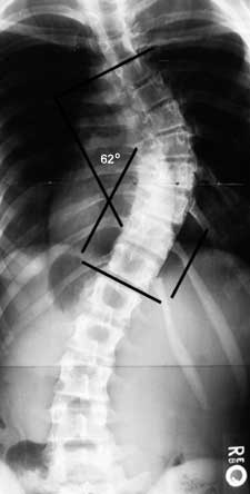
The Cobb method of measuring the degree of scoliosis. The physician chooses the most tilted vertebrae above and below the apex of the curve. The angle between intersecting lines drawn perpendicular to the top of the superior vertebrae and the bottom of the inferior vertebrae is the Cobb angle (here, 62 degrees).
The risk of curve progression can be estimated by taking into account the patient's sex, time of menarche and growth potential (Tanner stage and Risser grade), and the magnitude of the curve. This is key information to help with decisions about the need for referral to an orthopedic surgeon and, in those who are not referred, about examination frequency for curves at lower risk of progression. It is important to remember that this information does not conclusively determine whether a specific curve will progress, only the general risk of a curve progressing. Table 2 summarizes the results of multiple studies done to assist in predicting the risk of curve progression in adolescents; the table can also assist in patient counseling.5,8–11
TABLE 2 Risk of Curve Progression
| Curve (degree) | Growth potential (Risser grade) | Risk* |
|---|---|---|
| 10 to 19 | Limited (2 to 4) | Low |
| 10 to 19 | High (0 to 1) | Moderate |
| 20 to 29 | Limited (2 to 4) | Low/moderate |
| 20 to 29 | High (0 to 1) | High |
| >29 | Limited (2 to 4) | High |
| >29 | High (0 to 1) | Very high |
*—Low risk = 5 to 15 percent; moderate risk = 15 to 40 percent; high risk = 40 to 70 percent; very high risk = 70 to 90 percent.
The risk that an untreated adolescent with scoliosis will have a curve progress into adulthood has been determined. Curves less than 30 degrees at bone maturity are unlikely to progress, whereas curves measuring from 30 to 50 degrees progress an average of 10 to 15 degrees over a lifetime. Curves greater than 50 degrees at maturity progress steadily at a rate of 1 degree per year.1 In most patients, life-threatening effects on pulmonary function do not occur until the scoliotic curve is 100 degrees or greater.1
Of equal significance is the fact that significant psychologic illness has been found in up to 19 percent of females who have curves greater than 40 degrees as adults.2 Social isolation, limited job opportunities and lower marriage rates are possible consequences.
Pathophysiology
Many studies have attempted to uncover the pathophysiologic process underlying idiopathic scoliosis. Multiple abnormalities have been found, yet none has been conclusively linked to all cases.
Studies of twins12 have given the firmest indication that the most significant factor is genetic. Indeed, a recent meta-analysis13 showed that not only is the risk for scoliosis greater in monozygotic twins than in dizygotic twins, the rate of curve progression is nearly identical among twins subjected to a variety of environmental influences. Current theorists believe that scoliosis is a multigene dominant condition with variable phenotypic expression.5 Therefore, even though scoliosis is typically present in most members of the same family, its severity can vary widely from parent to child and sibling to sibling. When both parents have scoliosis, the risk that their children will require treatment is 50 times that in the general population.7
Screening
Screening for scoliosis was common in schools and communities in past years. Over-referral of adolescents with insignificant curves led to a marked decrease in many such programs. Recent studies have demonstrated that over-referral is common even when multiple diagnostic modalities are used.14,15 The American Academy of Orthopedic Surgeons recommends screening girls at ages 11 and 13, and screening boys once at age 13 or 14 years of age. The American Academy of Pediatrics has recommended scoliosis screening with the Adam's forward bending test at routine health visits at 10, 12,14 and 16 years of age, although evidence does not exist to support these recommendations.2
In 1996, the U.S. Preventive Services Task Force released its opinion on screening for adolescent idiopathic scoliosis. The Task Force noted that “there is insufficient evidence for or against routine screening of asymptomatic adolescents for idiopathic scoliosis. Clinicians should remain alert for large spinal curvatures when examining adolescents.”2
HISTORY AND PHYSICAL EXAMINATION
Adolescent idiopathic scoliosis is primarily a diagnosis of exclusion. The history and physical examination are intended to exclude secondary causes for the spinal deformity. The patient should be asked about a family history of scoliosis, menstrual onset, and the presence of pain and neurologic changes, including bowel and bladder dysfunction. The presence of severe pain or neurologic symptoms would be atypical for idiopathic scoliosis.
The physical examination should include an assessment of Tanner stage and a complete neurologic examination. Peak curve progression occurs during Tanner stage 2 or 3. Any abnormal neurologic findings should raise concern for spinal cord pathology. Although there is no ideal screening test, the Adam's forward bend test requires no additional equipment (such as a scoliometer or humpometer) and can help to identify scoliosis.5 The child bends forward at the waist until the spine becomes parallel to the horizontal plane, while holding palms together with arms extended. The examiner looks along the horizontal plane of the spine from the back and side to detect an asymmetry in the contour of the back known as a “rib hump” (Figure 3). A rib hump is a hallmark of scoliotic curves greater than 10 degrees and should prompt radiographic evaluation.
The direction of curves in adolescent idiopathic scoliosis is remarkably consistent. Ninety percent of thoracic curves are to the right.16 Therefore, left thoracic curves should raise a red flag and prompt more extensive evaluation.
Additional red flags include markedly painful scoliosis, untoward stiffness, deviation to one side during the forward bend test, sudden rapid progression in a previously stable curve, extensive progression in a patient after skeletal maturity, abnormal neurologic findings and the stigmata of other clinical syndromes associated with spinal curvature.5,16,17
Radiography and Additional Tests
Abnormalities on physical examination require radiographic evaluation with a single standing posteroanterior radiograph to allow measurement of the curve using the Cobb method and Risser grading of the iliac apophysis. Magnetic resonance imaging is indicated whenever there is a left thoracic curve, unusual pain or abnormalities on neurologic examination, or other red flags, to evaluate for spondylolisthesis, tumors or syringomyelia.18
Referral Guidelines and Treatment
Treatment options for patients with scoliosis range from the unproven or harmful to the beneficial. Physical therapy, chiropractic care, biofeedback and electric stimulation have not been shown to alter the natural history of scoliosis.2,7,12 In contrast, bracing and spinal surgery have been proved to alter the natural history of curve progression. Bracing techniques have also improved markedly; braces are more comfortable and better tolerated than in the past, when studies had shown that adolescents wore their braces only 65 percent of the time they were intended to use them.19
In addition, most modern braces are of the underarm thoracolumbar-sacral orthosis type, which can be worn under the clothing. A recent study showed that bracing had a 74 percent success rate at halting curve progression.20 It is important to counsel adolescents and their parents that bracing does not correct scoliosis but may prevent significant progression of the spinal curvature. Orthosis use is usually continued until the patient reaches Risser grade 4 or 5.5
Spinal surgery with instrumentation corrects a significant part of the deformity and hopefully stops further progression of the scoliotic curve. Current consensus is that surgery should be performed for curves greater than 40 to 45 degrees when there is growth remaining.5,21 Many implants are available to provide excellent stability and strong corrective forces to the spinal column.22 Modern surgery is accompanied by spinal cord monitoring using somatosensory and motor-evoked potentials, thereby decreasing the rate of neurologic injury to one in 7,000 procedures.5
FIGURE 3.
Adam's forward bend test. (Left) As the patient bends over, the examiner looks from behind and from the side, horizontally along the contour of the back. (Right) A rotational deformity known as a rib hump (arrow) can be easily identified.
TABLE 3 Treatment and Referral Guidelines for Patients with Scoliosis
| Curve (degrees) | Risser grade | X-ray/refer | Treatment |
|---|---|---|---|
| 10 to 19 | 0 to 1 | Every 6 months/no | Observe |
| 10 to 19 | 2 to 4 | Every 6 months/no | Observe |
| 20 to 29 degrees | 0 to 1 | Every 6 months/yes | Brace after 25 |
| 20 to 29 | 2 to 4 | Every 6 months/yes | Observe or brace* |
| 29 to 40 | 0 to 1 | Refer | Brace |
| 29 to 40 | 2 to 4 | Refer | Brace |
| >40 | 0 to 4 | Refer | Surgery† |
*—If the patient is Risser grade 4, probably only observation is warranted.
†—If the patient is Risser grade 4, surgery can be delayed.
Adolescent scoliosis can be followed by the family physician if the curve has a low risk of progression and an underlying cause has been excluded. Curves that demonstrate progression in patients with continued growth remaining and curves with a high risk of progression should be referred to an orthopedic subspecialist. Referral is always indicated when red flags are present on physical or radiographic examination. Recommendations for radiograph frequency, treatment and referral are summarized in Table 3.5,7,8,10,11,20,21
