The routine newborn assessment should include an examination for size, macrocephaly or microcephaly, changes in skin color, signs of birth trauma, malformations, evidence of respiratory distress, level of arousal, posture, tone, presence of spontaneous movements, and symmetry of movements. A newborn with one anatomic malformation should be evaluated for associated anomalies. Total and direct bilirubin levels should be measured in newborns with jaundice, and a complete blood count should be obtained in those with pallor or a ruddy complexion. Neurosurgical consultation is necessary in infants with craniosynostosis accompanied by restricted brain growth or hydrocephalus, cephaloceles, or exophytic scalp nodules. Neck masses can be identified by their location and include vascular malformations, abnormal lymphatic tissue, teratomas, and dermoid cysts. Most facial nerve palsies resolve spontaneously. Conjunctivitis is relatively common in newborns. Infants with chest abnormalities may need to be evaluated for Poland's syndrome or Turner's syndrome. Murmurs in the immediate newborn period are usually innocent and represent a transition from fetal to neonatal circulation. Because cyanosis is primarily secondary to respiratory or cardiac causes, affected newborns should be evaluated expeditiously, with the involvement of a cardiologist or neonatologist.
A careful examination at delivery helps the physician detect anomalies, birth injuries, and cardiorespiratory disorders that may compromise a newborn's successful adaptation to extrauterine life. A detailed examination should also be performed after the newborn has completed the transition from fetal to neonatal life. The examination may begin with an evaluation of neonatal size (Table 1). The presence of one anatomic malformation should prompt an evaluation for associated anomalies. Part I of this two-part article focuses on anomalies and disorders involving the skin, head and neck, chest, and respiratory and cardiovascular systems.
TABLE 1 Factors to Consider in Evaluating Size in Newborns
| Small for gestational age (birth weight below 10th percentile) | |
| Symmetric | |
| Features: onset early in gestation; brain size corresponding with body size; glycogen and fat content corresponding with body size (hence, lower risk of hypoglycemia) | |
| Etiology: environmental factors such as smoking or drugs (heroin, methadone, ethanol, phenytoin [Dilantin]); genetic factors such as small maternal size or chromosomal disorder (trisomy 13, 18, and 21 syndromes, Turner's syndrome); intrauterine infections such as TORCH, bacterial (tuberculosis), or spirochetic (syphilis); metabolic disorders such as phenylketonuria | |
| Asymmetric | |
| Features: onset late in gestation; no effect or minimal effect on fetal brain growth; reduced glycogen and fat content relative to body size (hence, increased risk of hypoglycemia); increased risk of perinatal asphyxia and polycythemia (hyperviscosity) | |
| Etiology: uteroplacental insufficiency with chronic fetal hypoxia | |
| Large for gestational age (birth weight above 90th percentile) | |
| Features: increased incidence of perinatal asphyxia and birth injuries; respiratory distress syndrome; hypoglycemia | |
| Etiology: maternal diabetes (increased likelihood of large birth size, respiratory distress syndrome, and hypoglycemia) | |
TORCH =toxoplasmosis,other viruses,rubella,cytomegaloviruses,herpes [simplex] viruses.
Skin
Erythema toxicum neonatorum, transient neonatal pustular melanosis, sucking blister, miliaria, and mongolian spots are among the many benign skin conditions that can occur in newborns. Total and direct bilirubin levels should be measured in newborns with jaundice, and various causes for this condition should be considered (Table 2). The American Academy of Pediatrics1 has published guidelines on the management of hyperbilirubinemia in healthy term infants. A complete blood count should be obtained in newborns with pallor or a ruddy complexion.
TABLE 2 Causes of Hyperbilirubinemia in Newborns
| Unconjugated hyperbilirubinemia |
| Physiologic hyperbilirubinemia (most common cause) |
| Breastfeeding and breastmilk jaundice |
| Increased production of bilirubin: hemolysis (immune or nonimmune), sequestered blood (subdural hematoma, cephalhematoma, hemangioma, ecchymosis), polycythemia, sepsis |
| Decreased hepatic uptake or conjugation: hypothyroidism, Gilbert syndrome, Crigler-Najjar syndrome (types I and II), transient familial neonatal hyperbilirubinemia (Lucey-Driscoll syndrome) |
| Conjugated hyperbilirubinemia |
| Hepatobiliary disorders: neonatal idiopathic hepatitis, infections (TORCH, echovirus, syphilis, systemic infections), prolonged parenteral nutrition, severe hemolytic disease, metabolic disorders (galactosemia, glycogen storage diseases) |
| Ductal disturbances in bilirubin excretion: biliary atresia, choledochal cyst, bile plug syndrome |
TORCH =toxoplasmosis,other viruses,rubella,cytomegaloviruses,herpes [simplex] viruses.
The diagnosis and treatment of cutaneous vascular lesions in newborns are reviewed elsewhere.2
Head and Neck
Head circumference and fontanelle size can indicate a congenital disorder or head trauma (Tables 3 and 43). Craniosynostosis, or premature fusion of cranial sutures, results in growth restriction perpendicular to the affected suture(s) and compensatory overgrowth in unrestricted regions4 (Figure 1). This anomaly may suggest a genetic disorder such as Apert's syndrome or Crouzon's disease. If the synostosis is accompanied by restricted brain growth or hydrocephalus, neurosurgical intervention is necessary.
TABLE 3 Head Circumference and Fontanelle Size in Newborns*
| Macrocephaly: as an isolated anomaly, is often familial, with autosomal dominant inheritance; may be a manifestation of other anomalies, including hydrocephalus and skeletal disorders such as achondroplasia |
| Microcephaly: can be familial, with autosomal dominant or recessive inheritance; may be associated with infections (viruses such as cytomegalovirus) and syndromes such as trisomy 13 and 18, Cornelia de Lange's, Rubinstein-Taybi, Prader-Willi, and fetal alcohol |
| Large fontanelles: may be associated with hypothyroidism, trisomy 13, 18, and 21 syndromes, and bone disorders such as cleidocranial dysostosis or hypophosphatasia |
*—The size of the head and the anterior and posterior fontanelles should be compared with appropriate standards. Head size varies with age, sex, and ethnicity and has a general correlation with body size.
TABLE 4 Common Forms of Head Trauma in Newborns
| Caput succedaneum |
| Commonly observed after prolonged labor |
| Secondary to accumulation of blood or serum above the periosteum |
| Clinical features: poorly demarcated soft tissue swelling that crosses suture lines; accompanying pitting edema and overlying petechiae, ecchymoses and purpura |
| Treatment: none needed because condition usually resolves within days |
| Cephalhematoma |
| Less common than caput succedaneum but may occur after prolonged labor and instrumentation |
| Secondary to rupture of blood vessels that traverse skull to periosteum |
| Clinical features: well-demarcated, often fluctuant swelling that does not cross suture lines; no overlying skin discoloration; possibly, skull fractures; sometimes, elevated ridge of organizing tissue |
| Complications: intracranial hemorrhage with resultant shock; hyperbilirubinemia |
| Treatment: none recommended for uncomplicated lesions, which usually reabsorb in 2 weeks to 3 months; for suspected or detected fracture, radiographs again at 4 to 6 weeks to ensure closure of linear fractures and to exclude formation of leptomeningeal cysts, which can be detected by radiography (if there is doubt, cranial computed tomographic scanning can be helpful)3; for depressed skull fractures, immediate neurosurgical consultation |
FIGURE 1.
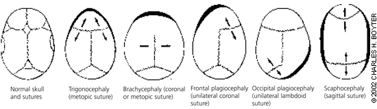
Skull shapes associated with premature closure of single sutures. Arrows denote directions of continued growth across sutures that remain open. Heavy lines indicate areas of maximal skull flattening. When combinations of sutures remain closed, more complex skull shapes occur.
Large meningoceles or encephaloceles are usually diagnosed prenatally or at birth. Smaller defects may be mistaken for cutaneous lesions such as hemangiomas or dermoid cysts. Congenital exophytic scalp nodules should always be evaluated further, because 20 to 37 percent of these lesions connect to the underlying central nervous system.5 Cutaneous signs of cranial dysraphism include the “hair collar sign” (darker, coarser hair encircling the scalp nodule), vascular malformations, and cutaneous dimples and sinuses. Cephaloceles and exophytic scalp nodules should be assessed by magnetic resonance imaging (MRI), and a neurosurgical consultation should be obtained.5
The most common neck masses are vascular malformations, abnormal lymphatic tissue, teratomas, and dermoid cysts. Neck masses can be identified based on their location (Figure 2).6 Thyroglossal duct cysts, one of the most frequent congenital anomalies of the neck, are typically midline and inferior to the hyoid bone. Surgical consultation is required in newborns with thyroglossal duct cysts.
FIGURE 2.
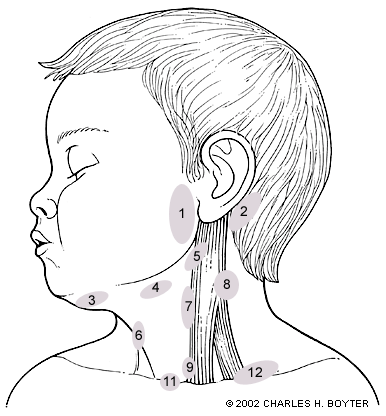
Identification of neck masses based on their location.
KEY:
1 = Preauricular area (parotid gland): congenital lesions-cystic hygroma, hemangioma, venous malformation; inflammatory condition-lymphadenitis secondary to infection in upper face and/or anterior scalp
2 = Postauricular area: congenital lesions-branchial cleft I (cystic, inflamed, or both); inflammatory condition-lymphadenitis secondary to inflammation of posterior scalp
3 = Submental area: congenital lesions-thyroglossal duct cyst, cystic hygroma, dermoid cyst, venous malformation; inflammatory condition-lymphadenitis secondary to inflammation in perioral area, anteriour oral area, or nasal cavity
4 = Submandibular area: congenital lesions-cystic hygroma, hamangioma, ranula; inflammatory condition-lymphadenitis of submandibular gland secondary to inflammation in cheek and/or mid-oral cavity; in cystic fibrosis, enlartement of submandibular gland without inflammation
5 = Jugulodiagastric area (tonsil node; normal structures include transverse process of C2 and styloid process): congenital lesions-bronchial cleft I or II, hemangioma, cystic hygrom; inflammatory condition- lymphadenitis secondary to oropharyngeal inflammation
6 = Area of neck midline (normal structures include hyoid, thyrouid isthmus, and thyroid cartilage): congenital lesions-thyroglossal duct cyst, dermoid cyst; inflammatory condition-lymphadenitis
7 = Area at anterior border of sternocleidomastoid muscle (normal structures include hyoid, thyroid cartilage, and carotid bulb): congenital lesions-branchial cleft I, II, or III (IV is rare), laryngocele, hemangioma, lymphangioma, hematoma (fibroma of sternocleidomastoid muscle)
8 = Spinal accessorry: inflammatory condition-lymphadenitis secondary to nasopharyngeal inflammation
9 = Paratracheal area: thyroid mass, parathyroid mass, esophageal diverticulum, metastatic lesion
10 = Supraclavicular area (normal structures include fat pad, pneumatocele from apical lobe related to defect in Gibson fascia[prominent mass with Valsalva's maneuver]): congenital lesion-cystic hygroma; neoplastic lesion-lipoma.
11 = Suprasternal area: thyroid mass, lipoma, dermoid cyst, thymis mass, mediastinal mass
Information from May M. Neck masses in children: diagnosis and treatment. Pediatr Ann 1976;5:518–35.
Clavicular fractures are the most common broken bones in newborns, especially large neonates. Of these, greenstick fractures are the most frequent and are usually asymptomatic. Newborns may present with decreased or absent movement and pain or tenderness on movement of the arm on the affected side, deformity and discoloration over the fracture site, and crepitus or irregularity along the clavicle. Treatment is directed at minimizing the newborn's pain or discomfort. If the newborn with clavicular fracture is in pain, the affected arm should be immobilized, with the arm abducted more than 60 degrees and the elbow flexed more than 90 degrees.3
FACE
Facial nerve paralysis is caused by compression of the nerve against the sacral promontory or by trauma resulting from the use of forceps during delivery. Paralysis is usually apparent on the first or second day of life. The nasolabial fold on the paralyzed side is obliterated, and the corner of the mouth droops; with crying, the mouth is drawn to the normal side4 (Figure 3). With peripheral facial nerve paresis, the forehead and eyes may be affected.
FIGURE 3.
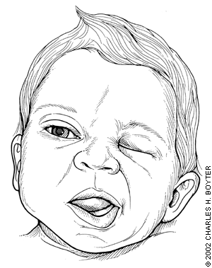
Asymmetry caused by facial nerve paralysis, with inability to close eye, nasolabial fold flattening, and inability to move lips on the affected side. Newborns with facial nerve paralysis have difficulty effecting a seal around the nipple and consequently exhibit drooling of milk or formula from the paralyzed side of the mouth.
Most facial nerve palsies resolve spontaneously within days, although full recovery may require weeks to months. A persistently open eye should be protected from corneal drying. Electrodiagnostic testing may be necessary if no improvement occurs within seven to 10 days; rarely, surgical intervention is needed.3 Congenital absence or hypoplasia of the depressor anguli oris muscle may simulate facial palsy.
Erupted teeth are present in approximately one of 2,000 newborns.7 Although natal teeth are frequently found in normal infants, they are more often present in newborns with cleft palate. They are also commonly associated with Ellis–van Creveld syndrome, Hallermann-Streiff syndrome, and pachyonychia congenita syndrome. Most erupted teeth, particularly if loose, require removal.
Isolated cleft palate differs genetically from cleft lip. Mild forms of cleft palate include sub-mucosal clefts, pharyngeal incompetence and bifid uvula. Cleft lip, with or without cleft palate, is found in newborns with trisomy 13 syndrome, holoprosencephaly (median cleft lip), and amnion rupture sequence. Newborns with a cleft lip or palate require genetic evaluation and plastic surgery. Because of feeding difficulties, the mothers of these infants may benefit from lactation consultation and occupational therapy.
EYES
Marked lid edema often results in eversion of the upper lid when force is applied to open the eye. Examination should be postponed until the edema resolves. Subconjunctival hemorrhages, which are common after vaginal delivery, usually do not represent ocular trauma. Conjunctivitis is relatively common in newborns (Table 5).8,9
TABLE 5 Conjunctivitis in Newborns
| Chemical conjunctivitis |
| Usually occurs within 24 hours of instillation of eye prophylaxis after birth |
| Clinical features: mild lid edema with sterile discharge from eyes |
| Treatment: none needed because condition usually resolves within 48 hours after birth |
| Gonorrheal conjunctivitis |
| Usually occurs within 24 to 48 hours after birth |
| Clinical features: profound lid edema, chemosis, intensely purulent exudates, corneal ulceration |
| Treatment: for proven penicillin-susceptible organisms, aqueous crystalline penicillin G, 100,000 units per kg per day IV given in four divided doses for 7 days; because of emergence of resistant strains of Neisseria gonorrhoeae, recommended therapy is ceftriaxone (Rocephin), 25 to 50 mg per kg IV or IM (not to exceed 125 mg) given once, or cefotaxime (Claforan), 100 mg per kg IV or IM given once; until discharge is eliminated, frequent eye irrigations with saline; gonorrheal treatment for the mother and her sexual partner(s) |
| Chlamydial conjunctivitis |
| Usually occurs within 7 to 14 days after birth |
| Clinical features: watery discharge that later becomes copious and purulent; if untreated, may result in corneal scarring and pannus formation |
| Treatment: orally administered erythromycin, 50 mg per kg per day in four divided doses for 2 weeks |
| HSV conjunctivitis |
| Usually occurs within 2 weeks after birth |
| Eyes involved in 5% to 20% of HSV-infected infants |
| Clinical features: infants may present with keratitis, cataracts, chorioretinitis, or optic neuritis; imperative to rule out disseminated herpes |
| Treatment: both topical and systemic antiviral agents, because HSV-infected neonates do not present with isolated conjunctivitis; systemic therapy—acyclovir (Zovirax), 60 mg per kg per day in three divided doses for 14 days if disease is limited to skin, eyes, and mouth; topical therapy—1% trifluridine (Viroptic) or 3% vidarabine (Vira-A); referral to subspecialist |
IV = intravenous; IM = intramuscular; HSV = herpes simplex virus.
Information from Meisler DM, Beauchamp GR. Disorders of the conjunctiva. In: Nelson LB, Harley RD, eds. Harley's Pediatric ophthalmology. 4th ed. Philadelphia: Saunders, 1998:199–214, and Pickering LK, ed. 2000 Red book: report of the Committee on Infectious Diseases. 25th ed. Elk Grove Village, Ill.: American Academy of Pediatrics, 2000.
Coloboma (absence or defect of some ocular tissue) may involve the eyelid margin, as in Treacher Collins syndrome, or the iris and retina, as in the CHARGE association (syndrome of coloboma, heart disease, choanal atresia, postnatal growth retardation, genital hypoplasia and ear anomalies). Aniridia (absence of the iris) is usually bilateral and is almost always associated with poor vision and nystagmus. Newborns with aniridia or coloboma should have a formal eye examination.
The red reflex normally shows no dullness or irregularities. A white pupil (cat's eye reflex) denotes an abnormality of the lens, vitreous, or fundus. One of the most common presenting signs of a cataract is a white pupillary reflex. Congenital cataract is present in 0.4 percent of newborns. These infants should be tested for TORCH (toxoplasmosis, other viruses, rubella, cytomegaloviruses, herpes [simplex] viruses) infections. Newborns with monocular congenital or dense cataracts are at risk for developing deprivation amblyopia. Newborns with cataracts should be evaluated by an ophthalmologist.10
Congenital glaucoma, while uncommon, may present at birth. More often, signs of glaucoma develop during the first several weeks or months of life and include corneal cloudiness and enlargement, tearing, blepharospasm, and photophobia. Corneal edema can also occur secondary to the malpositioning of forceps during delivery. Prompt referral to an ophthalmologist is indicated.11
Chest
Although pectus carinatum and pectus excavatum are of concern to parents, these physical anomalies are rarely of clinical significance. Unilateral absence or hypoplasia of the pectoralis major muscle suggests the diagnosis of Poland's syndrome (sometimes called Poland's sequence). Common associated findings in this syndrome include rib defects, hypoplasia of the upper extremities, and syndactyly. Occasionally, more severe limb reduction deformities, hemivertebrae, renal anomalies, and dextrocardia may be present.
Widely spaced nipples, excessive nuchal skin, and lymphedema are findings associated with Turner's syndrome. The evaluation of newborns suspected of having this syndrome should include chromosomal analysis, echocardiography to detect cardiac lesions, and a genetic consultation.
A small thorax suggests pulmonary hypoplasia. A bell-shaped thorax is often present in newborns with neurologic abnormalities or some dwarfing syndromes.
Respiratory and Cardiovascular Systems
Newborns with choanal atresia present with cyanosis that is relieved by crying. The diagnosis is usually established by the inability to pass a catheter through the nostril(s). Unilateral choanal atresia may remain undiagnosed for years. Infants and children with this congenital anomaly may present with mucus or foul-smelling secretions from the affected nares and respiratory distress associated with upper respiratory infection.
Newborns with significant cyanosis should be evaluated expeditiously. Depending on the clinical findings, consultation with a neonatologist may be required. Respiratory disease is more likely in newborns who are tachypneic and using accessory muscles of respiration. Newborns with heart disease generally breathe normally, except for mild tachypnea or hyperpnea. The differential diagnosis and evaluation of cyanosis in infants are presented in Table 6.
The normal heart rate in newborns is 120 to 160 beats per minute. Some term newborns have a resting heart rate below 90 beats per minute. If the heart rate does not increase appropriately with stimulation, serum electrolyte levels should be checked, and an electrocardiogram should be obtained to rule out heart block.
TABLE 6 Differential Diagnosis of Cyanosis in Infants
| Pulmonary disorders |
| Respiratory distress syndrome |
| Aspiration syndromes: blood, meconium, amniotic fluid |
| Infections: pneumonia |
| Pneumothorax, pleural effusion |
| Diaphragmatic hernia |
| Persistent pulmonary hypertension of the newborn |
| Choanal atresia |
| Pierre Robin syndrome* |
| Abdominal distension: elevation of |
| diaphragm |
| Cardiac disorders |
| Fallot's tetrology |
| Transposition of the great arteries |
| Tricuspid atresia |
| Pulmonary atresia with intact ventricular septum |
| Truncus arteriosus |
| Ebstein's anomaly |
| Double-outlet right ventricle |
| Single ventricle with pulmonary stenosis |
| Total anomalous pulmonary venous drainage |
| Coarctation of the aorta† |
| Interrupted aortic arch† |
| Hypoplastic left heart syndrome† |
| Others |
| Central nervous system disorders: neuromuscular infections, asphyxia, seizures, hemorrhage, apnea |
| Hematologic disorders: polycythemia |
| Metabolic and endocrine disorders: hypoglycemia, hypocalcemia, hypermagnesemia |
| Hypothermia |
*—Pierre Robin syndrome includes micrognathia, glossoptosis, and cleft of the soft palate; the primary defect is early mandibular hypoplasia.
†—Coarctation of the aorta, interrupted aortic arch, and hypoplastic left heart syndrome cause systemic hypoperfusion with mild or no cyanosis.
Diminished pulses in all extremities indicate poor cardiac output or peripheral vasoconstriction. Absent or diminished femoral pulses suggest the presence of ductaldependent cardiac lesions (e.g., coarctation of the aorta). Although hypertension is uncommon in newborns, it is rarely idiopathic. An approach to determining the cause of neonatal hypertension is presented in Figure 4.12
Palpation and auscultation may reveal a shift in the position of the heart from normal, as occurs in dextrocardia. In newborns, a murmur does not always signify the presence of heart disease, nor does the absence of a murmur provide reassurance of normalcy. Newborns with relatively benign lesions, such as small ventricular septal defects, often have the loudest murmurs, whereas newborns withsevere heart disease may have no murmurs. The most commonly auscultated murmurs in the immediate newborn period are flow murmurs that represent a transition from fetal to neonatal circulation (e.g., tricuspid regurgitation, patent ductus arteriosus). Further evaluation is required if a murmur persists beyond several weeks in a healthy newborn or if a murmur is present in a critically ill infant.
