Given the high rate of morbidity and mortality associated with abdominal aortic aneurysms (AAAs), accurate diagnosis and preoperative evaluation are essential for improved patient outcomes. Ultrasonography is the standard method of screening and monitoring AAAs that have not ruptured. In the past, aortography was commonly used for preoperative planning in the repair of AAAs. More recently, computed tomography (CT) has largely replaced older, more invasive methods. Recent advances in CT imaging technology, such as helical CT and CT angiography, offer significant advantages over traditional CT. These methods allow for more rapid scans and can produce three-dimensional images of the AAA and important adjacent vascular structures. Use of endovascular stent grafts has increased recently and is less invasive for the repair of AAAs in selected cases. Aortography and CT angiography can precisely determine the size and surrounding anatomy of the AAA to identify appropriate candidates for the use of endovascular stent grafts. Helical CT and CT angiography represent an exciting future in the preoperative evaluation of AAAs. However, this technology is not the standard of care because of the lack of widespread availability, the cost associated with obtaining new equipment, and the lack of universal protocols necessary for acquisition and reconstruction of these images.
Recently, significant technologic advances have been made in the imaging of abdominal aortic aneurysms (AAAs). Ultrasonography, with its wide availability and low cost, remains the principal tool for diagnosing suspected aneurysms and monitoring the progress of known aneurysms.
Over the years, the use of aortography has changed significantly. In the past, it has been used extensively in the preoperative evaluation of AAAs. Most recently, aortography has been used more selectively to address specific clinical issues not resolved by less invasive means, such as the presence of other nearby vessel stenoses (e.g., renal, mesenteric, or iliac lesions) that might alter plans for surgical repair. In addition, aortography now has a role in the use and placement of endovascular stent grafts for the repair of AAAs.
Recently, aortography has been replaced with less invasive technologies such as computed tomography (CT) and magnetic resonance imaging (MRI). While MRI has a limited role in the pre-operative work-up of AAAs because of its higher cost, limited availability, and susceptibility to artifact, CT technology has emerged as the most commonly used imaging modality. Advantages of CT include minimal invasiveness, widespread availability, consistently reproducible results, and a relative cost savings compared with aortography and MRI. Advancements in CT technology, such as the advent of helical CT and CT angiography, will likely increase the role of CT imaging in the evaluation and treatment of AAAs.
Helical CT technology can provide superior three-dimensional anatomic detail and is particularly useful when repair with endovascular stent graft is being considered. The current drawbacks of this technology include cost, less availability of equipment and software, and the need for standardization of methods to construct the three-dimensional images.
Epidemiology
AAAs are a significant health care burden and a cause of growing concern. From 1951 to 1980, the reported incidence of AAAs rose from 8.7 per 100,000 persons to 36.5 per 100,000—a threefold increase.1,2 Several factors, including a growing elderly population, increased life expectancy, and improvements in diagnostic capabilities, have influenced this increase in incidence. The prevalence of AAAs varies according to race and gender and increases with age. The estimated prevalence of AAAs is 2 to 5 percent in populations older than 60 years.3 For any given patient, aortic rupture carries an overall mortality rate of 80 to 90 percent.4 In contrast, the operative mortality rate for unruptured, electively repaired AAAs is 8.4 percent.5
Unruptured aneurysms that are not repaired often gradually enlarge. The majority of aneurysms discovered in screening are small and do not require surgical repair. The risk of rupture generally increases as the diameter of the aneurysm increases. According to results of two recently published large AAA screening trials,5,6 the U.K. Small Aneurysm Trial (UKSAT) and the Aneurysm Detection and Management (ADAM) study, the rupture risk of aneurysms that were 4.0 to 5.5 cm in diameter was 1.0 percent and 0.5 percent per year, respectively. In comparison, other older studies reported an annual rupture risk of 3.4 percent for aneurysms that were 5.0 to 5.9 cm in diameter.7
Risk Factors
Identifying patients at risk for AAAs can lead to early detection with subsequent repair before rupture occurs. Numerous risk factors, including male sex, tobacco use, and increased age, are associated with AAAs. Population-based studies have demonstrated that AAAs occur four to five times more often in men than in women.8,9 In a large-scale screening study,10 more than 73,400 veterans were screened for the presence of AAAs. Smoking was identified as the most closely associated risk factor.10 Other risk factors include atherosclerosis, hypertension, chronic obstructive pulmonary disease, family history of aneurysms, white race, and the presence of other aneurysms.8,11
Pathogenesis
An aneurysm is commonly defined as a 50 percent increase in normal blood vessel diameter. Atherosclerosis has long been considered the primary cause of aneurysm formation. However, recent findings12 suggest a multifactorial degenerative process with atherosclerosis possibly representing a response to other nonspecific types of vessel wall injury. These other types of injury include infection, inflammatory disease, increased protease activity within the arterial wall, genetically regulated defects in collagen and fibrillin, and mechanical factors.1,12,13 The clinical presentation of AAAs varies. Approximately 75 percent of aneurysms are asymptomatic on identification.13 Symptomatic aneurysms often appear as a pulsatile abdominal mass, and cause pain in the back, testicles, or groin, and sometimes can even cause shock.1 Aneurysms tend to become symptomatic when they rapidly expand, leak, or rupture.13
Imaging Modalities
The choice of imaging modality depends on the clinical presentation of the patient (Table 1). Screening and serial monitoring are performed most efficiently with ultrasonography. Symptomatic patients with stable vital signs are usually evaluated with CT in an emergent situation, whereas those who are hemodynamically unstable typically go directly to surgery.
TABLE 1 Comparison of Available Imaging Modalities
| Imaging modality | Advantages | Disadvantages |
|---|---|---|
| Ultrasonography | Lower cost Widely available Noninvasive | Suboptimal in obese patients |
| Suboptimal in patients with increased bowel gas | ||
| Increased interobserver variation | ||
| Aortography | Visualize renovascular disease Identifies anomalous vessels Aids placement of endovascular stent grafts | Invasive |
| Higher cost | ||
| Increased patient morbidity | ||
| Underestimates aneurysm size | ||
| Exposure to iodinated contrast | ||
| MRI | Noninvasive Lack of ionizing radiation | Higher cost |
| Motion artifact | ||
| Contraindications with metal clips and pacemakers | ||
| Patient claustrophobia | ||
| Availability of scanner and software | ||
| CT | Noninvasive Highly predictive of aneurysm size Localize proximal extent of aneurysm Identify other abdominal pathology Procedure of choice for suspected rupture | Use of ionizing radiation |
| Higher cost compared with ultrasonography | ||
| Limited information regarding arterial anatomy | ||
| Helical CT and CTA | Noninvasive Faster scanning time Use in conjunction with endovascular stent grafts | Higher cost |
| Lack of availability of scanner and software | ||
| Use of ionizing radiation |
MRI = magnetic resonance imaging; CT = computed tomography; CTA = computed tomographic angiography.
ULTRASONOGRAPHY
Ultrasonography is the examination of choice for screening and monitoring the rate of AAA enlargement. Figures 1 and 2 are longitudinal and transverse sonograms, respectively, of an AAA. Ultrasonography has a sensitivity of nearly 100 percent in the diagnosis of AAAs.12–15 This procedure is largely preferred for screening and monitoring because of its relatively low cost, availability, and noninvasive nature. Ultrasonography is accurate to within 0.3 cm in estimating aneurysm diameter on serial scans and has little interobserver variation.3,16 However, the procedure does have limitations. Images tend to be suboptimal in patients who are obese or who have excessive bowel gas. Ultrasonography cannot reliably identify the presence of periaortic disease, or the proximal and distal extent of the aneurysm. It also cannot determine the patency of visceral vessels, the relationship of the aneurysm with respect to renal vessels, or the presence of iliac aneurysms.12–14,17 As a result, ultrasonography alone cannot fully provide the necessary information required in the preoperative evaluation of a patient undergoing elective aneurysm repair.
FIGURE 1.
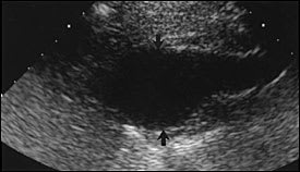
Longitudinal view of the abdominal aorta demonstrating a focal area of enlargement (arrows) consistent with an abdominal aortic aneurysm.
FIGURE 2.
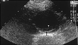
Transverse sonogram of the abdominal aorta demonstrating an abdominal aortic aneurysm (arrows) with a small amount of mural thrombus (arrowhead).
AORTOGRAPHY
Conventional aortography now has a limited role in the preoperative evaluation of AAAs. Aortography is often reserved for special situations, such as when patients have suspected renovascular stenosis, chronic mesenteric ischemia, occlusive iliac disease, juxtarenal or suprarenal AAAs, horseshoe kidneys, previous colectomy, and thoracoabdominal extension, and is also used in endovascular graft placement.18 Advantages of aortography include consistent visualization of the renal artery origins, renal and visceral artery stenoses, accessory renal arteries, and extension of an aneurysm into the iliac arteries.18 Figures 3a and 3b show a bilobed infrarenal AAA.
FIGURE 3A.
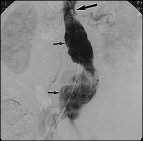
Early arterial phase of a posteroanterior abdominal aortogram showing a bilobed aneurysm (arrows) originating just below the renal arteries.
FIGURE 3B.
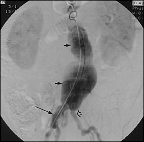
Late arterial phase of the same posteroanterior abdominal aortogram, further defining the bilobed abdominal aortic aneurysm. The aneurysm extends to, but does not involve, the left common iliac artery (outline arrow). On the contralateral side, the aneurysm extends to and involves the right common iliac artery (long arrow).
The use of aortography in the imaging of AAAs also has several drawbacks that prevent its more routine use. For instance, aortography tends to underestimate the size of the aneurysm because it only demonstrates the patent lumen, and typically there is a circumferential organized thrombus within most aneurysms. In some cases, an aneurysm is missed altogether. Other disadvantages of aortography include its invasive nature, cost, and risk of exposure to large amounts of iodinated contrast.
Recently, endovascular stent grafts have been developed as a less invasive treatment for selected patients with AAA. Aortic angiography and fluoroscopy are used for preoperative planning and placement of the endovascular stent grafts. The stent graft is loaded within a large catheter that enters via the femoral artery and is deployed within the abdominal aorta over a guidewire at the level of the aneurysm. Once the stent graft is in place, it is expanded with an angioplasty balloon to hold it tightly against the abdominal aorta and exclude the aneurysm from aortic blood flow. Over time, the aneurysm should shrink.
MRI
MRI is minimally invasive and, when combined with magnetic resonance angiography (MRA), can provide excellent details for the preoperative evaluation of AAAs. MRI with MRA has 100 percent sensitivity in detecting aneurysms, and successfully identifies the proximal and distal extent of the aneurysms, the number and origins of renal arteries, and the presence of inflammation.18 Renal artery stenoses greater than 50 percent can be detected with a sensitivity of 84 to 100 percent.13,19 Advantages of MRI with MRA include lack of ionizing radiation and iodinated contrast medium exposure. Disadvantages include increased cost, patient claustrophobia during longer scanning times, and difficulties with patient compliance in lying still during the procedure to avoid motion artifact.
CT
CT has a broad array of uses in the imaging of AAAs. It is used as a screening test when ultrasound images are suboptimal; as a diagnostic test when a hemodynamically stable, ruptured AAA is suspected; and in the preoperative work-up for the repair of AAAs. CT is a superior diagnostic modality compared with ultrasonography because it offers the clinician valuable information about not only the aneurysm but also the surrounding anatomy (Figure 4). Unlike ultrasonography, there is no concern regarding reproducibility of results, interobserver technical error, or limitations caused by body size or the presence of bowel gas. CT technology is widely available, noninvasive, and can be performed quickly when needed to rule out a suspected AAA rupture. While CT has numerous advantages, there are limitations to traditional CT. CT images contain limited information on arterial anatomy. Complex cases involving AAAs may require additional imaging modalities to obtain the necessary road map of nearby vessels for planning surgical repair.
FIGURE 4.
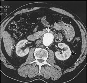
Computed tomographic scan of the abdominal aorta at the level of the kidneys (outline arrows) showing an aneurysm (arrows) with a small amount of mural thrombus along the posterior and lateral walls. The entire abdomen is also clearly visualized, providing useful information about the surrounding anatomy.
HELICAL CT
The development of helical CT and CT angiography will likely lead to wide use of these modalities in the preoperative evaluation of AAAs. Also, the use of endovascular stent grafts in the repair of AAAs requires a technologically advanced modality, such as helical CT and CT angiography, to provide the necessary detailed preoperative anatomic information. Helical CT is accomplished by rotation of the scanning device coupled with continuous table feed of the patient.20 When compared with conventional CT, helical CT has faster scanning time (30 to 60 seconds) and the ability to obtain all images in one breath hold, thereby eliminating the risk of respiratory artifact. Dual-slice helical CT is a newer form of helical CT technology that allows the volume of data scanned within a given time to double, thus achieving even faster scanning times.21
CT angiography is accomplished by combining all of the axial slices to produce a three-dimensional reconstructed image of the AAA. This image can be rotated into any plane that best demonstrates the relevant anatomy (Figures 5a and5b). Dual-slice helical CT correlates well with surgical findings in measuring the proximal and distal extent of the aneurysm. CT and CT angiography are not only less invasive than conventional aortography but also allow for more rapid scanning times and evaluation of the rest of the abdomen.
FIGURE 5A.
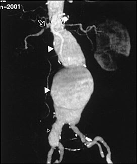
Reconstructed computed tomographic angiogram in the frontal projection demonstrating the bilobed infrarenal aortic aneurysm (arrow heads). The renal arteries are well visualized (small arrows), revealing a severe stenosis of the proximal right renal artery (outline arrow) and the proximity of the aneurysm neck to the renal arteries. The relationship of the aneurysm to the iliac arteries and the aneurysmal dilatation of the right common iliac artery (outline arrow head) are also well displayed.
FIGURE 5B.
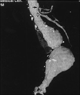
Reconstructed computed tomographic angiogram in the lateral projection showing the bilobed nature of the aneurysm. The celiac artery (arrow) and the superior mesenteric artery (outline arrow) are well demonstrated.
A complete evaluation of the abdomen is important in identifying relevant associated abnormalities, such as a horseshoe kidney, venous or arterial anomalies that would alter the surgical approach, or inflammatory/fibrotic changes within the aneurysm. Other details, including assessment for patency of the inferior mesenteric artery and possible involvement of the iliac arteries, are also reliably achieved with CT and CT angiography. Notably, even patients being considered for endovascular repair with a stent graft sometimes have preoperative evaluation with CT and CT angiography instead of aortography.
