Am Fam Physician. 2003;68(8):1601-1602
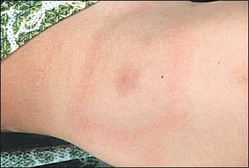
A five-year-old boy was brought to the office in mid-July by his parents. He had a nine-day history of fever as high as 39.4°C (103°F), headache, and malaise. During the past two days, he complained of pain in his right lower extremity. He could bear full weight and walked without an appreciable limp. He had a sick contact with an upper respiratory infection and productive cough for the past two weeks. No unusual travel history was elicited. Physical examination revealed an alert, well-developed, and well-nourished child. His vital signs were significant for a temperature of 39.2°C (102.6°F) orally, a pulse of 136, and a respiratory rate of 28. His head and neck examination was unremarkable. Lungs were clear to auscultation and cardiac examination was normal. On palpation of his abdomen, mild hepatomegaly was appreciated. The boy had full range of motion of his lower extremities and no focal tenderness or erythema. A neurologic examination was nonfocal. Laboratory analysis revealed a white blood cell count of 6,800 per mm3, normal chemistries, and an elevated erythrocyte sedimentation rate of 64 mm per hr. A chest radiograph was normal, and a plain film of the right lower extremity revealed only a small effusion of the knee. Four days after initial presentation, an erythematous rash appeared on the patient's right hip area (see accompanying figure).

Question
Discussion
The answer is B: Borrelia infection. The patient in this case has Lyme disease, and the annular rash depicted is erythema chronicum migrans (ECM), characteristic of this disorder. The diagnosis was based on the clinical presentation. Enzyme-linked immunosorbent assay (ELISA) serology and Western blot analysis corroborated a diagnosis of early Lyme disease. The patient was treated with oral amoxicillin, and his symptoms dramatically improved after 96 hours.
Lyme disease is caused by the spirochete Borrelia burgdorferi and is transmitted to human hosts by ticks of the Ixodes genus. Most cases in the United States are reported in the Northeast, upper Midwest, and northern California, from May through September. There are three stages of Lyme disease that have been described: early localized, early disseminated, and late disease.
Early localized disease is seen days to weeks after a tick bite and is characterized by ECM. Fever, headache, malaise, myalgias, and arthralgias also may be seen. The early disseminated stage, on the other hand, occurs days to months after a tick bite and can involve many different organ systems. Neurologic manifestations include meningitis and cranial nerve palsies. Patients with cardiac involvement may present with pericarditis, myocarditis, or cardiac conduction blocks. Hepatitis, iritis, lymphadenopathy, and renal abnormalities also can be seen. Late Lyme disease is characterized by a chronic monoarticular or asymmetric oligoarticular arthritis involving large joints, especially the knee. Neurologic sequelae such as subacute encephalopathy and axonal polyneuropathy also can be seen as a part of the late stage of Lyme disease.1
The diagnosis of Lyme disease is generally based on clinical presentation. Serologic tests such as ELISA and Western blot analysis may be used to support the clinical diagnosis but have limited sensitivity and specificity. Polymerase chain reaction (PCR) testing of a skin biopsy from the wound site may detect Borrelia DNA. Treatment options for ECM include two to three weeks of oral amoxicillin for children younger than eight years and doxycycline for older children and adults. The vaccine for prevention of Lyme disease (Lyme RIX) has been withdrawn from the market by the manufacturer.
Osteomyelitis also may present with fever, and elevated sedimentation rate and limb pain. Children usually present with an abnormal gait (inability or unwillingness to bear weight) and focal tenderness to palpation. There also may be erythema and purulent drainage from the site. Periosteal elevation may be noted on plain film and leukocytosis is generally present. The diagnosis can be confirmed with a bone scan, and treatment consists of intravenous antibiotics and surgical drainage.
Loxosceles reclusa is a tan to brown spider found mostly in the southeastern and south-central United States. Stinging pain usually develops within hours at the site of the spider bite and a dark blister evolves over days. Systemic symptoms may occur in the very young, old, and debilitated. Unlike the spreading erythema of ECM, the skin lesion in Loxosceles envenomation usually remains localized and may progress to necrotic ulceration in some cases. There is no clearly efficacious treatment.
Ehrlichiosis is a tick-borne illness that also is seen chiefly in the southeastern and south-central United States. The Lone Star and Ixodes ticks are the vectors for this disease. Patients typically present with an influenza-like illness. A maculopapular or petechial rash may be seen, but this occurs in less than one half of patients. Leukopenia, thrombocytopenia, and elevated liver function tests may be demonstrated on laboratory analysis, and PCR and antibody testing may be used to confirm the diagnosis.2 The disease can progress to multiorgan system failure, coma, and death if not recognized and treated early with doxycycline.
Ewing's sarcoma is a childhood malignancy of bone and soft tissue. Presenting symptoms include pain, swelling, limited range of motion, and tenderness over the involved site. Fever and weight loss also may be seen. Radiographically, a lytic bone lesion with periosteal reaction (“onion-skinning”) may be present. Treatment requires a multidisciplinary approach involving chemotherapy, radiation, and surgery.