
Am Fam Physician. 2005;71(6):1175-1176
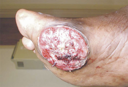
A 63-year-old man presented with a two-year history of a worsening verrucous lesion on his right foot. It had ulcerations with partial healing, and weight bearing caused pain (see accompanying figure). On physical examination, the patient was found to have a 4- ×6-cm ulcerated verrucous plaque on the plantar surface of his forefoot. No other verrucous lesions were noted on the foot. Previous treatments included multiple excisions and topical agents for verruca vulgaris, which produced only limited improvement.
Question
Discussion
The answer is B: squamous cell carcinoma. A biopsy confirmed the diagnosis of squamous cell carcinoma (verrucous carcinoma) of the foot. Malignancy should be considered in the differential diagnosis of a nonhealing ulcerative verrucous lesion on the foot. Because the foot has thin skin, subcutaneous tissue, and small muscles, palpation of the tumor is relatively easy.1
Malignant lesions are especially uncommon in the foot and ankle, but they often are confused with common benign lesions and, therefore, misdiagnosed (e.g., plantar sarcomas as fibromatosis, dorsal synovial sarcomas as ganglions, malignant melanomas as chronic ulceration, primary bone tumors as stress fractures).2
Verrucous carcinoma is found most commonly on the sole or ball of the foot, but has been described in the web spaces of the toe and on the toes, leg, and knee.4–6 Foul-smelling keratogenous material may be excreted through multiple sinus openings. Most tumors have been treated as recalcitrant warts or corns for some time.6 Excision is the treatment of choice because of local aggressiveness and infrequent metastasis. In more serious cases, amputation may be necessary for cure.7
Actinomycosis, a chronic bacterial infection caused by gram-positive bacilli (Actinomyces sp.), can present with fistulous tract lesions. Sulfur granules similar to the keratogenous material in the verrucous carcinoma may be present.
Verruca plantaris are common solitary lesions on the sole of the foot. The overlying skin is typically a thickened cornified layer.
Pseudoepitheliomatous hyperplasia is a pathologic reaction pattern of squamous epithelium that usually occurs in association with certain neoplasms or over a chronic inflammatory process (i.e., following trauma or irritation). The reactive epithelium may extend into the superficial reticular dermis, simulating a carcinoma.
Plantar fibromatosis is a rare, benign neoplasm of the plantar aponeurosis. It is a well-circumscribed, indurated mass usually detectable with palpation. It only invades locally, but has a tendency to recur after adequate surgical or corticosteroid therapy.8
| Condition | Characteristics |
|---|---|
| Actinomycosis (Madura foot) | Chronic bacterial infection that can present with fistulous tract lesions |
| Squamous cell carcinoma (verrucous carcinoma) | Hyperkeratotic ulcerated verrucous plaque, multiple sinus openings draining foul-smelling keratogenous material |
| Verruca plantaris (plantar wart) | Solitary painful lesions with thickened cornified layer |
| Pseudoepitheliomatous hyperplasia | Single layer nodules, usually in the plantar arch |
| Plantar fibromatosis | Firm subdermal indurated mass of the plantar aponeurosis |