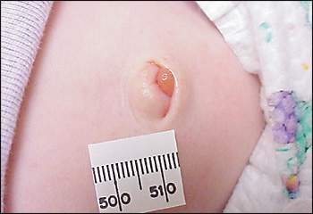
A six-week-old male infant was brought to the office by his mother. During a diaper change, she noted a lesion in the umbilicus (see accompanying figure). The infant was born at term by primary cesarean section. He was feeding well and gaining weight appropriately. The lesion was not noted during a previous office visit when the infant was 10 days old and still had the umbilical cord attached. On examination, there was a soft, red mass at the base of the umbilicus. Although the lesion was slightly moist, no foul discharge or purulent drainage was present. The surrounding skin was not red, warm, or swollen.
Question
Based on the history and physical examination, which one of the following is the most likely diagnosis?
A. Umbilical hernia.
B. Umbilical pyogenic granuloma.
C. Omphalitis.
D. Omphalocele.
E. Patent urachus.
Discussion
The answer is B: umbilical pyogenic granuloma. A solid, red umbilical mass having a soft, velvety appearance without a fistulous tract suggests a granuloma. An umbilical pyogenic granuloma is a common, benign abnormality in neonates. It develops within the first few weeks of life and should not be present at birth. It forms from excess granulation tissue persisting at the base of the umbilicus after cord separation.1 Umbilical granulomas often appear pedunculated and can secrete a small amount of serous or serosanguineous drainage.
Umbilical hernias, omphaloceles, and gastroschisis usually are apparent on gross inspection. Umbilical hernias form as a result of a fascial defect that allows viscera to protrude when the child strains or cries. Umbilical hernias are covered with skin, which helps distinguish them from granulomas and polyps. Although considered congenital, umbilical hernias developing during infancy usually are not apparent at birth. Umbilical hernias occur more often in premature infants and those of African American descent.2 Umbilical hernias spontaneously close in most children by approximately three years of age and rarely become incarcerated. Taping or strapping the hernia does not hasten resolution.3
An omphalocele is a serious congenital condition in which viscera enclosed in a sac protrude from a midline defect at the base of the umbilicus. Gastroschisis is characterized by lack of a membranous sac. In gastroschisis, bowel contents protrude lateral to an intact umbilical cord.4 It is important to distinguish these two abdominal wall defects because an omphalocele is associated with a higher incidence of other anomalies.
Less common conditions include umbilical polyps, urachal tract, and omphalomesenteric duct remnants.5 These require surgical correction. Umbilical polyps are firm masses comprised of intestinal or urinary tract tissue. They tend to be larger than granulomas, and do not respond to silver nitrate. A patent urachus is an embryonic duct that extends from the bladder to the umbilicus and intermittently leaks urine. Umbilical drainage containing bilious or fecal material should prompt a work-up to exclude a persistent omphalomesenteric duct, which is a residual communication between the ileum and the umbilicus.
Neonatal umbilical infection, or omphalitis, is characterized by redness, induration, and purulent or malodorous drainage from the umbilical stump. Symptoms of omphalitis begin two to three days after birth and may progress to necrotizing fasciitis or systemic infection.6 Omphalitis must be treated aggressively with antibiotics.
Several noninvasive measures are available to treat umbilical pyogenic granulomas. Granuloma formation is favored when cord separation is delayed and there is inflammation. Thus, applying topical antibiotics and eliminating the friction of a wet diaper may allow the granulation tissue to epithelialize. Conventional management has been to dry the umbilical stump and carefully cauterize the granuloma with a 75 percent silver nitrate stick. Because of the risk of drainage, the granuloma can be dried with gauze to avoid chemical burns or discoloration to the surrounding skin.7 Furthermore, caution should be exercised if silver nitrate is used for large granulomas because chemicals from repeated applications can leak onto healthy tissue. A small randomized controlled study concluded that conservative measures such as air drying with alcohol wipes should be tried before cauterizing with silver nitrate.8 Persistence of a presumed umbilical pyogenic granuloma after repeat applications of silver nitrate warrants further evaluation to rule out other pathology.
Cryosurgery, electrocautery, salt, and ligature are other treatment options. In one small study, cryosurgery was associated with skin depigmentation, but was favored because repeat applications were unnecessary.9 Cryotherapy also offered more rapid healing compared with the use of chemicals and electrocautery. Twice daily application of common table salt to umbilical pyogenic granulomas for three days is a simple, cost-effective, and curative method that can be performed by parents at home.10 The application of a double-ligature can be considered for pedunculated umbilical pyogenic granulomas.11 Proponents of this technique caution against using double ligatures on broad-based or friable lesions and emphasize the importance of first excluding more critical diagnoses. Larger granulomas or those that do not resolve with the above measures may require surgical excision.
Selected Differential Diagnosis of Neonatal Umbilical Masses
| Condition | Characteristics |
|---|---|
| Umbilical pyogenic granuloma | Small, red, moist, velvety mass |
| Umbilical polyp | Firm, red mass; does not resolve after application of silver nitrate |
| Umbilical hernia | Covered with skin, usually reducible |
| Patent urachus | Discharge (urine) from the umbilicus |
| Omphalomesenteric duct remnant | Bilious or fecal discharge from umbilicus |
| Omphalocele | Membranous sac containing viscera herniates through the umbilicus |
| Gastroschisis | Loops of bowel protrude lateral to an intact umbilicus |
