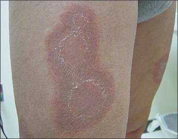
A 57-year-old man presented with a three-week history of a red, ring-like rash on the thighs, hips, and flexor surfaces of his arms (see accompanying figure). The lesions began as pinkish papules and expanded centrifugally. The center of the lesions began clearing and left an inner trailing scale. Upon further questioning, the patient revealed that he was treated previously for tinea pedis. A potassium hydroxide preparation from these lesions was negative.
Question
Given the history and physical appearance of the lesions, which one of the following conditions is the most appropriate diagnosis?
A. Tinea corporis.
B. Urticaria.
C. Subacute cutaneous lupus erythematosus.
D. Erythema annulare centrifugum.
E. Granuloma annulare.
Discussion
The answer is D: erythema annulare centrifugum. This patient’s condition is characterized by nonindurated, annular patches of centrifugally expanding erythema and desquamation at the inner margin (i.e., trailing scale). Occasionally, there may be minimal edema causing elevation of the annular lesion into a plaque. The annular lesions expand over a few weeks and affect the trunk, buttocks, thighs, and legs. The hands, feet, and face usually are spared.
Erythema annulare centrifugum belongs to the class of skin lesions known as the gyrate or figurate erythemas. These include erythema marginatum (associated with rheumatic heart disease), erythema gyratum repens (a paraneoplastic eruption often associated with lung carcinoma), and erythema chronicum migrans (associated with Lyme disease)
The etiology and pathogenesis of erythema annulare centrifugum are unknown, but it may represent a cutaneous hypersensitivity reaction pattern to a separate underlying immunologic insult. In this way, it can be considered similar to erythema nodosum. Such “insults” include fungal, viral, and parasitic infections; medications (diuretics, antimalarials, and gold); particular foods (blue cheese)1; and occasionally malignancies (lymphomas).2
The diagnosis is largely clinical, and in most cases a biopsy is unnecessary. If the clinical diagnosis is unclear, a small punch biopsy usually will show the characteristic histologic findings of tight perivascular “coat-sleeving” of lymphocytes in the dermis. Papillary dermal edema, spongiosis, and parakeratosis also may be present.3The rash can last for weeks to months, depending on the nature and duration of the underlying illness. It usually is self-limited and resolves with treatment of the underlying disorder. Lesions typically heal without scarring.3
Treatments include identifying and treating the underlying cause of the erythema. For example, this patient was treated for tinea pedis, and the rash resolved. Symptomatic rashes can be treated locally with topical corticosteroids or tar ointments.2 Antihistamines can be prescribed if there is associated pruritus
Tinea corporis presents as well-demarcated, erythematous papules or plaques on the skin.3 Patients often note gradual enlargement. Typically, the erythema and scale are most prominent at the borders, and this represents the active edge of the lesion and the best site for performing a skin scraping. Treatment includes topical or systemic antifungal agents and education about avoiding future contact with dermatophytes.4
Urticaria is characterized by pruritic, edematous, well-circumscribed, erythematous plaques that are transient over several hours. Also known as “wheals,” or “hives,” urticaria usually blanches with pressure.
Subacute cutaneous lupus erythematosus presents as diffuse, symmetric, smaller (typically less than 3 cm), annular pink to red papules or plaques with scale, typically in areas of sun-exposed skin. Scaling typically occurs on the surface of the papules or plaques.2
Granuloma annulare is characterized by smooth, skin-colored papules coalescing into annular plaques, and is often found on the hands, feet, wrists, and ankles. The plaques have a raised, nonscaly appearance, occasionally with erythematous borders.5
Selected Differential Diagnosis of Annular Lesions
| Condition | Characteristics |
|---|---|
| Tinea corporis | Pink annular plaques with central clearing and scale at the active (leading) border; positive result on potassium hydroxide preparation |
| Urticaria | Pink to red pruritic, round, edematous, blanchable plaques that change morphology over several hours or with stroking |
| Subacute cutaneous lupus rythematosus | Pink to red papules and polycyclic plaques with thin scale and usually telangiectasia and dyspigmentation; occur in sun-exposed areas |
| Erythema annulare centrifugum | Pink to red polycyclic plaques on trunk, buttocks, and inner thighs with trailing (inner border) scale |
| Granuloma annulare | White to pink flat-topped papules without scale that coalesce to form an annular plaque on extensor surfaces |
