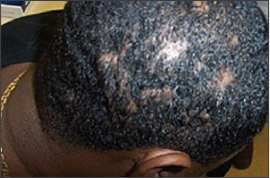
A 32-year-old man complained of persistent, painful, draining scalp lesions that had been present for the past five years. These lesions caused progressively worsening alopecia accompanied by drainage that often was purulent. The lesions had been incised and drained in the past, and a recent culture grew only rare gram-positive cocci. Treatments over the preceding years involved various oral and topical medications, including antibiotics, antifungals, isotretinoin (Accutane), dapsone, and intralesional steroids. Oral steroids controlled the lesions, but symptomatic flares occurred when these were weaned. The patient’s scalp (Figures 1 and 2) had numerous cysts and sinuses. There was diffuse alopecia about these cysts. The axillae were without lesions, and there was no evidence of acne. A 1.5-cm left posterior auricular cervical lymph node was palpable.
Figure 1.
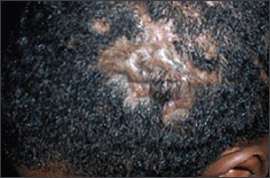
Figure 2.
Question
Based on the patient’s history and physical examination, which one of the following is the correct diagnosis?
A. Dissecting cellulitis of scalp.
B. Kerion.
C. Folliculitis.
D. Discoid lupus erythematosus.
E. Acne keloidalis nuchae.
Discussion
The answer is A: dissecting cellulitis of scalp. Dissecting cellulitis of scalp, also known as perifolliculitis capitis abscedens et suffodiens, is a chronic, progressive, inflammatory disease similar to cystic acne. It typically begins as a simple folliculitis of the vertex and occiput, with clusters of perifollicular pustules. This progresses to abscess and sinus formation and the development of scarring alopecia. Nodules may be firm or fluctuant and may express serosanguineous fluid when gentle pressure is applied.
These lesions are thought to result from the retention of keratinous material, with subsequent inflammation after follicular dilation and rupture.1 Lesions may persist for years at different stages. Bacterial superinfection appears to be an outcome rather than a cause. Whereas bacterial superinfection is common, systemic disease is not. Most often, these lesions are resistant to therapy and are progressive. Extensive scarring of the scalp develops and may be hypertrophic or keloidal.2
Dissecting cellulitis of scalp is a rare disease, typically occurring between 18 and 40 years of age. Black men are affected almost exclusively, although there have been some case reports of dissecting cellulitis in white patients.3 This disease may be associated with acne conglobata and hidradenitis suppurativa, forming the “follicular occlusion triad.” When also associated with pilonidal sinuses, the follicular tetrad is present.
Potassium hydroxide preparations and fungal and bacterial cultures should be completed to rule out more common conditions such as kerion and folliculitis. Lesional biopsy reveals an extensive dermal lymphohistiocytic infiltrate, numerous giant cells, and follicular plugging.
In mild and limited cases, scalp hygiene involving the use of local (chlorhexidine [Peridex], benzoyl peroxide, clindamycin [Cleocin]) and systemic (e.g., tetracycline) antibiotics and local incision and drainage of cysts may be effective.1 Oral zinc sulfate has been found to be effective, and oral isotretinoin therapy has been reported to induce remission.4,5 Other treatment options may involve oral or intralesional steroids, radiation treatment, laser treatment, or surgery.1 Given the potential for recalcitrant disease and the nature of the treatments, dermatology consultation should be considered.
Kerion is an inflammatory tinea capitis appearing as one or more inflamed, boggy, tender areas of alopecia and pustules. These lesions may be accompanied by fever, leukocytosis, and occipital adenopathy.6
Folliculitis, which is an inflammation of the hair follicle, is characterized by dome-shaped pustules surrounded by a small area of erythema arising in the center of the follicle.
Discoid lupus erythematosus is more common in women and in the fourth decade of life. Lesions typically are well-defined, flat-topped plaques with scale firmly attached. Follicular plugging may be prominent.
Acne keloidalis nuchae also occurs in younger black men. Lesions begin as follicular papules; evolve into firm, keloidal nodules; and typically are located on the neck and occipital scalp.
Selected Differential Diagnosis of Scalp Lesions and Hair Loss
| Condition | Characteristics |
|---|---|
| Dissecting cellulites of scalp | Folliculitis progressing to abscess and sinus formation with scarring alopecia |
| Kerion | Inflamed, boggy areas of alopecia caused by tinea capitis |
| Folliculitis | Pustules with erythema around the hair follicle |
| Discoid lupus erythematosus | Well-defined, flat-topped plaques with firmly attached scale; follicular plugging may be prominent. |
| Acne keloidalis nuchae | Papules and nodules, most often on the nape of the neck |
