
Am Fam Physician. 2006;74(11):1909-1911
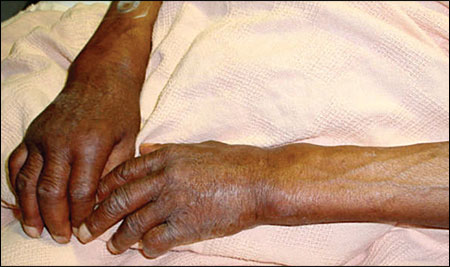
A 67-year-old man presented with pain and swelling of both wrists that began a few weeks earlier. He also reported a poor appetite and a 12-lb (5.4-kg) weight loss, as well as a productive cough and shortness of breath. He denied experiencing any morning joint stiffness, paresthesias, or other constitutional symptoms. On examination, the patient appeared ill and dyspneic. His lung examination revealed decreased breath sounds in his right upper lung field. There was broadening of the wrists with tenderness, warmth, and mild erythema along both distal forearms and wrists. Bilateral nail clubbing also was noted (Figure 1).
Question
Discussion
The answer is E: secondary hypertrophic osteoarthropathy. Hypertrophic osteoarthropathy is a syndrome of clubbing, periostitis, and synovitis.1,2 It is characterized by new subperiosteal bone formation at the distal ends of long bones. More than 95 percent of cases are secondary (i.e., they are associated with an underlying systemic disease, most often a neoplasm or infection). Primary hypertrophic osteoarthropathy describes a case in which the cause is familial or idiopathic.2–4
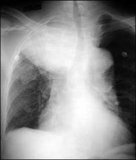
The most common cause of secondary hypertrophic osteoarthropathy is pulmonary neoplasm, typically a bronchogenic carcinoma (Figure 2). Less common causes include cystic fibrosis, bronchiectasis, cyanotic congenital heart disease, infective endocarditis, hepatoma, ulcerative colitis, and acquired immunodeficiency syndrome.4
Secondary hypertrophic osteoarthropathy can present with acute onset of redness, swelling, pain, and limited range of motion of the affected joints and bones. It is typically symmetric and associated with clubbing.1
The results of laboratory tests such as rheumatoid factor and antinuclear antibodies are normal.1 Periosteal new bone formation causes an elevated serum alkaline phosphatase level. If a joint effusion is present, the joint aspirate shows a noninflammatory fluid (cell count of less than 500 cells per μ L) with a lymphocytic and monocytic predominance.
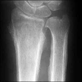
Periostitis on long bone radiography is seen as new bone formation with periosteal elevation (Figure 3). A radioisotope bone scan shows diffusely increased uptake in the periosteum and is valuable in doubtful cases or when the radiographic images are normal.
Because hypertrophic osteoarthropathy is more often secondary than primary, an underlying cause must be sought. Furthermore, the only definitive therapy for secondary hypertrophic osteoarthropathy is treatment of the underlying condition. For example, secondary hypertrophic osteoarthropathy regresses in two to four weeks and may disappear by three to six months after surgical resection of the underlying causative tumor.5 Nonsteroidal anti-inflammatory drugs, steroids, or colchicine are treatment options if the hypertrophic osteoarthropathy is associated with pain.1
Rheumatoid arthritis is a chronic, progressive disorder that symmetrically involves the synovial and articular surfaces of multiple joints. In rheumatoid arthritis, clubbing is absent and radiography shows periosteal sparing.
Primary hypertrophic osteoarthropathy, also known as pachydermoperiostosis or Touraine-Solente-Golé syndrome, is a rare autosomal dominant disorder occurring mainly in young males, characterized by hypertrophic osteoarthropathy with thickening and furrowing of facial skin (i.e., “leonine faces”).1
Carpal tunnel syndrome is associated with insidious onset of hand paresthesias without any visible clinical swelling, erythema, or wrist deformity. Nerve conduction studies often help confirm the diagnosis.
Acute osteomyelitis is more common in children and immunosuppressed adults. Constitutional symptoms such as fever and malaise usually are present. Skin over the affected bone may be erythematous. Positive blood cultures with an elevated erythrocyte sedimentation rate aid in the diagnosis. Radiographic images can be normal early in the disease course, in which case a nuclear bone scan or magnetic resonance imaging can assist in the diagnosis.
| Condition | Characteristics |
|---|---|
| Acute osteomyelitis | Pain, erythema at affected site, and constitutional symptoms; elevated erythrocyte sedimentation rate, possibly positive blood cultures; abnormal results on bone scan, magnetic resonance imaging, or bone biopsy |
| Carpal tunnel syndrome | Typically nocturnal or positional paresthesias; can be bilateral; nerve conduction studies demonstrate median nerve neuropathy |
| Pachydermoperiostosis (primary hypertrophic osteoarthropathy) | Familial triad of clubbing, hypertrophic osteoarthropathy, and coarsening of facial features; unassociated with underlying systemic disorder |
| Rheumatoid arthritis | Joint involvement is symmetric and polyarticular, with sparing of distal interphalangeal joints; subcutaneous nodules and other deformities (e.g., swan neck deformity) occur; clubbing is absent. |
| Secondary hypertrophic osteoarthropathy | Painful enlargement of the wrists with clubbing; caused by underlying systemic illness, typically neoplastic or infectious; radiographic and bone scans show diffuse periosteal process. |