Second trimester pregnancy loss is uncommon, but it should be regarded as an important event in a woman's obstetric history. Fetal abnormalities, including chromosomal problems, and maternal anatomic factors, immunologic factors, infection, and thrombophilia should be considered; however, a cause-and-effect relationship may be difficult to establish. A thorough history and physical examination should include inquiries about previous pregnancy loss. Laboratory tests may identify treatable etiologies. Although there is limited evidence that specific interventions improve outcomes, management of contributing maternal factors (e.g., smoking, substance abuse) is essential. Preventive measures, including vaccination and folic acid supplementation, are recommended regardless of risk. Management of associated chromosomal factors requires consultation with a genetic counselor or obstetrician. The family physician can play an important role in helping the patient and her family cope with the emotional aspects of pregnancy loss.
Pregnancy loss during the second trimester (i.e., 13 to 27 weeks' gestation) is rare1 and often is not distinguished from first trimester pregnancy loss.2 However, a true second trimester loss should be considered a unique entity, and an appropriate evaluation is indicated. Pregnancy loss is considered a miscarriage when it occurs before 20 weeks' gestation; after this time it is considered a stillbirth. Nevertheless, there is considerable overlap between these definitions, and definitions vary by state.3,4
SORT: KEY RECOMMENDATIONS FOR PRACTICE
| Clinical recommendation | Evidence rating | References |
|---|---|---|
| Hysteroscopic metroplasty for septate uterus is associated with favorable pregnancy outcomes. | C | 3 |
| Cervical cerclage can be considered in women with three or more unexplained second trimester losses or preterm deliveries and cervical change before viability. | B | 21–23 |
| Combined unfractionated heparin and aspirin may reduce pregnancy loss by 54 percent in women with antiphospholipid antibody syndrome and previous pregnancy loss. | B | 26 |
| Providing support and counseling regarding typical reactions, available resources, and support groups is recommended to help the patient and her family adjust to a second trimester pregnancy loss. | C | 35,36 |
A = consistent, good-quality patient-oriented evidence; B = inconsistent or limited-quality patient-oriented evidence; C = consensus, disease-oriented evidence, usual practice, expert opinion, or case series. For information about the SORT evidence rating system, see page 1262 orhttps://www.aafp.org/afpsort.xml.
Rates of pregnancy loss decrease as the pregnancy progresses. Overall, about 10 to 20 percent of all recognized pregnancies and 30 to 40 percent of all conceptions end in pregnancy loss.4–6 Miscarriage that occurs at 13 to 14 weeks' gestation usually reflects a pregnancy loss that happened one to two weeks earlier.7 Approximately 1 to 5 percent of pregnancies are lost at 13 to 19 weeks' gestation, whereas stillbirth occurs in 0.3 percent of pregnancies at 20 to 27 weeks' gestation, a rate similar to that of third trimester stillbirth (Figure 1).1
Figure 1. Estimated Fetal Death Rate by Gestational Age
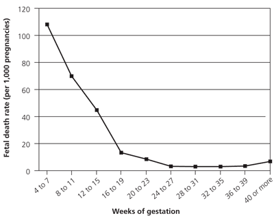
Estimated rates of fetal mortality by weeks of gestation.
Adapted with permission from French FE, Bierman JM. Probabilities of fetal mortality. Public Health Rep 1962;77:843.
Factors Associated with Pregnancy Loss
Table 1 outlines the conditions associated with pregnancy loss by trimester.4,8–10 Establishing a cause-and-effect relationship is difficult; causation is well established only for chromosomal and fetal structural problems. Overall, the cause of pregnancy loss remains unexplained in up to 50 percent of cases.11
Table 1 Factors Associated with Pregnancy Loss by Trimester
| Trimester | Factors | |||
|---|---|---|---|---|
| Fetal | Maternal | Fetomaternal | Other | |
| First | Chromosomal abnormalities |
| — |
|
| Congenital abnormalities | ||||
| Second | Chromosomal abnormalities |
| — |
|
| Congenital abnormalities | ||||
| Third | Chromosomal abnormalities |
|
|
|
| Congenital abnormalities | ||||
At least one half of first trimester pregnancy losses are caused by chromosomal abnormalities.12 Results from chorionic villus sampling suggest that this type of abnormality causes up to 85 percent of all miscarriages.8 Less common causes of first trimester loss include maternal illness, luteal phase defect and other hormonal problems (e.g., polycystic ovary syndrome), and intrauterine adhesions.8
The incidence of disorders associated with third trimester pregnancy losses varies greatly from study to study because of differences in the populations surveyed. In addition, many causes are interdependent; for example, maternal hypertension can cause uteroplacental insufficiency and growth restriction.9,10
Etiologies of Second Trimester Pregnancy Loss
Conditions associated with second trimester pregnancy loss overlap those of the first and third trimesters to a certain extent, but some are characteristic of second trimester losses.2 Table 2 lists clinical clues that suggest potential etiologies of second trimester loss, and it suggests further testing and treatments.
Table 2 Diagnostic Approach to the Patient with a Second Trimester Pregnancy Loss
| Clinical clues | Possible causes | Further evaluation | Potential action |
|---|---|---|---|
| Recurrent first trimester losses | Chromosomal abnormalities | Fetal autopsy; karyotype analysis | Genetic counseling |
| Fetal malformation; stillbirth | Chromosomal abnormalities; maternal teratogen exposure; maternal illness | Fetal autopsy; karyotype analysis; maternal blood tests (i.e., glucose, thyroid, liver); maternal history | Genetic counseling; avoidance of teratogens; treatment of maternal illness |
| Painless rupture of membranes | Cervical insufficiency | Ultrasonography; hysterography | Cervical cerclage |
| Unexplained loss | Maternal anatomic factors (müllerian duct anomalies, intrauterine adhesions) | Physical examination; ultrasonography; hysterosalpingography; hysteroscopy; pelvic magnetic resonance imaging | Metroplasty or other uterine surgical procedures |
| Placental abruption | Maternal hypertension; cocaine use; smoking; thrombophilias; physical abuse, trauma | Directed history and physical examination; laboratory evaluation (e.g., blood tests for factor V Leiden, activated protein C resistance, prothrombin G20210A mutation, protein S deficiency); pelvic ultrasonography; toxicology screen | Treatment of maternal illness; substance abuse counseling or intervention |
| Maternal systemic lupus erythematosus or primary Sjögren's syndrome; cutaneous thrombosis | Antiphospholipid antibody syndrome | Blood tests for lupus anticoagulant and anticardiolipin antibody | Referral; combination aspirin and heparin therapy |
| Normal fetus, possible maternal history of thrombosis | Thrombophilias | Blood tests for factor V Leiden, activated protein C resistance, prothrombin G20210A mutation, protein S deficiency | Referral; combination aspirin and heparin therapy |
| Maternal fever, preterm labor, pathologic evidence of placental inflammation | Maternal infection | Maternal (and fetal, if possible) cultures; assays for infectious agents | Antimicrobial agents; avoidance of cat litter and sexually transmitted infections; rubella immunization |
CHROMOSOMAL ABNORMALITIES
In addition to their role in first trimester miscarriage, chromosomal abnormalities also cause pregnancy loss in the second trimester. About 24 percent of pregnancy losses in the second trimester are caused by chromosomal abnormalities, and about 12 percent of late second trimester losses are attributed to this cause.12 Chromosomal abnormalities found in second trimester losses are similar to those found in live births; the most common are trisomies 13, 18, and 21, monosomy X (i.e., Turner syndrome), and sex chromosome polysomies.8
FETAL AND MATERNAL ANATOMIC FACTORS
Pregnancy loss can also be caused by structural abnormalities resulting from uncontrolled maternal diabetes at conception, neural tube defects, amniotic band syndrome, or maternal exposure to teratogens. Major uterine anomalies have traditionally been associated with second trimester pregnancy loss.13–15 One case-control study showed a significant association between some müllerian duct anomalies and second trimester loss, as well as low birth weight, breech presentation, and maternal hemorrhage.16 The role of surgical correction of these anomalies is controversial, with most studies showing no difference in fetal survival rates with surgical correction compared with uncorrected anomalies. For more severe anomalies (e.g., complete septate uterus), surgical repair may be the only realistic option for a woman to carry her pregnancy to viability. Hysteroscopic metroplasty for septate uterus is associated with favorable pregnancy outcomes.2,17–20 A decision regarding surgical procedures should be made in consultation with an obstetrician. Surgical correction of uterine leiomyomata has not been shown to improve reproductive outcomes.19
Cervical insufficiency or incompetence is classically associated with second trimester loss after painless cervical dilation (i.e., without initial labor). Typically the membranes balloon into the vagina; this is followed by rupture of membranes, contractions, and expulsion of a premature fetus. There is usually a history of second or third trimester loss. Patients should be questioned about cervical trauma during previous vaginal deliveries and any history of cone biopsy. The diagnosis suggested by history and physical examination can be confirmed with hysterography or transvaginal ultrasonography.8,20 The most recent reviews of randomized trials, as well as a report from the American College of Obstetricians and Gynecologists, conclude that there is insufficient evidence to recommend cerclage for a shortened cervix detected by ultrasonography.21–23 There is better support for cervical cerclage in women with three or more second trimester losses or preterm deliveries and cervical change before fetal viability.
THROMBOPHILIA
A meta-analysis of 31 studies on the effect of thrombophilic disorders in pregnancy loss showed that a nonrecurrent pregnancy loss after 20 to 24 weeks' gestation is associated with factor V Leiden, protein S deficiency, and the prothrombin G20210A mutation.24 Antiphospholipid antibodies, specifically lupus anticoagulant and anticardiolipin antibody, can occur in women with systemic lupus erythematosus or other immunologic conditions, can occur as an isolated syndrome, and can be transient. These antibodies cause placental thrombosis and have emerged as well-established risks for second and third trimester pregnancy loss.25 Work-up of thrombophilia is, therefore, recommended in women with a pregnancy loss after 20 weeks' gestation. In patients who have had a pregnancy loss before 20 weeks, there is insufficient evidence to recommend for or against such a work-up after the loss.
Management of thrombophilic conditions and antiphospholipid antibodies is done after a pregnancy loss and is typically beyond the scope of most family physicians; however, combination therapy with heparin and aspirin may reduce rates of pregnancy loss by 54 percent in women with antiphospholipid antibody syndrome who have had a previous loss.26 Heparin is often used to treat thrombophilia in pregnant women; however, there are no trials showing its effectiveness in this population.27
INFECTION
Infection has been implicated in 10 to 25 percent of second trimester pregnancy losses.28 Many infectious agents have been suggested, including bacteria, spirochetes, protozoa, viruses, and fungi.29,30 Most authorities acknowledge that infection plays a role in some cases of late pregnancy loss, but they think that positive cultures and pathologic inflammation are most often postmortem findings. Infection is more closely linked to pregnancy loss in developing countries.28,31 Bacterial vaginosis infection has been linked to second trimester loss, but not first trimester loss.32 As with other etiologies that are difficult to confirm, there is limited proven benefit of intervention. Nevertheless, rubella and influenza vaccination is prudent in all pregnant women, and treating bacterial vaginosis may prevent premature labor in women with a history of preterm birth.33
General Assessment and Follow-up
HISTORY AND PHYSICAL EXAMINATION
After a second trimester loss, all patients warrant a thorough history and physical examination to look for factors that might predispose them to another loss. Ideally, this work-up should be done during preconception counseling.
The history should include symptoms and signs of pregnancy loss, chronic maternal medical conditions that may contribute to pregnancy loss, family history that suggests genetic problems, medication use as an indication of underlying illness, environmental exposures, substance abuse, trauma, and obstetric history. A detailed review of the pregnancy should be performed, including vital signs, weight gain, dating parameters, ultrasonography, and laboratory tests.
If the loss is a stillbirth, pathologic examination of the fetus and placenta is advocated; chromosomal analysis should also be performed, if possible.34 Cultures should be ordered only if the patient has clinical symptoms of a specific infection. Particularly, asymptomatic patients should not be treated for bacterial vaginosis.
DISEASE MANAGEMENT AND COUNSELING
Detailed work-up and management of many of the maternal factors associated with second trimester pregnancy loss often require referral to an obstetrician or perinatologist; however, the family physician can still play an important role. If a maternal medical illness appears to have contributed to the pregnancy loss, the family physician should optimize management of the patient's diabetes, thyroid disease, or hypertension. Nutritional education and folic acid supplementation can improve maternal illness and help prevent neural tube defects.
Smoking, alcohol consumption, and substance abuse have been implicated in poor fetal outcomes; therefore, patients should be offered counseling and treatment, even if the roles of these activities in the pregnancy loss are not well established. Although trauma is an uncommon cause of pregnancy loss, advocacy for the prevention of physical abuse can be initiated and coordinated by the family physician.
Patients who have had an unexplained pregnancy loss should be offered genetic counseling with an option for karyotype analysis, even though these interventions have few measurable outcomes.
PSYCHOLOGICAL FACTORS
The family physician is in an ideal position to address psychological factors in women who have had a second trimester pregnancy loss. After an early pregnancy loss, women experience the same emotional and psychological reactions as those who have experienced any type of death; however, the duration of the distress is typically shorter. Patients initially go through recognizable emotions, including shock, searching, and yearning. Often, the patient will have intense preoccupation with seeing or hearing the infant, and there may be a period of disorganization, with features similar to those of depression, before she gradually adjusts and is able to move on.35
Many patients must also cope with their emotional responses during a subsequent pregnancy. Women who have had a pregnancy loss often have a strong impetus to become pregnant again. During the next pregnancy, these patients may have intense anxiety and ambivalence, with little emotional attachment. They may also be overprotective of the child after birth.35 Awareness of common and expected responses to pregnancy loss can help the family physician in providing guidance to these patients, who need information, reassurance, and encouragement.
Support groups for parents who have had a pregnancy loss can be invaluable. There are many Internet resources available. However, there is no evidence from randomized trials to indicate any benefit from providing specific psychological support or counseling after a pregnancy loss.36
