Photo Quiz
Scalded Mouth with Headache
Am Fam Physician. 2008 May 1;77(9):1307-1309.
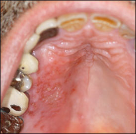
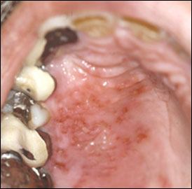
A 60-year-old man presented after burning the roof of his mouth on hot soup five days earlier. The burned sensation resolved after one day; however, a couple of days later, he noticed tingling on the right side of the hard palate that progressed to a scalded sensation. This was accompanied by tenderness along the right temporal area and discomfort below the right ear. He denied a history of oral lesions.
Examination revealed erythema that was confined to the right side of the hard palate with multiple ulcerations and vesicles (Figures 1 and 2). Although mild redness and swelling were noted just below the right ear, no skin lesions were present.

Figure 1.

Figure 2.
Question
Based on the patient's history and physical examination, which one of the following is the most likely diagnosis?
A. Herpes zoster (shingles).
B. Intraoral herpes simplex virus infection.
C. Primary syphilis.
D. Recurrent aphthous stomatitis.
E. Ulcers from thermal trauma.
Discussion
The answer is A: herpes zoster (shingles). The presence of multiple vesicles that vary in size and are confined to the right side of the hard palate (i.e., cranial nerve V2 distribution) accompanied by neuralgia and erythema in ipsilateral cutaneous portions of the right trigeminal nerve suggests herpes zoster over oral herpes simplex virus (HSV) infection. HSV infection is also less likely in this patient because he has no history of oral ulcers. However, herpes zoster cannot be differentiated from HSV infection based on clinical findings alone because HSV lesions can occur unilaterally and be indistinguishable from those caused by the varicella zoster virus. A more definitive clinical diagnosis of herpes zoster requires the presence of an associated cutaneous herpetiform rash in the appropriate distribution of the affected nerve.
Laboratory testing of affected tissue or fluid from the vesicles is needed for a conclusive herpes zoster diagnosis. A positive Tzanck smear result showing multinucleated giant cells on a scraping from a vesicle base rules out other causes of oral lesions, but does not differentiate herpes zoster from HSV.1 A polymerase chain reaction (PCR) assay is the most sensitive test for identifying HSV and varicella zoster virus; it can detect viral DNA from vesicle fluid swabs, crusts, Tzanck smear debris, and tissue specimens fixed in formalin. Compared with culture, PCR assays are 22 percent more sensitive for HSV and 91 percent more sensitive for varicella zoster virus, with 100 percent specificity for each virus.2,3 Additionally, real-time PCR assays now offer rapid turnaround times at costs similar to culture.2–4
The primary therapeutic goals for herpes zoster are to shorten the duration of initial symptoms and to decrease the incidence and extent of postherpetic neuralgia. Acyclovir (Zovirax), valacyclovir (Valtrex), and famciclovir (Famvir) are available for the treatment of herpes zoster. Current evidence supports using these agents to hasten resolution of lesions and acute pain, as well as to attenuate the development of postherpetic neuralgia. Evidence from randomized, double-blind trials suggests that valacyclovir and famciclovir are more effective and require less frequent dosing than acyclovir.5–9 However, generic acyclovir, 800 mg five times daily, may be considered when antiviral therapy is precluded because of cost.
Antiviral therapy should be initiated within 72 hours of the onset of the rash or if new lesions continue to form. The benefit of adding oral steroids to antiviral treatment is unclear and potentially harmful. Acyclovir combined with steroids does not reduce the incidence or duration of postherpetic neuralgia.9
The oral lesions in this patient differ significantly from those typically associated with primary syphilis. With primary syphilis, a papule develops on loose, nonkeratinized mucosa, then breaks down into a large, painless, solitary ulcer with a raised border.
Recurrent aphthous stomatitis (canker sores), HSV, or varicella zoster virus can appear as grouped or clustered oral ulcerations. However, recurrent aphthous stomatitis does not have a vesicular phase and usually appears as a single ulcer or a small number of ulcers. Aphthous stomatitis usually occurs on loose, nonkeratinized mucosal surfaces such as buccal or labial mucosa, the floor of the mouth, and the ventral surface of the tongue.10 Conversely, herpetic ulcers, which are usually multiple and have a vesicular stage, affect immovable, keratinized mucosa, including the hard palate, attached gingivae, and dorsal tongue.
Keratinized mucosa is an integral part of the masticatory apparatus; thus, it is routinely subject to oral trauma, such as burns from hot food. Ulcers from thermal injuries often affect the keratinized surfaces, but are unlikely to cause multiple, discrete ulcerations.
Characteristics of Different Types of Acute Oral Ulcerations
Type of ulceration | Location | Size | Morphology | Vesicular phase | Pain | Frequency | Recurrence | Dermatomal pattern |
---|---|---|---|---|---|---|---|---|
Herpes zoster (shingles) | Hard or soft palate(cranial nerve V) or inner cheek (V3) | Usually small and discrete (1 to 2 mm) | Multiple, usually grouped | Initially | More severe than HSV ulcers | Rare | Rare | Typical |
HSV infection | Immovable, keratinized (masticatory) mucosa | Usually small and discrete (1 to 2 mm) | Multiple, usually grouped | Initially | Mild | Uncommon intraorally | In up to 40 percent of patients | No |
Primary syphilis | Loose, nonkeratinized mucosa | Large (3 to 20 mm) | Usually solitary with a raised border | None; begins as a papule | None | Uncommon | Rare | No |
Recurrent aphthous stomatitis | Loose, nonkeratinized mucosa | Typically larger than an HSV ulcer | Usually solitary or few in number; may be clustered (herpetiform) | None | Sometimes intense | Common | Common | No |
Minor (80 percent of cases), 1 to 5 mm; major (10 percent of cases), 5 to 15 mm; herpetiform, 1 to 2 mm | ||||||||
Thermal trauma | Usually keratinized mucosa | Usually larger and nondiscrete | Variable | None | Mild | Common | With recurrent trauma | No |
HSV = herpes simplex virus.
Information from Hairston BR, Bruce AJ, Rogers RS III. Viral diseases of the oral mucosa. Dermatologic Clin. 2003;21(1)17–32; Fauci AS, Braunwald E, Kasper DL, et al., eds. Harrison's Principles of Internal Medicine. 17th ed. New York, NY: McGraw-Hill; 2006; and Habif TP. Clinical Dermatology. A Color Guide to Diagnosis and Therapy. 4th ed. Philadelphia, Pa.: Mosby; 2004.
REFERENCES
show all references1. Hairston BR, Bruce AJ, Rogers RS III. Viral diseases of the oral mucosa. Dermatologic Clin. 2003;21(1):17–32....
2. Espy MJ, Uhl JR, Mitchell PS, et al. Diagnosis of herpes simplex virus infections in the clinical laboratory by LightCycler PCR. J Clin Microbiol. 2000;38(2):795–799.
3. Espy MJ, Teo R, Ross TK, et al. Diagnosis of varicellazoster virus infections in the clinical laboratory by Light-Cycler PCR. J Clin Microbiol. 2000;38(9):3187–3189.
4. Espy MJ, Uhl JR, Sloan LM, et al. Real-time PCR in clinical microbiology: applications for routine laboratory testing [published correction appears in Clin Microbiol Rev. 2006;19(3):595]. Clin Microbiol Rev. 2006;19(1):165–256.
5. Beutner KR, Friedman DJ, Forszpaniak C, Andersen PL, Wood MJ. Valaciclovir compared with acyclovir for improved therapy for herpes zoster in immunocompetent adults. Antimicrob Agents Chemother. 1995;39(7):1546–1553.
6. Tyring S, Barbarash RA, Nahlik JE, et al., for the Collaborative Famciclovir Herpes Zoster Study Group. Famciclovir for the treatment of acute herpes zoster: effects on acute disease and postherpetic neuralgia. A randomized, double-blind, placebo-controlled trial. Ann Intern Med. 1995;123(2):89–96.
7. Wood MJ, Kay R, Dworkin RH, Soong SJ, Whitley RJ. Oral acyclovir therapy accelerates pain resolution in patients with herpes zoster: a meta-analysis of placebo-controlled trials. Clin Infect Dis. 1996;22(2):341–347.
8. Jackson JL, Gibbons R, Meyer G, Inouye L. The effect of treating herpes zoster with oral acyclovir in preventing postherpetic neuralgia. A meta-analysis. Arch Intern Med. 1997;157(8):909–912.
9. Wareham D. Postherpetic neuralgia. http://www.clinicalevidence.bmj.com/ceweb/conditions/ind/0905/0905.jsp (subscription required). Accessed February 20, 2008.
10. Bruce AJ, Rogers RS III. Acute oral ulcers. Dermatol Clin. 2003;21(1):1–15.
The editors of AFP welcome submissions for Photo Quiz. Guidelines for preparing and submitting a Photo Quiz manuscript can be found in the Authors' Guide at https://www.aafp.org/afp/photoquizinfo. To be considered for publication, submissions must meet these guidelines. E-mail submissions to afpphoto@aafp.org.
Copyright © 2008 by the American Academy of Family Physicians.
This content is owned by the AAFP. A person viewing it online may make one printout of the material and may use that printout only for his or her personal, non-commercial reference. This material may not otherwise be downloaded, copied, printed, stored, transmitted or reproduced in any medium, whether now known or later invented, except as authorized in writing by the AAFP. Contact
afpserv@aafp.org for copyright questions and/or permission requests.
Want to use this article elsewhere? Get Permissions
More in AFP
Editor's Collections
Related Content
More in Pubmed
MOST RECENT ISSUE
Email Alerts
Don't miss a single issue. Sign up for the free AFP email table of contents.