
Am Fam Physician. 2009;80(2):191-193
Author disclosure: Nothing to disclose.
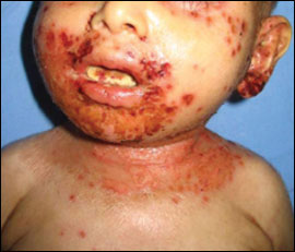
An 11-month-old boy presented with recurrent fever, petechial and infected cutaneous lesions that sometimes decreased in number, diarrhea, and purulent otitis. These symptoms had persisted since his first month of life. The patient was in poor general condition with erythematous, desquamative, plaque-like lesions on his entire body, including his scalp; hemorrhagic lesions on the mucosal sites; and candidiasis on his tongue (see accompanying figure). The membrane of his right ear was perforated, and purulent discharge was detected on the external auditory canal.
The child's height was 26 in (65 cm); his weight was 11 lb, 4 oz (5.1 kg); and his head circumference was 16 in (41 cm). Laboratory studies revealed a hemoglobin level of 9 g per dL (90 g per L), platelet count of 45,000 to 52,000 per mm3 (45 to 52 × 109 per L), and mean platelet volume of 4.6 to 7.0 fL. The C-reactive protein level was 1,910 mg per L (18,190 nmol per L), the erythrocyte sedimentation rate was 73 mm per hour, and the albumin level was 1.9 g per dL (19 g per L). Immunoglobulin tests showed a lower IgM level, and an elevated IgE level of 2,380 mcg per L (2.38 mg per L). The complement C3 and C4 levels were within normal limits. The prothrombin time was 15.5 seconds, the activated partial thromboplastin time was 33.8 seconds, and the serum zinc level was within the normal limit. Staphylococcus aureus was isolated in the blood culture.
The patient had two healthy siblings, but one male sibling with the same symptoms who died at 11 months of age. The patient also died after four days of hospitalization.
Question
Discussion
The correct answer is E: Wiskott-Aldrich syndrome. Wiskott-Aldrich syndrome is an X-linked, primary immunodeficiency disease with a prevalence of about four in 1 million. The syndrome was originally described as a clinical triad of immunodeficiency, eczema, and thrombocytopenia with small platelets.1 Bleeding episodes or symptoms of infection typically begin during the first six months of life. Although many patients with Wiskott-Aldrich syndrome present with all three of the typical clinical manifestations, others have a partial or variant phenotypic presentation. Because of the wide spectrum of clinical findings, the diagnosis relies on genetic analysis or the determination of protein expression with a specific antibody.2
The patient was definitively diagnosed with Wiskott-Aldrich syndrome because he is a boy; his presentation included recurrent infections, eczematiform skin lesions, thrombocytopenia, decreased mean platelet volume, and increased IgE levels; and his brother, who also died, had similar clinical findings. Wiskott-Aldrich syndrome should be considered in boys who have atopic dermatitis–like lesions and recurrent infections.
Acrodermatitis enteropathica is a rare hereditary disorder affecting zinc metabolism. It is characterized by dermatitis, alopecia, gastrointestinal disturbances, eye infections, and failure to thrive.5
Atopic dermatitis is a chronic, recurrent skin disorder. Pruritus, inflammation, crusted lesions, and lichenification are typical characteristics.6 It is common in flexural areas and on the cheeks and buttocks. Atopic dermatitis is not consistent with severe recurrent infections, immunodeficiency, and thrombocytopenia.
Job syndrome is a rare immune disorder characterized by atopic dermatitis–like skin lesions, elevated serum IgE level, recurrent skin and respiratory tract infections, and skeletal abnormalities. An elevated IgE level and peripheral eosinophilia are the most common findings in patients with this disorder.7
Omenn syndrome is an autosomal recessive, fatal condition characterized by profound susceptibility to infection with T-cell infiltration of the skin, intestines, liver, and spleen. This leads to an exfoliative erythroderma, lymphadenopathy, hepatosplenomegaly, alopecia, profound eosinophilia, and persistent diarrhea.8,9
| Disease | Age of onset | Localization | Lesions | Thrombocytopenia | IgE level | Systemic symptoms | Course |
|---|---|---|---|---|---|---|---|
| Acrodermatitis enteropathica | Between a few weeks and20 months | Mouth and nose, perineum, acral surfaces | Vesiculation and erosions | No | Normal | No | Generally good |
| Atopic dermatitis | Usually during the first five years | Infants: cheeks, wrists, extensor surfaces of the arms and legs Children: flexor surfaces | Erythematous, papulovesicular | No | Increased | No | Generally good |
| Job syndrome | Early childhood | Scalp, face, groin, flexural areas | Erythematous, papulovesicular, abscess | No | Very high | Yes | Generally good |
| Omenn syndrome | Median age is four weeks | Generalized dermatitis | Erythematous, scaly rash | No | Increased | Yes | Generally poor |
| Wiskott-Aldrich syndrome | During the first six months (generally occurs in boys) | Usually the face, scalp, and flexural areas | Erythematous, papulovesicular | Yes | Increased | Yes | Generally poor |