Cat-scratch disease is a common infection that usually presents as tender lymphadenopathy. It should be included in the differential diagnosis of fever of unknown origin and any lymphadenopathy syndrome. Asymptomatic, bacteremic cats with Bartonella henselae in their saliva serve as vectors by biting and clawing the skin. Cat fleas are responsible for horizontal transmission of the disease from cat to cat, and on occasion, arthropod vectors (fleas or ticks) may transmit the disease to humans. Cat-scratch disease is commonly diagnosed in children, but adults can present with it as well. The causative microorganism, B. henselae, is difficult to culture. Diagnosis is most often arrived at by obtaining a history of exposure to cats and a serologic test with high titers (greater than 1:256) of immunoglobulin G antibody to B. henselae. Most cases of cat-scratch disease are self-limited and do not require antibiotic treatment. If an antibiotic is chosen, azithromycin has been shown in one small study to speed recovery. Infrequently, cat-scratch disease may present in a more disseminated form with hepatosplenomegaly or meningoencephalitis, or with bacillary angiomatosis in patients with AIDS.
Cat-scratch disease (CSD) is the most common human infection caused by Bartonella species. CSD has worldwide distribution and has been described in all areas of North America. In northern temperate zones, it occurs more often in August through October, usually in humid, warm locales. There are an estimated 22,000 new cases of CSD per year in the United States.1
Bartonella henselae is the microorganism responsible for CSD. It is found in feline erythrocytes and fleas, which can contaminate saliva and then be introduced into humans through biting and clawing by cats. The cat flea, Ctenocephalides felis, is the vector responsible for horizontal transmission of the disease from cat to cat, and its bite can also infect humans.2 In addition, tick bites may transmit the bacterium to humans. Approximately 50 percent of cats harbor B. henselae and are entirely asymptomatic.3
SORT: KEY RECOMMENDATIONS FOR PRACTICE
| Clinical recommendation | Evidence rating | References |
|---|---|---|
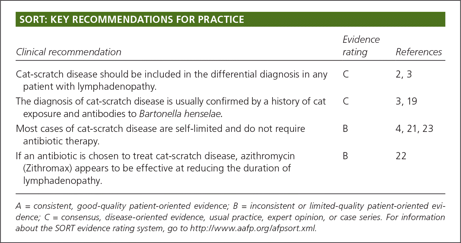
| Cat-scratch disease should be included in the differential diagnosis in any patient with lymphadenopathy. | C | 2, 3 |
| The diagnosis of cat-scratch disease is usually confirmed by a history of cat exposure and antibodies to Bartonella henselae. | C | 3, 19 |
| Most cases of cat-scratch disease are self-limited and do not require antibiotic therapy. | B | 4, 21, 23 |
| If an antibiotic is chosen to treat cat-scratch disease, azithromycin (Zithromax) appears to be effective at reducing the duration of lymphadenopathy. | B | 22 |
A = consistent, good-quality patient-oriented evidence; B = inconsistent or limited-quality patient-oriented evidence; C = consensus, disease-oriented evidence, usual practice, expert opinion, or case series. For information about the SORT evidence rating system, go to https://www.aafp.org/afpsort.xml.
Clinical Presentation
CSD is commonly diagnosed in children, but adults may also present with the disease. It should be suspected in patients with tender regional unilateral lymphadenopathy, especially if there is a history of exposure to kittens or cats. CSD causes local lymphadenopathy in 85 to 90 percent of patients.4 The differential diagnosis includes other causes of unilateral lymphadenopathy (Table 1). Although a history of exposure to cats is important, it is not absolutely necessary to make the diagnosis.
Table 1. Select Common Diseases That May Be Confused with Cat-scratch Disease Unilateral Lymphadenopathy
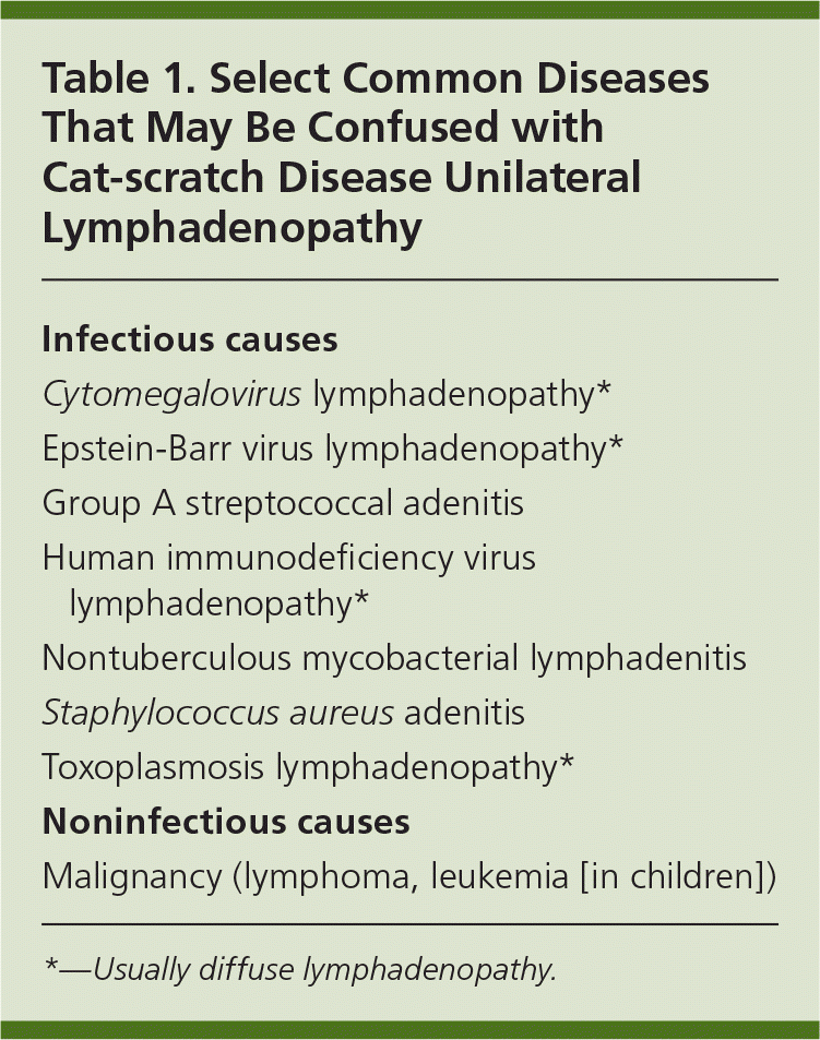
| Infectious causes |
| Cytomegalovirus lymphadenopathy* |
| Epstein-Barr virus lymphadenopathy* |
| Group A streptococcal adenitis |
| Human immunodeficiency virus lymphadenopathy* |
| Nontuberculous mycobacterial lymphadenitis |
| Staphylococcus aureus adenitis |
| Toxoplasmosis lymphadenopathy* |
| Noninfectious causes |
| Malignancy (lymphoma, leukemia [in children]) |
*—Usually diffuse lymphadenopathy.
After contact with an infected kitten3 or cat, patients can develop a primary skin lesion that starts as a vesicle at the inoculation site. A small number of patients do not recall contact with cats or having skin lesions. Regional lymphadenopathy develops one to two weeks later and is usually ipsilateral. According to one study, 46 percent of patients develop lymphadenopathy of the upper extremities, 26 percent develop lymphadenopathy of the neck and jaw, 18 percent develop lymphadenopathy of the groin, and 10 percent develop lymphadenopathy of other areas (pre- and postauricular, clavicular, and chest).4 In these patients, lymph nodes are swollen, tender, and may eventually suppurate.4 Seventy-five percent of patients develop aching, malaise, and anorexia, and 9 percent develop low-grade fever.4 Lymphadenopathy can persist for several months. Musculoskeletal manifestations, especially myalgia, arthralgia, and arthritis, are common and occur in more than 10 percent of patients.5 Visceral involvement has been reported6 and usually presents as hepatosplenomegaly with or without lymphadenopathy.7 Prolonged fever of unknown origin in children has been described.8,9
Rare cases of meningoencephalitis, endocarditis, and eye involvement have occurred in immunocompetent patients.10–12 One neurologic manifestation of CSD is encephalopathy, which manifests as severe headache and acute confusion one to six weeks after the onset of lymphadenopathy.10 Seizures may occur, and occasionally patients have focal neurologic deficits that are self-limiting, but can last up to one year. Parinaud oculoglandular syndrome is the most common ocular manifestation12 and consists of granulomatous conjunctivitis and ipsilateral periauricular lymphadenopathy. Neuroretinitis can occur in CSD and manifests as acute unilateral visual field loss secondary to optic nerve edema and star-shaped macular exudates.
In immunosuppressed patients, B. henselae can cause bacillary angiomatosis and peliosis.13 Bacillary peliosis is caused only by B. henselae and involves the liver and sometimes the spleen. Bacillary angiomatosis can be caused by B. henselae and Bartonella quintana, and usually involves the skin and lymph nodes, but can also involve bone and internal organs. Lesions consist of single or multiple red to purple papules.13 Bacillary angiomatosis was first described in patients with AIDS with very low CD4 cell counts.14 Evidence for previous Bartonella infection is common in patients with human immunodeficiency virus infection living in Rio de Janeiro, Brazil,15 and Bartonella infection was detected in 18 percent of febrile patients with human immunodeficiency virus infection living in San Francisco, Calif.16
Diagnostic Testing
The Bartonella species are difficult to culture, and culture is not routinely recommended. Serology is the best initial test and can be performed by indirect fluorescent assay or enzyme-linked immunosorbent assay. Although more sensitive than culture, serologic tests lack specificity because many asymptomatic persons have positive serology because of previous (often asymptomatic) exposure.17 The percentage of the general population that has a positive serologic test varies widely, but appears to be higher in cat owners.17 Immunoglobulin G titers less than 1:64 suggest the patient does not have current Bartonella infection. Titers between 1:64 and 1:256 represent possible infection; repeat testing should be performed in these patients in 10 to 14 days. Titers greater than 1:256 strongly suggest active or recent infection.18,19 A positive immunoglobulin M test suggests acute disease, but production of immunoglobulin M is brief. Immunoglobulin G has significant cross-reactivity between B. henselae and B. quintana. Polymerase chain reaction can detect different Bartonella species; specificity is very high, but the sensitivity is lower than with serology.
Consequently, when a child or adult presents with unilateral lymphadenopathy,3 the physician should consider the differential diagnoses provided in Table 1. A history of cat exposure should be sought and appropriate tests ordered, including serology for CSD. A history of cat exposure, lymphadenopathy, and elevated antibodies to B. henselae detected by enzyme-linked immunosorbent assay or indirect fluorescent assay confirms the diagnosis.
Lymph node biopsy is not indicated for most patients; however, it is appropriate in patients whose lymph nodes fail to involute and in whom diagnosis is uncertain. Lymph node specimens in patients with CSD show lymphoid hyperplasia and stellate granulomas. B. henselae is a small, curved, aerobic gram-negative bacillus that stains with silver. In bacillary angiomatosis, lobular proliferation of small blood vessels occurs with the presence of bacilli in adjacent connective tissue and blood vessels. In a series of 786 lymph node specimens from patients in whom CSD was suspected, only 245 (31.2 percent) had evidence of CSD. Thirteen of the 245 patients had concurrent mycobacteriosis or neoplasm. It is prudent that physicians follow up with patients who have unilateral lymphadenopathy, even those with confirmed CSD.20
Treatment
Treatment of CSD depends on the disease presentation. Most patients, especially children, have self-limited lymphadenopathy lasting two to eight weeks and do not require antibiotics. Up to 14 percent of persons develop dissemination to the liver, spleen, eye, or central nervous system4 and antibiotics may help.21
In a study from 1985, a single investigator evaluating 1,200 patients with lymphadenopathy who were believed to have CSD found that antibiotics were rarely used.4 Physicians today occasionally employ antibiotics in CSD. The results of one randomized trial support the use of oral azithromycin (Zithromax) for mild to moderate disease for five days (500 mg on day 1, followed by 250 mg daily for four more days for patients weighing more than 100 lb [45.5 kg]; or 10 mg per kg on day 1, followed by 5 mg per kg for four more days for patients weighing 100 lb or less).22 In this small study of 29 adult patients, the use of azithromycin led to a more rapid resolution of lymphadenopathy than placebo; eight of 14 patients taking azithromycin had more than 80 percent resolution at 30 days compared with one of 15 patients in the control group.22 The Infectious Diseases Society of America guidelines regarding CSD are equivocal about the routine use of antibiotics,23 whereas another panel of authorities recommended against the use of antibiotics in patients with mild or uncomplicated disease.21 Other antibiotics that have been used in CSD include rifampin, ciprofloxacin (Cipro), trimethoprim/sulfamethoxazole (Bactrim, Septra), and gentamicin.24
Treatment of bacillary angiomatosis and peliosis, which have high rates of relapse, with oral erythromycin or doxycycline for a prolonged course of three to four months has benefited patients. Treatment with cell wall–active antibiotics has not.13,23 Treatment of neurologic disease has not been evaluated, but a combination of erythromycin or doxycycline plus rifampin for four to six weeks may be effective as suggested by case reports of neuroretinitis.10
