
Am Fam Physician. 2011;83(3):307-308
Author disclosure: Nothing to disclose.
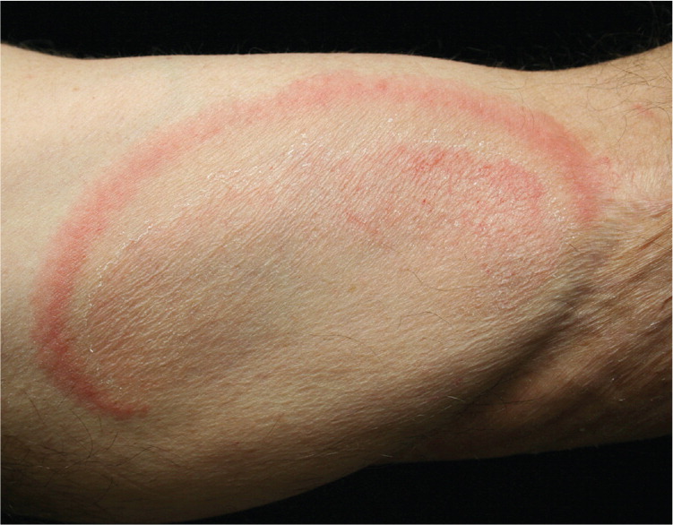
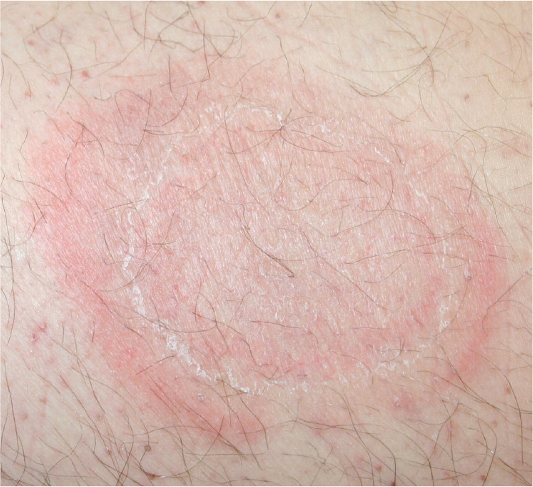
A 66-year-old man presented with a red, scaly, mildly pruritic skin rash that had persisted for five years. It started on his left big toe and progressed to his legs and inner arms. The rash did not respond to topical or systemic terbinafine (Lamisil) or itraconazole (Sporanox). He was otherwise healthy without constitutional symptoms, and had not started any new medications recently. Physical examination revealed large annular, erythematous patches with central clearing and an inner ring of scaling. Lesions were present on the left inner arm (Figure 1) and left medial thigh (Figure 2). Two smaller patches were present on the left buttock and right posterior thigh. The soles, palms, and nails were unaffected. Findings from potassium hydroxide (KOH) skin preparations were negative. Skin biopsy revealed epidermal spongiosis, superficial perivascular lymphocytic infiltrate without epidermotropism, and negative findings on a periodic acid-Schiff stain.
Question
Discussion
The answer is A: erythema annulare centrifugum. Patients with erythema annulare centrifugum classically present with erythematous, annular or arcuate (curved) patches on the trunk or proximal extremities. The lesions expand with central clearing, and scaling is present at the trailing edge of the erythema. The disorder is often pruritic and tends to spare the palms, soles, and mucosa. Patients with erythema annulare centrifugum have negative findings on KOH preparations, and the condition typically does not respond to antifungal therapy.1
Erythema annulare centrifugum is usually associated with epidermal spongiosis and superficial perivascular lymphocytic infiltrates. The histology is relatively nonspecific. Biopsy can rule out other diagnoses, such as tinea corporis, subacute cutaneous lupus erythematosus, or mycosis fungoides.1 Erythema annulare centrifugum may have a histologic reaction pattern suggestive of tumid lupus; spongiotic dermatitis; or pseudolymphoma, which has been associated with Borrelia species in Europe.2 Erythema annulare centrifugum is usually idiopathic.1 Treatment with potent topical steroids is the mainstay of therapy, but calcipotriol (called calcipotriene [Dovonex] in the United States)3 and metronidazole (Metrogel)4 have also been used.
Granuloma annulare presents as smooth, nonscaling, annular or arcuate, indurated plaques.5 The plaques typically occur on the extremities, alone or in groups.
Mycosis fungoides often presents as erythematous patches or plaques on the trunk or sun-protected skin, but usually lacks central clearing. Lesions often persist for many years.
Pityriasis rosea begins with a herald patch that can appear as an erythematous annular lesion, but the subsequent patches develop in a symmetric “Christmas tree” pattern.5 Patients with the condition often have a widespread truncal distribution of oval, salmon-colored patches with collarettes of scaling. Pityriasis rosea is more common in patients younger than 35 years and occurs along skin tension lines on the central trunk. It usually resolves within months.
Tinea corporis is much more common than erythema annulare centrifugum. The condition leads to erythematous, annular patches on the trunk or extremities. Scaling occurs at the leading edge of the lesions, as opposed to the trailing edge with erythema annulare centrifugum. A KOH preparation or fungal culture should be performed to confirm dermatophyte infection. Extensive tinea corporis may require oral terbinafine.5 When suspected dermatophyte infections do not respond to standard treatments, alternative diagnoses should be considered. A skin biopsy may be required to rule out the other conditions.
| Condition | Characteristics |
|---|---|
| Erythema annulare centrifugum | Erythematous, annular or arcuate (curved) patches on trunk or proximal extremities; central clearing; scaling at the trailing edge of the erythema |
| Granuloma annulare | Smooth, nonscaling, annular or arcuate, indurated plaques; lesions typically occur on the extremities, alone or in groups |
| Mycosis fungoides | Erythematous patches or plaques on trunk or sun-protected skin; usually lacks central clearing; often persists for many years |
| Pityriasis rosea | Widespread truncal distribution of oval, salmon-colored patches with collarettes of scaling; lesions occur along skin tension lines on the central trunk |
| Tinea corporis | Erythematous, annular patches on trunk or extremities; central clearing; scaling at leading edge |
