
Am Fam Physician. 2012;86(8):763-764
Author disclosure: No relevant financial affiliations to disclose.
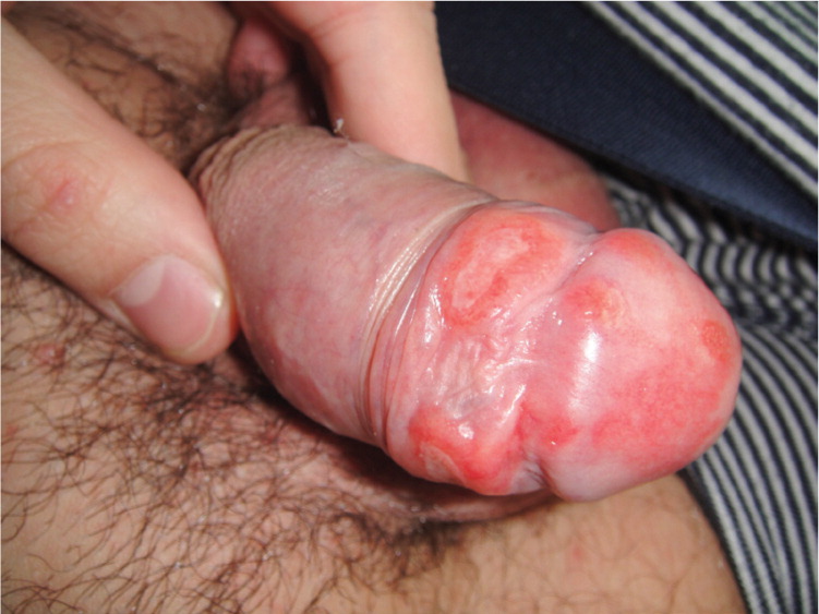
A 24-year-old man presented with multiple painless lesions on the shaft and glans of his penis that were accompanied by a rash on his trunk. He had never had a similar outbreak. The lesions were not painful or pruritic. He had no fever or joint pain. He was not taking any medications and had no history of sexually transmitted infections.
Physical examination revealed well-defined, erythematous, nonconfluent, ellipsoid erosions on the penis (Figure 1). The lesions had a smooth and polished surface, with induration. There were no vesicles or bullae. He had no urethral discharge. There was also a nonspecific rash on his trunk (Figure 2). Results of a complete blood count and routine analysis were within normal limits.

Question
Discussion
The answer is E: secondary syphilis. Syphilis is a sexually transmitted infection caused by Treponema pallidum. The highest incidence is in men who have sex with men.1 Because there was a strong suspicion of syphilis based on this patient's physical examination, serology testing was performed. A rapid plasma reagin test (1/32 titer) and fluorescent treponemal antibody absorption test were positive for the infection. Results of nontreponemal tests, such as the rapid plasma reagin test, will be positive four to six weeks after infection or one to three weeks after the chancre appears. Treponemal antigen tests are used to confirm the diagnosis.2 Histology may help in difficult cases.
The first stage (primary syphilis) presents as a chancre, which occurs at the site of inoculation 10 to 90 days after contact. Initially, the lesion is a round or ellipsoid, superficial, painless ulcer. It has a smooth and polished surface and may not be recognized as syphilis. At least 5 percent of syphilitic chancres are extragenital, usually on the oral mucosa, due to unprotected orogenital contact.3,4 Secondary syphilis can present with many different features that coexist with the primary ulcers, including lymphadenopathy and an early rash.
The coinfection of syphilis and human immunodeficiency virus (HIV) tends to have an aggressive course and a variety of cutaneous presentations. HIV transmission appears to be enhanced in the presence of the syphilitic chancre.5
For early secondary syphilis (less than one year after infection), the Centers for Disease Control and Prevention recommends a single dose of intramuscular penicillin G benzathine (2.4 million units).6 Repeat serology should be performed at six and 12 months after treatment to confirm that it was successful.
Behçet syndrome is characterized by recurrent, painful aphthous ulcers on the mouth and genitals. Anterior and posterior uveitis may also develop, which can lead to serious vision problems.
Chancroid is a sexually transmitted infection caused by Haemophilus ducreyi. It causes a painful ulcer at the site of inoculation, and unilateral lymphadenopathy. The ulcers are painful and friable with soft, undermined edges and yellow-gray, purulent exudate.
Herpes simplex virus infection is characterized by groups of painful vesicles in the genital area. Regional lymph nodes may also be enlarged and tender.
Lymphogranuloma venereum is a sexually transmitted infection caused by Chlamydia trachomatis. It causes painless, shallow ulcers that heal quickly, and painful lymphadenopathy that develops two to six weeks after infection.
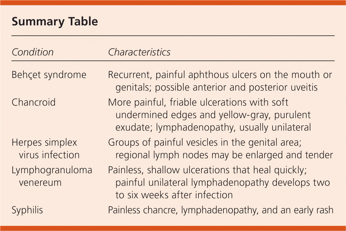
| Condition | Characteristics |
|---|---|
| Behçet syndrome | Recurrent, painful aphthous ulcers on the mouth or genitals; possible anterior and posterior uveitis |
| Chancroid | More painful, friable ulcerations with soft undermined edges and yellow-gray, purulent exudate; lymphadenopathy, usually unilateral |
| Herpes simplex virus infection | Groups of painful vesicles in the genital area; regional lymph nodes may be enlarged and tender |
| Lymphogranuloma venereum | Painless, shallow ulcerations that heal quickly; painful unilateral lymphadenopathy develops two to six weeks after infection |
| Syphilis | Painless chancre, lymphadenopathy, and an early rash |
