Approximately 1 percent of primary care office visits are for chest pain, and 1.5 percent of these patients will have unstable angina or acute myocardial infarction. The initial goal in patients presenting with chest pain is to determine if the patient needs to be referred for further testing to rule in or out acute coronary syndrome and myocardial infarction. The physician should consider patient characteristics and risk factors to help determine initial risk. Twelve-lead electrocardiography is typically the test of choice when looking for ST segment changes, new-onset left bundle branch block, presence of Q waves, and new-onset T wave inversions. For persons in whom the suspicion for ischemia is lower, other diagnoses to consider include chest wall pain/costochondritis (localized pain reproducible by palpation), gastroesophageal reflux disease (burning retrosternal pain, acid regurgitation, and a sour or bitter taste in the mouth), and panic disorder/anxiety state. Other less common but important diagnostic considerations include pneumonia (fever, egophony, and dullness to percussion), heart failure, pulmonary embolism (consider using the Wells criteria), acute pericarditis, and acute thoracic aortic dissection (acute chest or back pain with a pulse differential in the upper extremities). Persons with a higher likelihood of acute coronary syndrome should be referred to the emergency department or hospital.
Approximately 1 percent of all ambulatory visits in the primary care setting are for chest pain.1 Cardiac disease is the leading cause of death in the United States, yet only 1.5 percent of patients presenting to a primary care office with chest pain will have unstable angina or an acute myocardial infarction (MI).2 The most common causes of chest pain in the primary care population include chest wall pain (20 percent); reflux esophagitis (13 percent); and costochondritis (13 percent),2 although in practice, costochondritis is often included in the chest wall pain category. Other considerations include pulmonary (e.g., pneumonia, pulmonary embolism), gastrointestinal (e.g., gastroesophageal reflux disease [GERD]), and psychological (e.g., anxiety, panic disorder) etiologies, and cardiovascular disorders (e.g., acute congestive heart failure, acute thoracic aortic dissection). Table 1 lists the differential diagnosis of chest pain.3–15
SORT: KEY RECOMMENDATIONS FOR PRACTICE
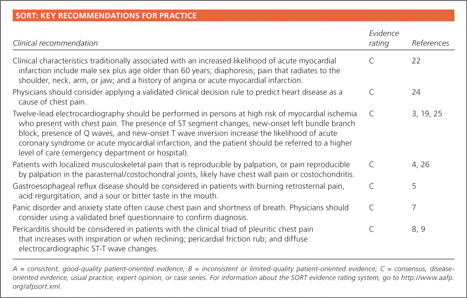
| Clinical recommendation | Evidence rating | References |
|---|---|---|
| Clinical characteristics traditionally associated with an increased likelihood of acute myocardial infarction include male sex plus age older than 60 years; diaphoresis; pain that radiates to the shoulder, neck, arm, or jaw; and a history of angina or acute myocardial infarction. | C | 22 |
| Physicians should consider applying a validated clinical decision rule to predict heart disease as a cause of chest pain. | C | 24 |
| Twelve-lead electrocardiography should be performed in persons at high risk of myocardial ischemia who present with chest pain. The presence of ST segment changes, new-onset left bundle branch block, presence of Q waves, and new-onset T wave inversion increase the likelihood of acute coronary syndrome or acute myocardial infarction, and the patient should be referred to a higher level of care (emergency department or hospital). | C | 3, 19, 25 |
| Patients with localized musculoskeletal pain that is reproducible by palpation, or pain reproducible by palpation in the parasternal/costochondral joints, likely have chest wall pain or costochondritis. | C | 4, 26 |
| Gastroesophageal reflux disease should be considered in patients with burning retrosternal pain, acid regurgitation, and a sour or bitter taste in the mouth. | C | 5 |
| Panic disorder and anxiety state often cause chest pain and shortness of breath. Physicians should consider using a validated brief questionnaire to confirm diagnosis. | C | 7 |
| Pericarditis should be considered in patients with the clinical triad of pleuritic chest pain that increases with inspiration or when reclining; pericardial friction rub; and diffuse electrocardiographic ST-T wave changes. | C | 8, 9 |
A = consistent, good-quality patient-oriented evidence; B = inconsistent or limited-quality patient-oriented evidence; C = consensus, disease-oriented evidence, usual practice, expert opinion, or case series. For information about the SORT evidence rating system, go to https://www.aafp.org/afpsort.xml.
Table 1. Differential Diagnosis of Chest Pain
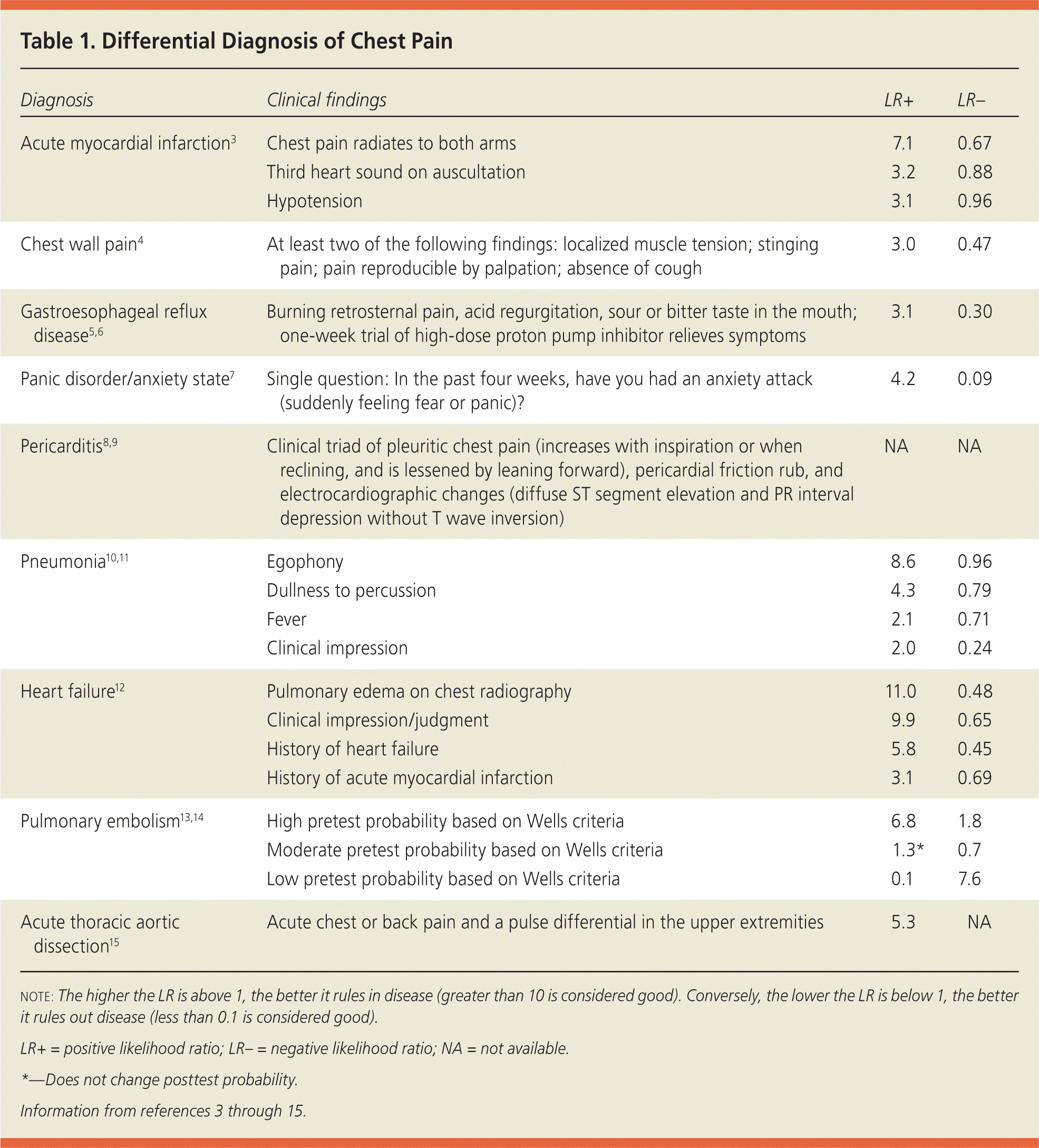
| Diagnosis | Clinical findings | LR+ | LR– |
|---|---|---|---|
| Acute myocardial infarction3 | Chest pain radiates to both arms | 7.1 | 0.67 |
| Third heart sound on auscultation | 3.2 | 0.88 | |
| Hypotension | 3.1 | 0.96 | |
| Chest wall pain4 | At least two of the following findings: localized muscle tension; stinging pain; pain reproducible by palpation; absence of cough | 3.0 | 0.47 |
| Gastroesophageal reflux disease5,6 | Burning retrosternal pain, acid regurgitation, sour or bitter taste in the mouth; one-week trial of high-dose proton pump inhibitor relieves symptoms | 3.1 | 0.30 |
| Panic disorder/anxiety state7 | Single question: In the past four weeks, have you had an anxiety attack (suddenly feeling fear or panic)? | 4.2 | 0.09 |
| Pericarditis8,9 | Clinical triad of pleuritic chest pain (increases with inspiration or when reclining, and is lessened by leaning forward), pericardial friction rub, and electrocardiographic changes (diffuse ST segment elevation and PR interval depression without T wave inversion) | NA | NA |
| Pneumonia10,11 | Egophony | 8.6 | 0.96 |
| Dullness to percussion | 4.3 | 0.79 | |
| Fever | 2.1 | 0.71 | |
| Clinical impression | 2.0 | 0.24 | |
| Heart failure12 | Pulmonary edema on chest radiography | 11.0 | 0.48 |
| Clinical impression/judgment | 9.9 | 0.65 | |
| History of heart failure | 5.8 | 0.45 | |
| History of acute myocardial infarction | 3.1 | 0.69 | |
| Pulmonary embolism13,14 | High pretest probability based on Wells criteria | 6.8 | 1.8 |
| Moderate pretest probability based on Wells criteria | 1.3* | 0.7 | |
| Low pretest probability based on Wells criteria | 0.1 | 7.6 | |
| Acute thoracic aortic dissection15 | Acute chest or back pain and a pulse differential in the upper extremities | 5.3 | NA |
note: The higher the LR is above 1, the better it rules in disease (greater than 10 is considered good). Conversely, the lower the LR is below 1, the better it rules out disease (less than 0.1 is considered good).
LR+ = positive likelihood ratio; LR– = negative likelihood ratio; NA = not available.
*—Does not change posttest probability.
Initial Evaluation
Algorithmic approaches to the diagnosis and workup of the patient presenting with chest pain in the office setting have not been specifically studied. Differentiating ischemic from nonischemic causes often is difficult, and patients with chest pain with an ischemic etiology often appear well. As such, the initial diagnostic approach should always consider a cardiac etiology for the chest pain, unless other causes are apparent.16
The first decision point for most physicians is whether or not the chest pain is caused by coronary ischemia.16 Acute coronary syndrome (ACS) is a constellation of clinical findings that suggests acute myocardial ischemia encompassing unstable angina and acute MI. Angina has been described as deep, poorly localized chest or arm discomfort (pain or pressure) that is reproducibly associated with physical exertion or emotional stress and is relieved promptly with rest or sublingual nitroglycerin.17 Unstable angina is defined as angina at rest, new-onset angina, or angina that has become more severe or longer in duration.18 Acute MI is defined as ST segment changes (elevation or depression) on electrocardiography (ECG) and positive laboratory markers of myocardial necrosis (e.g., troponin I).17 In office and ambulatory settings, the clinical impression is, in most cases, shaped by the presenting symptoms, physical examination, and initial ECG, combined with the patient's risk of ACS.16,19
The initial goal is to determine if the patient needs to be referred for further testing (e.g., troponin I or stress testing, coronary angiography) to rule in or out a potentially catastrophic ACS and acute MI. One recent meta-analysis concluded that the history and physical examination were mostly not helpful in diagnosing ACS or acute MI in patients presenting with chest pain, especially in a low prevalence setting.20
Although individual characteristics may not rule in or out a diagnosis, a combination of signs and symptoms may increase diagnostic accuracy.21 Characteristics traditionally associated with increased likelihood of acute MI include male sex plus age older than 60 years; diaphoresis; pain that radiates to the shoulder, neck, arm, or jaw; and a history of angina or acute MI.22 Predictability may be influenced by patient description of their symptoms. Patients often do not use the term pain to describe their symptoms, but frequently use other terms like discomfort, tightness, squeezing, or indigestion.16
Other clinical features that increase the likelihood of MI in patients with acute chest pain include pain that radiates to both arms (positive likelihood ratio [LR+] = 7.1), a third heart sound on auscultation (LR+ = 3.2), and hypotension (LR+ = 3.1). Clinical features that decrease the likelihood of acute MI include pleuritic chest pain (negative likelihood ratio [LR–] = 0.2), sharp or stabbing chest pain (LR– = 0.3), and chest pain reproduced by palpation (LR– = 0.2 to 0.4).3
The presence or absence of comorbidities, such as diabetes mellitus, tobacco use, hyperlipidemia, or hypertension, as cardiac risk factors weakly predict ACS in patients older than 40 years (LR+ = 2.1 in persons 40 to 65 years of age; LR+ = 1.1 in patients older than 65 years)23; however, evaluating for presence or absence of comorbidities is still an important component of the initial assessment.
One recently developed and validated clinical decision rule (Table 2) outlines five items that best predict coronary artery disease as the cause of chest pain: age/sex (55 years or older in men or 65 years or older in women); known coronary artery disease, occlusive vascular disease, or cerebrovascular disease; pain that is worse during exercise; pain not reproducible by palpation; and patient assumption that the pain is of cardiac origin.24 [corrected]
Table 2. Clinical Decision Rule for Identifying Patients with Chest Pain Caused by CAD
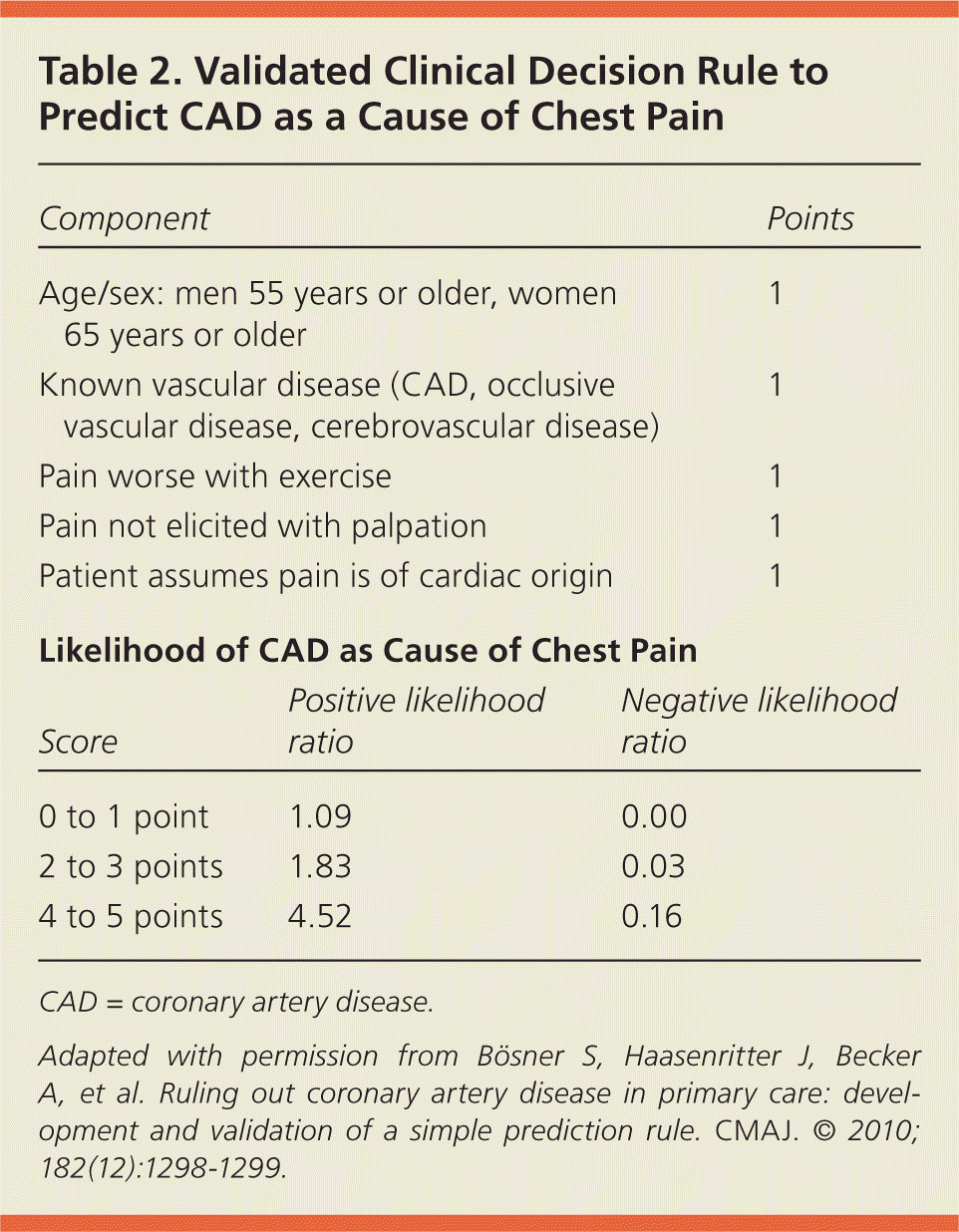
| Variable | Points |
|---|---|
| Age 55 years or older in men; 65 years or older in women | 1 |
| Known CAD or cerebrovascular disease | 1 |
| Pain not reproducible by palpation | 1 |
| Pain worse during exercise | 1 |
| Patient assumes pain is cardiogenic | 1 |
| Total points: | ______ |
CAD = coronary artery disease.
Information from reference 24.
Among those with none or one of these clinical factors, only 1 percent had coronary artery disease, whereas 63 percent of the patients with four or five of the factors had coronary artery disease. The study results suggest that patients with chest pain and four or five of these factors require urgent workup. Physicians should consider applying a validated clinical decision rule to predict heart disease as a cause of chest pain.24
Twelve-lead ECG is typically the test of choice in the initial evaluation of patients with chest pain.19 ST segment changes (elevation or depression), new-onset left bundle branch block, presence of Q waves, and new-onset T wave inversion increase the likelihood of ACS or acute MI.3,25 Concern based on the clinical impression (history, physical examination, risk factors, and 12-lead ECG) often will influence the physician's decision regarding whether to refer the patient to a higher level of care (emergency department or hospital) for further workup and treatment, or to look for other possible diagnoses for the chest pain.16,19
Other Diagnostic Considerations
If the initial evaluation indicates that a cardiac cause of ACS is less likely, other noncardiac causes of chest pain should be considered. Understanding that there are common conditions that often occur, with the clinical impression, will help lead to a correct diagnosis.
CHEST WALL PAIN
One prospective cohort study identified four clinical factors that predict a final diagnosis of chest wall pain in patients presenting to the primary care office with chest pain: localized muscle tension, stinging pain, pain reproducible by palpation, and the absence of a cough. Having at least two of these findings had a 77 percent positive predictive value for chest wall pain, and having none or one had an 82 percent negative predictive value.4
COSTOCHONDRITIS
Often considered a subset of chest wall pain, costochondritis is a self-limited condition characterized by pain reproducible by palpation in the parasternal/costochondral joints. It is sometimes called Tietze syndrome, which is distinguished from costochondritis by the presence of swelling over the affected joints.26 Costochondritis is a clinical diagnosis and does not require specific diagnostic testing in the absence of concomitant cardiopulmonary symptoms or risk factors.27
GERD
Classic symptoms of GERD include a burning retrosternal pain, acid regurgitation, and a sour or bitter taste in the mouth.5 No useful physical examination maneuvers exist to assist in establishing the diagnosis, and there is no standard test to rule it in or out. However, a one-week trial of a high-dose proton pump inhibitor is modestly sensitive and specific for GERD, with modest LRs (LR+ = 3.1; LR– = 0.3).6
PANIC DISORDER AND ANXIETY STATE
Panic disorder and anxiety state are common. One in four persons with a panic attack will have chest pain and shortness of breath.28 Yet, concomitant panic disorder and chest pain are often not recognized, leading to more testing, follow-up, and higher costs of care.28,29 Panic may cause chest pain and vice versa.28 Several validated brief questionnaires are used to diagnose panic disorder and anxiety state. One question (In the past four weeks, have you had an anxiety attack [suddenly feeling fear or panic]?) is sensitive (93 percent) and modestly specific (78 percent) in detecting panic disorder (LR+ = 4.2; LR– = 0.09).7
Less Common, but Important, Diagnostic Considerations
PERICARDITIS
Pericarditis is the clinical triad of pleuritic chest pain, pericardial friction rub, and diffuse electrocardiographic ST-T wave changes.8 ECG usually demonstrates diffuse ST segment elevation and PR interval depression without T wave inversion. Acute pericarditis should be considered in patients presenting with new-onset chest pain that increases with inspiration or when reclining, and is lessened by leaning forward.9
PNEUMONIA
Community-acquired pneumonia is a cause of chest pain and respiratory symptoms in the outpatient setting. Common symptoms include fever, chills, productive cough, and pleuritic chest pain.30 Fever, egophony heard during auscultation of the lungs, and dullness to percussion of the posterior thorax are suggestive of pneumonia.10 Clinical impression is modestly useful for ruling in or out pneumonia (LR+ = 2.0; LR– = 0.24).10 The test of choice for diagnosing pneumonia is chest radiography,11 although it has been more recently recommended that it be performed only if other diagnoses are being considered in the uncomplicated outpatient setting.31
HEART FAILURE
Most patients with heart failure present with dyspnea on exertion, although some will have chest pain.12 A history of heart failure or acute MI best predicts the presence of heart failure (LR+ = 5.8 and 3.1, respectively).12 Clinical impression/judgment is predictive of heart failure (LR+ = 9.9; LR– = 0.65), as is pulmonary edema on chest radiography (LR+ = 11.0).12
PULMONARY EMBOLISM
Diagnosing pulmonary embolism in the office based on signs and symptoms is difficult because of its highly variable presentation. Although dyspnea, tachycardia, and/or chest pain are present in 97 percent of those diagnosed with pulmonary embolism,32 there is no single clinical feature that effectively rules it in or out.33 The physician can estimate the patient's likelihood of pulmonary embolism by using a validated clinical decision rule, such as the Wells criteria (Table 313,34 ), to determine if further testing should be performed (e.g., d-dimer assay, ventilation-perfusion scan, helical computed tomography of the pulmonary arteries).13,14,35
Table 3. Wells Clinical Prediction Rule for PE
| Component | Points |
|---|---|
| Clinical signs of DVT (asymmetric leg swelling, palpable calf pain) | 3 |
| Diagnosis of PE is more likely than an alternative diagnosis | 3 |
| Heart rate greater than 100 beats per minute | 1.5 |
| Previous diagnosis of DVT or PE | 1.5 |
| Bed rest immobilization or surgery within the past four weeks | 1.5 |
| Hemoptysis | 1 |
| Malignancy within the past six months | 1 |
DVT = deep venous thrombosis; PE = pulmonary embolism.
Adapted with permission from Wells PS, Anderson DR, Rodger M, et al. Derivation of a simple clinical model to categorize patients probability of pulmonary embolism: Increasing the models utility with the SimpliRED d-dimer. Thromb Haemost. 2000;83(3):418, with additional information from reference 13.
ACUTE THORACIC AORTIC DISSECTION
Patients with acute thoracic aortic dissection may present with chest or back pain.36 History and physical examination are only modestly useful for ruling in or out the condition; acute chest or back pain and a pulse differential in the upper extremities modestly increase the likelihood of an acute thoracic aortic dissection (LR+ = 5.3).15
Data Sources: A PubMed search was completed using the key terms chest pain, diagnosis, clinical decision rule, and differential diagnosis. The search included meta-analyses, reviews, randomized controlled trials, and clinical trials. Also searched were the Cochrane Database of Systematic Reviews, the National Guideline Clearinghouse, Essential Evidence Plus, Database of Abstracts of Reviews of Effects, and UpToDate. Search date: the literature search was completed on several occasions; last date was June 20, 2012.
