Amblyopia is the leading cause of vision loss in children. It is treatable if diagnosed early, making identification of affected children critical. The American Association for Pediatric Ophthalmology and Strabismus and the American Academy of Pediatrics recommend that clinicians routinely perform age-appropriate vision chart testing, red reflex testing, and examination for signs of strabismus. The U.S. Preventive Services Task Force recommends vision screening for all children at least once between three and five years of age to detect the presence of amblyopia or its risk factors. Photoscreening may be a useful adjunct to traditional vision screening, but there is limited evidence that it improves visual outcomes. Treatments for amblyopia include patching, atropine eye drops, and optical penalization of the nonamblyopic eye. In children with moderate amblyopia, patching for two hours daily is as effective as patching for six hours daily, and daily atropine is as effective as daily patching. Children older than seven years may still benefit from patching or atropine, particularly if they have not previously received amblyopia treatment. Amblyopia recurs in 25 percent of children after patching is discontinued. Tapering the amount of time a patch is worn each day at the end of treatment reduces the risk of recurrence.
Amblyopia, from the Greek word for “dullness of vision,” is the leading cause of monocular childhood vision loss.1 It is defined as reduced best-corrected visual acuity caused by abnormal visual development. Its prevalence is estimated to be between 1 and 5 percent of the general population in the United States.2 Amblyopia is also a cause of permanent vision loss in 2.9 percent of adults.2 Although the ocular structures in affected children are usually normal, conditions such as strabismus (misalignment of the eyes) and unequal refractive error are highly associated with amblyopia. Bilateral amblyopia can also occur, particularly in patients with high refractive error in both eyes. Amblyopia may be diagnosed in children with cataracts, eyelid ptosis, and other eye conditions that impede the visual axis, if vision remains impaired after removal of the obstruction.
Although treatment ideally should begin before the patient reaches five years of age, a recent clinical trial demonstrated that children may benefit from treatment even at an older age.3 Other studies have shown that maximum hours of patching or atropine eye drops may not be necessary in all children.4,5 With early detection and appropriate, timely therapy, most affected children can have significant visual improvement.
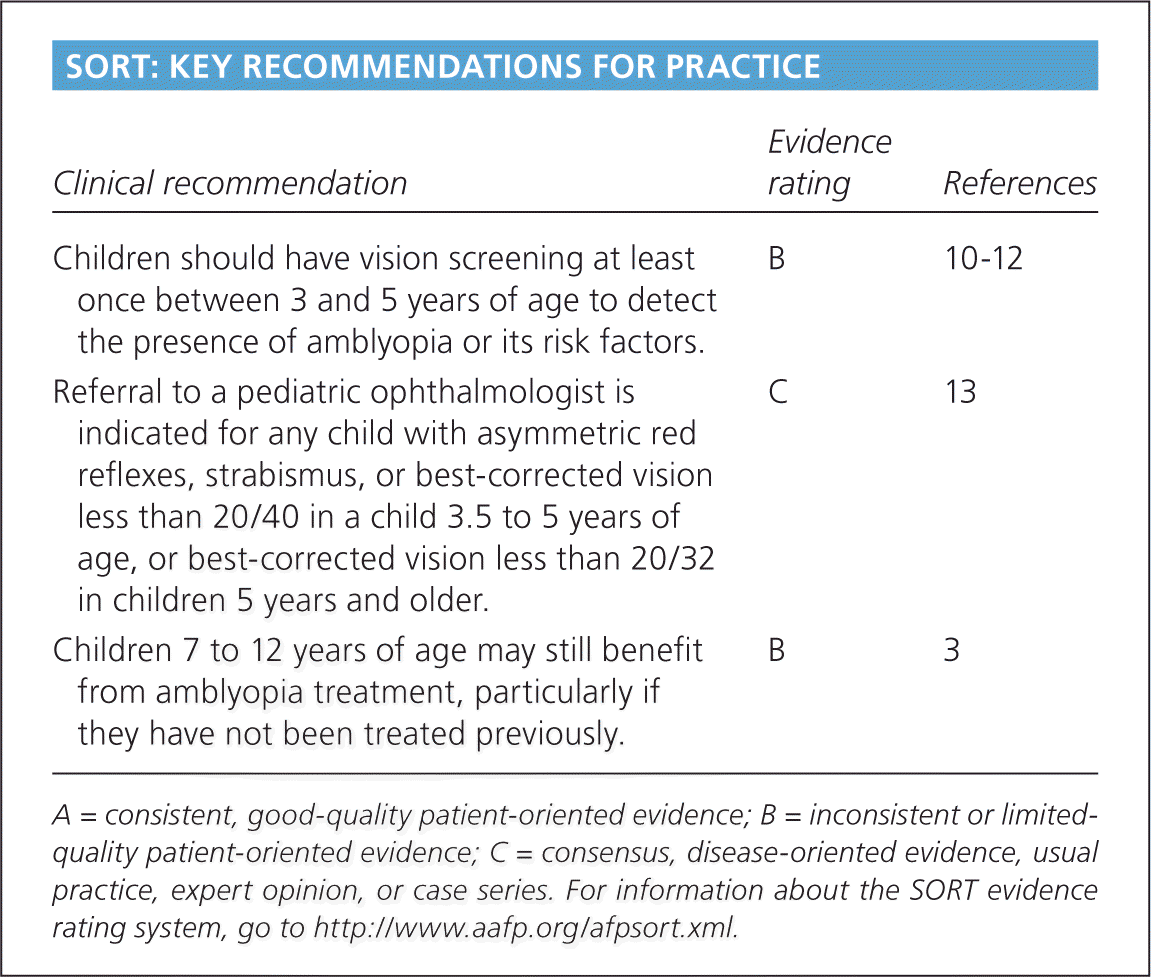
SORT: KEY RECOMMENDATIONS FOR PRACTICE
| Clinical recommendation | Evidence rating | References |
|---|---|---|
| Children should have vision screening at least once between 3 and 5 years of age to detect the presence of amblyopia or its risk factors. | B | 10–20 |
| Referral to a pediatric ophthalmologist is indicated for any child with asymmetric red reflexes, strabismus, or best-corrected vision less than 20/40 in a child 3.5 to 5 years of age, or best-corrected vision less than 20/32 in children 5 years and older. | C | 13 |
| Children 7 to 12 years of age may still benefit from amblyopia treatment, particularly if they have not been treated previously. | B | 3 |
A = consistent, good-quality patient-oriented evidence; B = inconsistent or limited-quality patient-oriented evidence; C = consensus, disease-oriented evidence, usual practice, expert opinion, or case series. For information about the SORT evidence rating system, go to https://www.aafp.org/afpsort.xml.
Etiology
Amblyopia is widely thought to develop in infancy, and continues through the first several years of life if there is visual blur at the level of the retina during a critical period of visual development. Such retinal blurring leads to disuse of the visual cortex, resulting in amblyopia.
The major types of amblyopia are listed in Table 1. Recent clinical trials suggest an equal distribution of strabismic, anisometropic (or refractive), and a combination of strabismic and refractive types.6
Table 1. Major Types of Amblyopia and Identifying Characteristics
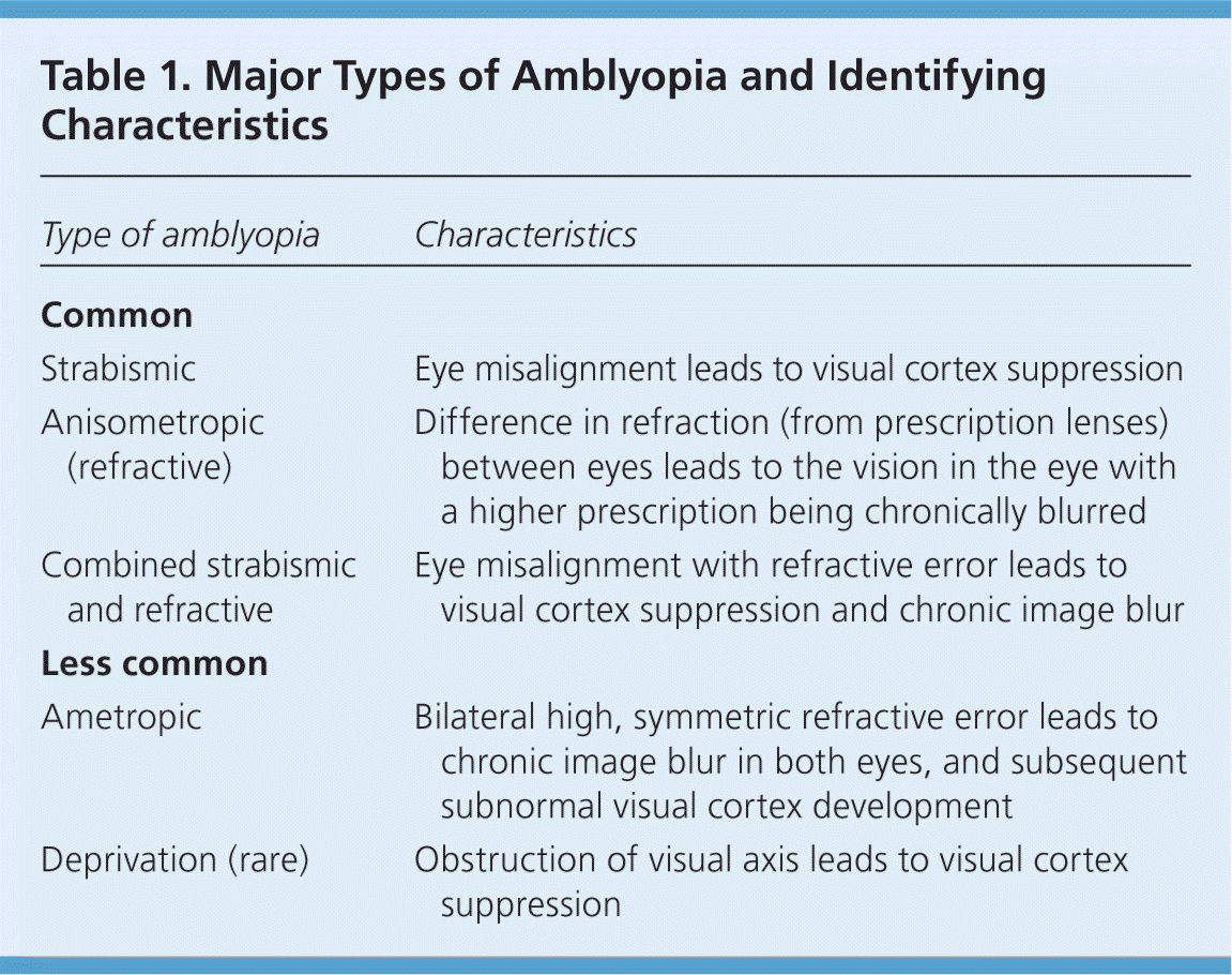
| Type of amblyopia | Characteristics |
|---|---|
| Common | |
| Strabismic | Eye misalignment leads to visual cortex suppression |
| Anisometropic (refractive) | Difference in refraction (from prescription lenses) between eyes leads to the vision in the eye with a higher prescription being chronically blurred |
| Combined strabismic and refractive | Eye misalignment with refractive error leads to visual cortex suppression and chronic image blur |
| Less common | |
| Ametropic | Bilateral high, symmetric refractive error leads to chronic image blur in both eyes, and subsequent subnormal visual cortex development |
| Deprivation (rare) | Obstruction of visual axis leads to visual cortex suppression |
Strabismic amblyopia occurs when eye misalignment leads to visual cortex suppression of the deviating eye. This occurs because each eye has a different image displayed on the retina, and the brain cannot fuse the two into a single image. Such central suppression leads to loss of depth perception (stereopsis) and binocular function.
Anisometropic, or refractive, amblyopia occurs when there is a difference in refractive error between the eyes, leading to vision in one eye being blurred. The eye with the higher refractive error requires greater focusing effort to form a clear retinal image, and often is left unfocused. Chronic blur leads to amblyopia. Hyperopic (farsighted) and astigmatic (astigmatism) anisometropia are more highly associated with vision loss than myopic (nearsighted) anisometropia. A subset of anisometropic amblyopia is associated with ocular conditions such as periorbital capillary hemangioma, eyelid ptosis, and mild cataracts. Although there may not be obstruction of the pupillary axis, induced astigmatism can occur, leading to amblyopia. In these cases, anisometropic amblyopia is more common than amblyopia caused by visual deprivation.
Ametropic and deprivation amblyopia are less common. Ametropic amblyopia occurs in children with bilateral, symmetric high refractive error. Vision loss occurs in both eyes because accommodation is often inadequate to provide the retina with a clear image, resulting in subnormal visual cortex development. Deprivation amblyopia is rare, and occurs with obstruction of the visual axis such as in congenital ptosis. Although this type of amblyopia accounts for less than 3 percent of cases, it is associated with the most severe vision loss, and can affect both eyes.1
Screening and Diagnosis
The American Academy of Pediatrics and the American Association for Pediatric Ophthalmology and Strabismus recommend vision screening in children three years and older with an eye chart–based test in the physician's office.7–9 Because treatment is more likely to succeed if initiated at a younger age,10 it is crucial to make the diagnosis of amblyopia as early as possible. A meta-analysis of four randomized clinical trials evaluating the effect of age on response to amblyopia treatment concluded that children seven years and older were less responsive to treatment than children three to six years of age.11 There was also a trend of greater responsiveness in children three to younger than five years, compared with children five to younger than seven years with severe amblyopia. Although the U.S. Preventive Services Task Force recommends vision screening for all children at least once between three and five years of age, it found insufficient evidence that vision screening for children younger than three years leads to improved visual outcomes.12
HISTORY
Children with a family history of strabismus or amblyopia have an increased risk of amblyopia. If a child has amblyopia, siblings should be observed for risk factors and undergo vision screening. If the child with amblyopia required glasses before two-and-a-half years of age, and did not have strabismus as a detectable risk factor, it is reasonable to refer a younger sibling for a comprehensive dilated eye examination.
Physicians should ask parents about torticollis (abnormal head posture), nystagmus, squinting of one eye, or strabismus. Torticollis, nystagmus, and squinting of one eye can all indicate strabismus. Torticollis with visual tasks may be a sign of strabismus, because the child may adopt a head position to better align his or her eyes. Torticollis is commonly associated with vertical strabismus, such as in congenital superior oblique palsy.
Children with nystagmus may adopt a head turn to find a visual area in which their nystagmus decreases (null zone), thus improving their vision. They may also demonstrate eye crossing while fixing on a near target as a result of converging their eyes to dampen the nystagmus. Any patient with strabismus is at risk of amblyopia, so a child with torticollis requires a comprehensive eye examination to rule out eye misalignment. Squinting of one eye, especially in bright light conditions, can be a sign of intermittent exotropia, or eye wandering.
The diagnosis of amblyopia is made when a child has decreased vision usually associated with an amblyogenic risk factor and without ocular structural abnormalities. It can also be diagnosed in a child after removal of a visual axis obstruction (i.e., cataract) with persistent reduced vision. The diagnosis of amblyopia is always determined with best-corrected vision.
EXAMINATION
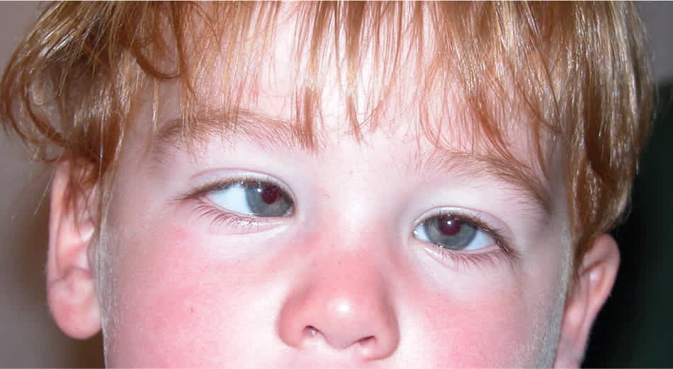
In infants and children, red reflex testing (in which a direct ophthalmoscope is used to compare the reflex in both eyes for asymmetry) is valuable in detecting risk factors for amblyopia, such as a cataract, refractive error, and retinal pathology (Figure 1). Fixation preference of one eye can signify amblyopia. In infants and children up to two-and-a-half years of age, using a toy or other near fixation target to hold the child's attention, one eye is occluded with an occluder or adhesive patch, and fixation behavior is observed. When the other eye is then occluded, a similar fixation behavior is expected. If a child consistently appears uncooperative when patching one eye, amblyopia is suspected. In children with straight eyes, fixation preference can be determined by using a prism over one eye to induce separate visual images (induced tropia test). In a child with strabismus, fixation preference is often easily determined in the nondeviating eye (Figure 2).
Figure 1.
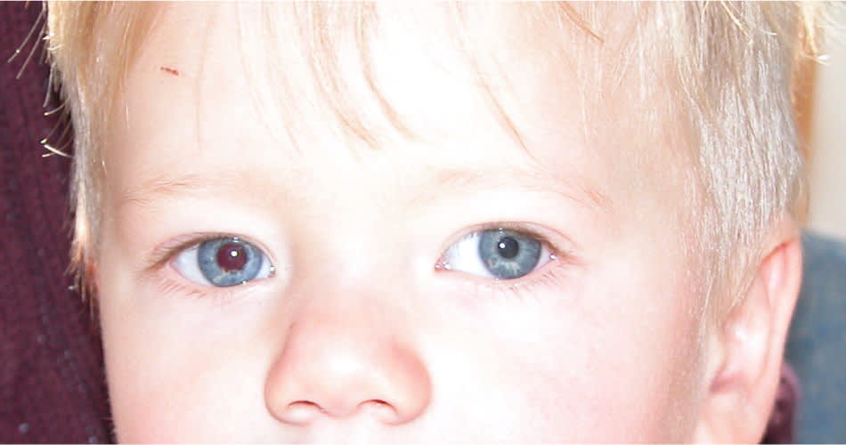
Patient with asymmetric red reflexes and left exotropia. The patient was found to have retinoblastoma of the left eye.
Figure 2.
VISION TESTING
In children three years and older, vision testing is performed monocularly with standard eye charts. The charts are labeled with the proper testing distance of 10 or 20 feet (approximately 3 or 6 m). The most advanced test that a child can perform is recommended, because picture testing may mildly overestimate vision compared with letter testing. The American Association for Pediatric Ophthalmology and Strabismus vision screening guidelines are provided in Table 2.13 Any child three-and-a-half to five years of age with best-corrected vision less than 20/40 in either eye, or five years and older with best-corrected vision less than 20/32 in either eye, or with a two-line difference between eyes, should be referred to a pediatric ophthalmologist.13 Children should be tested with lines of letters, and not a single letter, because the latter can overestimate vision. An adhesive patch to cover the eye is preferred for testing over a handheld occluder, because children may peek around the occluder unnoticed, and amblyopia can be missed. Patients with nystagmus should have their vision tested with both eyes first, which gives their true functioning visual acuity. When a patch or occluder is placed over one eye, increased nystagmus may occur, further decreasing the patient's vision. This results in falsely lowered monocular visual acuity.
Table 2. American Association for Pediatric Ophthalmology and Strabismus Vision Screening Recommendations
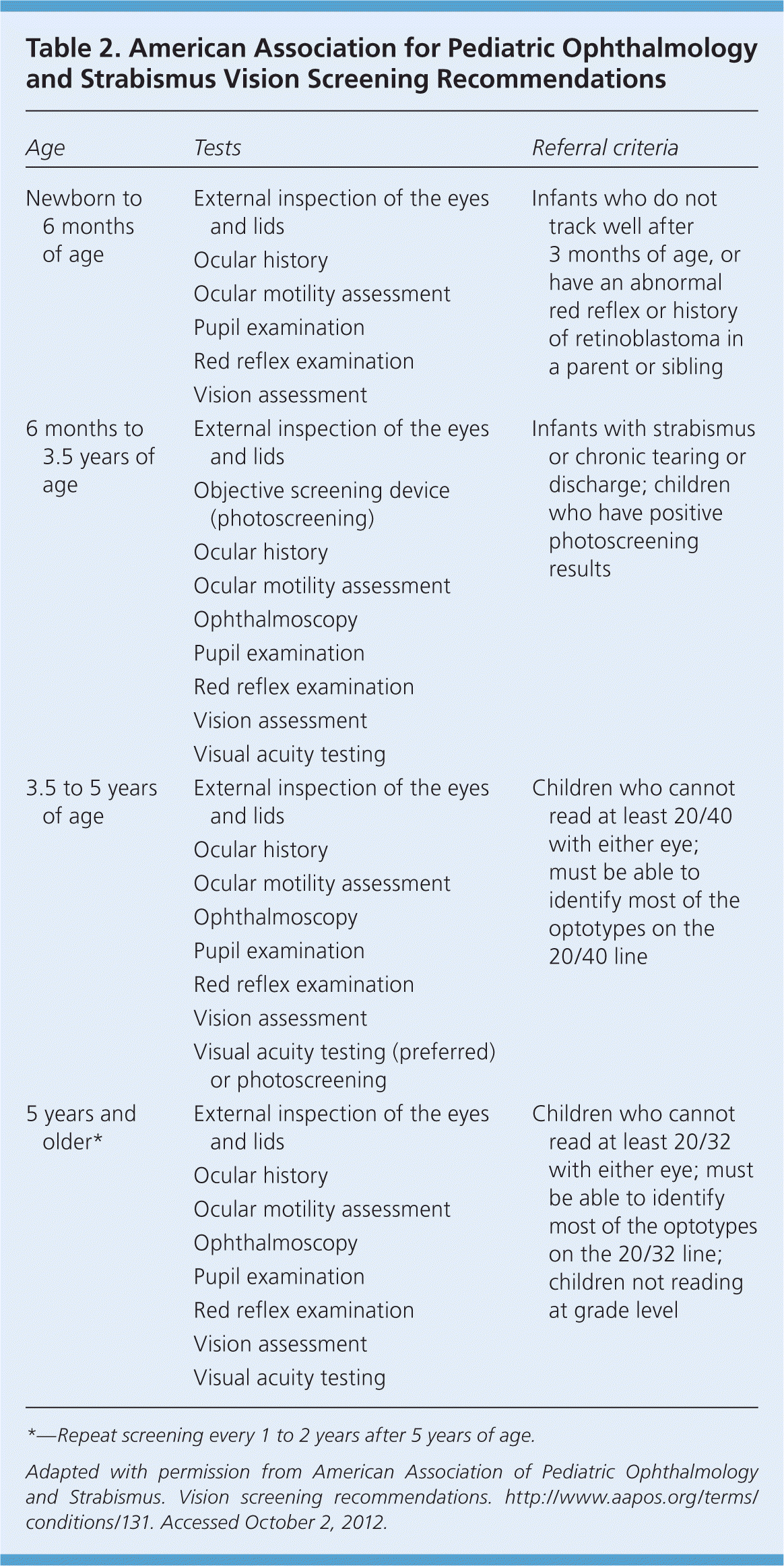
| Age | Tests | Referral criteria |
|---|---|---|
| Newborn to 6 months of age | External inspection of the eyes and lids | Infants who do not track well after 3 months of age, or have an abnormal red reflex or history of retinoblastoma in a parent or sibling |
| Ocular history | ||
| Ocular motility assessment | ||
| Pupil examination | ||
| Red reflex examination | ||
| Vision assessment | ||
| 6 months to 3.5 years of age | External inspection of the eyes and lids | Infants with strabismus or chronic tearing or discharge; children who have positive photoscreening results |
| Objective screening device (photoscreening) | ||
| Ocular history | ||
| Ocular motility assessment | ||
| Ophthalmoscopy | ||
| Pupil examination | ||
| Red reflex examination | ||
| Vision assessment | ||
| Visual acuity testing | ||
| 3.5 to 5 years of age | External inspection of the eyes and lids | Children who cannot read at least 20/40 with either eye; must be able to identify most of the optotypes on the 20/40 line |
| Ocular history | ||
| Ocular motility assessment | ||
| Ophthalmoscopy | ||
| Pupil examination | ||
| Red reflex examination | ||
| Vision assessment | ||
| Visual acuity testing (preferred) or photoscreening | ||
| 5 years and older* | External inspection of the eyes and lids | Children who cannot read at least 20/32 with either eye; must be able to identify most of the optotypes on the 20/32 line; children not reading at grade level |
| Ocular history | ||
| Ocular motility assessment | ||
| Ophthalmoscopy | ||
| Pupil examination | ||
| Red reflex examination | ||
| Vision assessment | ||
| Visual acuity testing |
*—Repeat screening every 1 to 2 years after 5 years of age.
Adapted with permission from American Association of Pediatric Ophthalmology and Strabismus. Vision screening recommendations. http://www.aapos.org/terms/conditions/131. Accessed October 2, 2012.
PHOTOSCREENING
Photoscreening provides another approach to vision screening. The equipment is designed for use with minimal personnel training and has been associated with successful amblyopia treatment in children identified with risk factors.14 A child's red reflex of both eyes taken simultaneously with digital or flash photography is evaluated for signs of uncorrected refractive error. The sensitivity in detecting amblyopia among photoscreeners is between 63 and 98 percent.15,16 However, photoscreeners have high false-positive rates, leading to higher rates of referral of children with normal vision. Although photoscreening should not replace traditional vision chart testing, it may be a useful adjunct in children who cannot cooperate for vision chart testing.
Treatment
Treatment for amblyopia includes one or more of the following: patching, atropine eye drops, and optical penalization of the unaffected eye to stimulate visual development of the amblyopic eye.17 Treatment is initiated only after any refractive errors are corrected. Atropine 1% eye drops block parasympathetic innervation to the ciliary muscle and pupil, causing temporary loss of accommodation (focusing at near) and pupillary dilation. Atropine is more effective in farsighted eyes, because removing the ability to accommodate in such eyes does not allow the child to focus at near. The goal of atropine use is to stimulate preferential near fixation of the amblyopic eye, leading to visual improvement. A less utilized therapy option is the Bangerter filter, in which a graded adhesive is applied to the child's glasses over the lens of the nonamblyopic eye, producing a blurred image. This is mostly used in older children who prefer a less noticeable option than patching.
Individual treatment plans depend on the age of the patient, the severity of amblyopia, and compliance with patching or atropine regimen and follow-up. For example, a seven-year-old patient with anisometropia who is initially diagnosed with dense amblyopia would likely benefit from a combination of glasses with patching instead of observation with glasses alone. A similar three-year-old patient may be treated with glasses alone, followed by patching after any improvement in vision stabilizes. Surveillance for occlusion amblyopia (decreased vision in the patched eye due to deprivation) and amblyopia recurrence after successful treatment are critical. Amblyopia recurs in 25 percent of children after patching is discontinued.18 Tapering the amount of time a patch is worn each day at the end of treatment reduces the risk of recurrence.
Recent clinical trials have discovered that in children who have moderate amblyopia (patient's vision: 20/40 to 20/80), patching two hours daily is as effective as patching six hours daily. The trials have also found that performing near activities does not enhance amblyopia treatment; that daily atropine is as effective as daily patching in moderate amblyopia; and that children seven to 12 years of age may benefit from amblyopia treatment that is sustained, particularly if they have not been treated for amblyopia before.3,19–21
Data Sources: A PubMed search was completed in Clinical Queries using the key terms amblyopia and vision screening. The search included meta-analyses, randomized controlled trials, clinical trials, and reviews. Search date: October 19, 2012.
