A 32-year-old woman presented with a pruritic skin rash that had appeared intermittently for two years. Initially, the rash involved multiple papules and small plaques on the face, neck, and trunk. The lesions progressed to large plaques with centrifugally spreading edges and central clearing. The patient did not have fever or joint pain, and was otherwise healthy. Sun exposure did not exacerbate the rash.
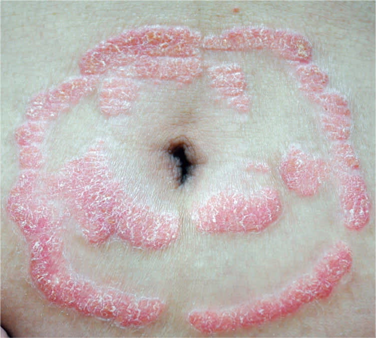
Physical examination revealed erythematous, annular plaques on the face, neck, back, scalp, chest, abdomen, and buttocks (Figures 1 and 2). There was a fine scale, but no vesicles or pustules. The palms, soles, nails, and mucosa were not affected. Results of a potassium hydroxide preparation of a skin scraping were negative. A skin biopsy was performed. No hyphae or spores were detected on a periodic acid–Schiff stain.
Figure 1.
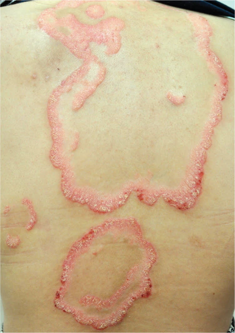
Figure 2.
Question
Based on the patient's history, physical examination, and laboratory findings, which one of the following is the most likely diagnosis?
A. Annular plaque psoriasis.
B. Erythema annulare centrifugum.
C. Erythema gyratum repens.
D. Subacute lupus erythematosus.
E. Tinea corporis.
Discussion
The answer is A: annular plaque psoriasis. Psoriasis is a chronic inflammatory skin disorder with a genetic predisposition. Triggers include infection, drug use, and trauma. Plaque psoriasis is characterized by annular, well-demarcated, erythematous plaques with adherent, silvery-white scales and central clearing. The elbows, knees, scalp, intergluteal region, lower back, periumbilical area, palms, and soles are often involved. Nail changes or arthritis may occur. Other types of psoriasis include guttate, inverse, pustular, and erythrodermic. The annular pattern occurs with plaque or pustular psoriasis.1
The histologic findings of annular plaque psoriasis are identical to those of typical plaque psoriasis. The classic features include marked acanthosis, confluent parakeratosis, diminution of the granular layer, thinning of the suprapapillary plate, tortuous and dilated capillaries in the papillary dermis, and perivascular lymphocytic infiltrates. Accumulation of neutrophils within the stratum corneum (Munro microabscess) is diagnostic.2
Topical corticosteroids or vitamin D analogues are effective for mild to moderate psoriasis. Phototherapy, systemic treatments, or biologic immunotherapy should be considered for more severe cases.3
Erythema annulare centrifugum is characterized by annular or arcuate, erythematous patches or plaques with central clearing and trailing scales behind the advancing margin on the trunk and extremities. The lesions slowly spread centrifugally. The condition causes sleeve-like, perivascular, lymphohistiocytic infiltrates with or without spongiosis and parakeratosis.4
Erythema gyratum repens appears as concentric, scaly bands of erythema in a gyrate pattern, forming a “wood grain” appearance. The bands spread rapidly (up to 1 cm per day). More than 80 percent of patients with the condition have an underlying malignancy, most commonly lung cancer.5
Subacute lupus erythematosus has two typical cutaneous manifestations: annular lesions and papulosquamous lesions. The erythematous, scaly plaques predominantly appear on sun-exposed areas, such as the shoulders, neck, upper back, anterior chest, and extensor surface of the upper limbs. Most patients with the condition have a history of photosensitivity and a positive anti-Ro antibody test result.6
Tinea corporis is a superficial dermatophyte infection that affects the trunk and extremities. It may appear as annular, erythematous, scaly, pruritic patches or plaques with central clearing. Septate hyphae and fungal spores on potassium hydroxide preparation or positive fungal culture results are helpful to confirm the diagnosis. In addition, hyphae can be accentuated on a periodic acid–Schiff stain of the skin biopsy.7
Summary Table
| Condition | Characteristics |
|---|---|
| Annular plaque psoriasis | Annular, well-demarcated, erythematous plaques with adherent, silvery-white scales and central clearing; accumulation of neutrophils within the stratum corneum (Munro microabscess); possible nail changes or arthritis |
| Erythema annulare centrifugum | Annular or arcuate, erythematous patches or plaques with central clearing and trailing scales behind the advancing margin; appears on the trunk and extremities |
| Erythema gyratum repens | Concentric, scaly bands of erythema in a gyrate pattern (“wood grain“ appearance); spreads rapidly; associated with underlying malignancy |
| Subacute lupus erythematosus | Annular or papulosquamous, erythematous, scaly plaques on sun-exposed areas; history of photosensitivity and a positive anti-Ro antibody test result |
| Tinea corporis | Annular, erythematous, scaly, pruritic patches or plaques with central clearing; appears on the trunk and extremities; hyphae and fungal spores on potassium hydroxide preparation |
