
Am Fam Physician. 2013;88(4):261-262
Author disclosure: No relevant financial affiliations.
Question
Discussion
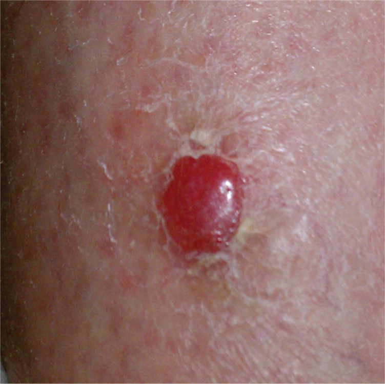
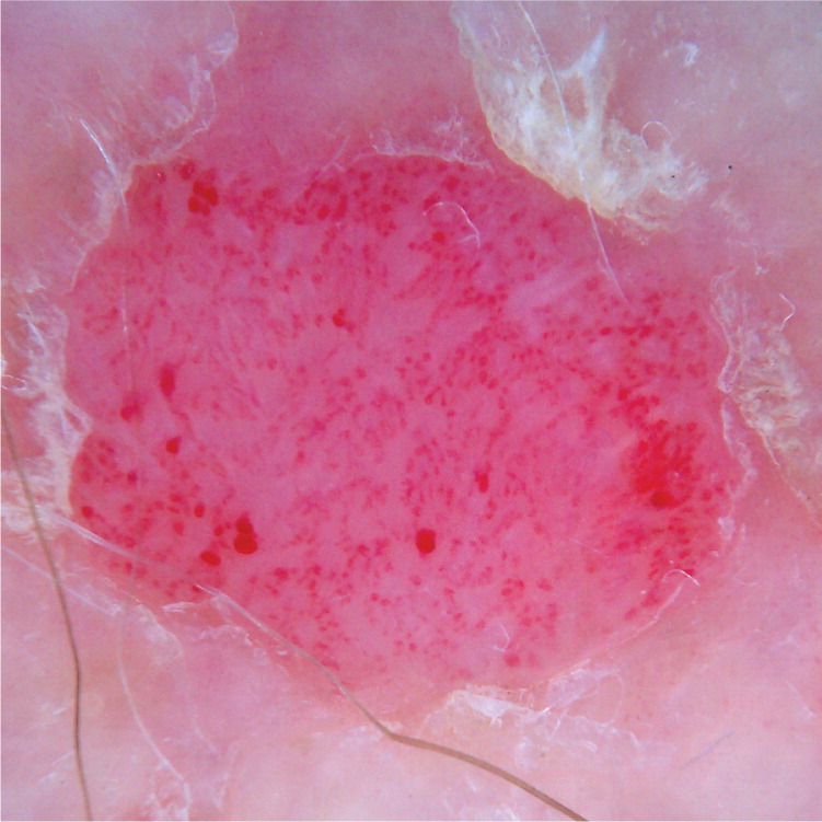
The correct answer is C: clear cell acanthoma. Clear cell acanthoma, also known as Degos acanthoma, is a benign epidermal tumor of unknown etiology.1 It typically presents as a solitary, pink to brown, glistening, round papule or plaque on the leg of an older person.1–3 It is slow growing and usually smaller than 2 cm. The margin is sharply demarcated, often with a thin rim of scale.1,4 Dermoscopy shows a smooth and stippled surface with pinpoint vessels appearing as tiny red dots.4 A highly specific feature is the linear arrangement of these vessels5 with a “string of pearls” appearance.6
On histology, clear cell acanthoma appears as psoriasiform epidermal hyperplasia, and a sharp demarcation from the adjacent normal epidermis. The glycogen content of the clear cells is highlighted with magenta on a periodic acid–Schiff stain, which is distinct from the surrounding normal epidermis that stains uniformly light blue.
Amelanotic melanoma is a malignant skin tumor that can present as a solitary featureless, pink papule or nodule, without the typical brown to black color of melanoma. Dermoscopic features include a blue-white veil (irregular area of confluent blue pigment with a ground-glass haze), which is caused by hyperkeratinization; a scar-like or irregularly shaped depigmentation; blue-gray dots; irregular brown dots or globules; multiple shades of color; and prominent central vessels.7
Bowen disease, or squamous cell carcinoma in situ of the skin, commonly presents as an erythematous scaling patch or a slightly raised plaque on sun-exposed skin of an older person. It may develop de novo or within a preexisting actinic keratosis.1 Dermoscopy shows dotted or linearly coiled vessels arranged in clusters, possibly with a white halo around the vessels and surface scales.5
Pyogenic granuloma is a rapidly growing, friable, vascular papule or polyp of the skin or mucosa most common in children and young adults. It may be associated with trauma or irritation.1 On dermoscopy, red homogeneous areas are surrounded by a white collarette and intersected by white “rail lines.”5
| Condition | Characteristics | Dermoscopic features |
|---|---|---|
| Amelanotic melanoma | A solitary pink, featureless papule or nodule, without the brown to black color of melanoma | Blue-white veil (irregular area of confluent blue pigment with a ground-glass haze), scar-like or irregularly shaped depigmentation, blue-gray dots, irregular brown dots or globules, multiple shades of color, prominent central vessels |
| Bowen disease | Erythematous scaling patch or slightly raised plaque on sun-exposed skin of an older person | Dotted or linearly coiled vessels arranged in small clusters |
| Clear cell acanthoma | Solitary slow-growing, pink to brown, glistening, round papule or plaque on the leg of an older person; sharply demarcated margin, often with a thin rim of scale; less than 2 cm in size | Smooth and stippled surface with pinpoint vessels appearing as tiny red dots in the characteristic “string of pearls” arrangement |
| Guttate psoriasis | Multiple small, discrete, scaly papules and plaques on the trunk; common in children; may follow a severe upper respiratory tract infection | Homogeneous dotted vessels |
| Pyogenic granuloma | Rapidly growing, friable, vascular papule or polyp of skin or mucosa, common in children and young adults; may be associated with trauma or irritation | Red homogeneous areas surrounded by a white collarette and intersected by white “rail lines” |
