
Am Fam Physician. 2015;92(1):61-62
Author disclosure: No relevant financial affiliations.
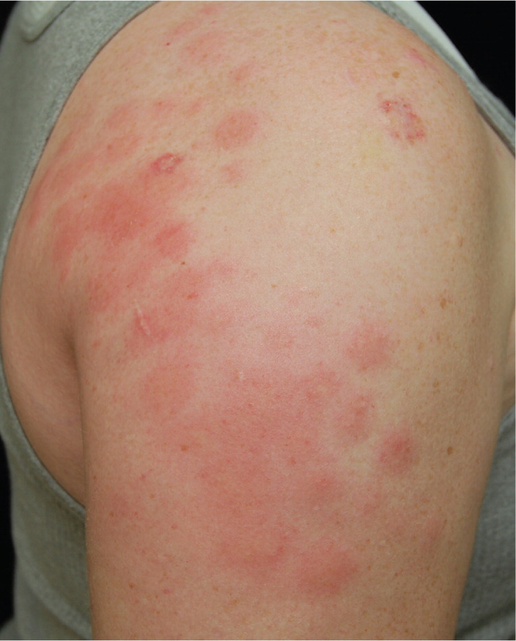
An otherwise healthy 26-year-old woman presented for a two-week follow-up after arthroscopic rotator cuff repair. She had a mildly pruritic eruption that began approximately one week after surgery and gradually worsened. The lesions were fixed and occurred in the area where she was using a cooling device. She was afebrile and feeling otherwise well.
Physical examination revealed multiple, round, erythematous, 1- to 2-cm plaques coalescing into larger plaques confined to the shoulder and proximal upper arm (Figure 1). There was no overlying scale and minimal induration.
Question
Discussion
The answer is B: cold panniculitis. Cold panniculitis is an inflammatory condition of the adipose tissue that results from prolonged exposure of the skin to cold temperatures. In this case, it was induced by a cooling compression system that was used postoperatively. These devices allow for continuous cooling and compression of the affected area, which helps to reduce inflammation after injuries or orthopedic surgery.1
Cold panniculitis is a subtype of traumatic panniculitis. It tends to primarily affect the subcutaneous fat. There are several manifestations depending on the severity of cold, length of exposure, and mechanism of cold injury. It most commonly occurs in children on the perioral region after eating frozen desserts (“popsicle panniculitis”).2 However, it has occurred in other settings, including after ice therapy in neonates with supraventricular tachycardias and on the thighs of women horseback riders.3,4 The mechanism of injury may consist of thermal damage or ischemia from cold-induced vasospasm.5
The key to diagnosing cold panniculitis is a history of prolonged exposure to cold with typical appearing lesions in an otherwise asymptomatic patient. Lesions usually appear within 48 hours of the cold exposure, but can occur anywhere from six to 72 hours after exposure.2,6 Lesions are erythematous to violaceous nodules or plaques with ill-defined margins. They can be pruritic or painful, and occasionally focally ulcerated or crusted. In most cases, lesions are localized to areas of subcutaneous adipose tissue that are exposed to cold temperatures. In infants, the cheeks and chin are most commonly involved, whereas the thighs, buttocks, and lower abdomen are most often affected in adults.2,7
The diagnosis is mainly clinical but can be confirmed with biopsy. Histologically, cold panniculitis causes lobular inflammation within the subcutaneous fat, with scattered lymphohistiocytic and eosinophilic infiltrates without vasculitis.1 Treatment consists of discontinuing the cold exposure.7 Lesions usually resolve within two to three weeks, typically without scarring, although hyper-pigmentation can persist within the lesions.2
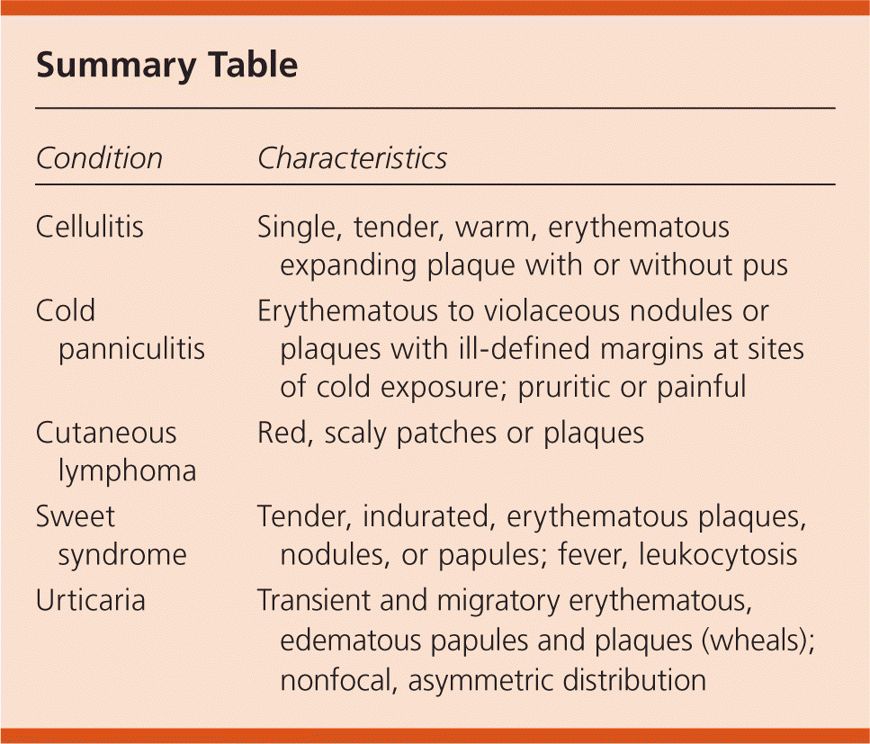
| Condition | Characteristics |
|---|---|
| Cellulitis | Single, tender, warm, erythematous expanding plaque with or without pus |
| Cold panniculitis | Erythematous to violaceous nodules or plaques with ill-defined margins at sites of cold exposure; pruritic or painful |
| Cutaneous lymphoma | Red, scaly patches or plaques |
| Sweet syndrome | Tender, indurated, erythematous plaques, nodules, or papules; fever, leukocytosis |
| Urticaria | Transient and migratory erythematous, edematous papules and plaques (wheals); nonfocal, asymmetric distribution |
Cellulitis usually presents as a single, tender, warm, erythematous, expanding plaque with or without pus.8 It is often preceded by a superficial break in the skin.
Cutaneous lymphoma usually presents as red, scaly patches or plaques. As with other malignancies, fever, night sweats, and weight loss are common.
Sweet syndrome (acute febrile neutrophilic dermatosis) presents as tender, indurated, erythematous plaques, nodules, or papules, which can look similar to cold panniculitis lesions. Sweet syndrome can be distinguished from cold panniculitis by the presence of fever and leukocytosis. It is associated with malignancy and medication use.9
Urticaria usually presents as erythematous, edematous papules and plaques (wheals) in a nonfocal, asymmetric distribution. They are transient and migratory. Individual lesions usually last less than 24 to 36 hours, have an asymmetric distribution, and are typically pruritic.10
The views expressed in this material are those of the authors and do not reflect the official policy or position of the U.S. government, the Department of Defense, or the Department of the Air Force.
