The most common cause of acute dysuria is infection, especially cystitis. Other infectious causes include urethritis, sexually transmitted infections, and vaginitis. Noninfectious inflammatory causes include a foreign body in the urinary tract and dermatologic conditions. Noninflammatory causes of dysuria include medication use, urethral anatomic abnormalities, local trauma, and interstitial cystitis/bladder pain syndrome. An initial targeted history includes features of a local cause (e.g., vaginal or urethral irritation), risk factors for a complicated urinary tract infection (e.g., male sex, pregnancy, presence of urologic obstruction, recent procedure), and symptoms of pyelonephritis. Women with dysuria who have no complicating features can be treated for cystitis without further diagnostic evaluation. Women with vulvovaginal symptoms should be evaluated for vaginitis. Any complicating features or recurrent symptoms warrant a history, physical examination, urinalysis, and urine culture. Findings from the secondary evaluation, selected laboratory tests, and directed imaging studies enable physicians to progress through a logical evaluation and determine the cause of dysuria or make an appropriate referral.
Dysuria is burning, tingling, or stinging of the urethra and meatus associated with voiding. It should be distinguished from other forms of bladder discomfort, such as suprapubic or retropubic pain, pressure, or discomfort that usually increases with bladder volume.1–3 Dysuria is present at least occasionally in approximately 3% of adults older than 40 years, according to a survey of roughly 30,000 men and women.4 Acute cystitis is the most common cause in women, accounting for 8.6 million outpatient visits in 2007 and 2.3 million emergency department visits in 2011.5,6 This article describes an evidence-based approach to the evaluation of adult outpatients with dysuria, focusing on the history, physical examination, and selected tests.
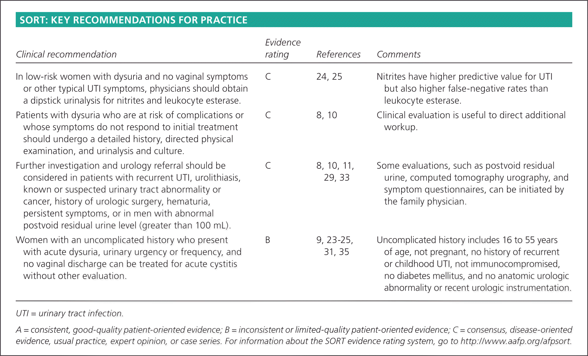
SORT: KEY RECOMMENDATIONS FOR PRACTICE
| Clinical recommendation | Evidence rating | References | Comments |
|---|---|---|---|
| In low-risk women with dysuria and no vaginal symptoms or other typical UTI symptoms, physicians should obtain a dipstick urinalysis for nitrites and leukocyte esterase. | C | 24, 25 | Nitrites have higher predictive value for UTI but also higher false-negative rates than leukocyte esterase. |
| Patients with dysuria who are at risk of complications or whose symptoms do not respond to initial treatment should undergo a detailed history, directed physical examination, and urinalysis and culture. | C | 8, 10 | Clinical evaluation is useful to direct additional workup. |
| Further investigation and urology referral should be considered in patients with recurrent UTI, urolithiasis, known or suspected urinary tract abnormality or cancer, history of urologic surgery, hematuria, persistent symptoms, or in men with abnormal postvoid residual urine level (greater than 100 mL). | C | 8, 10, 11, 29, 33 | Some evaluations, such as postvoid residual urine, computed tomography urography, and symptom questionnaires, can be initiated by the family physician. |
| Women with an uncomplicated history who present with acute dysuria, urinary urgency or frequency, and no vaginal discharge can be treated for acute cystitis without other evaluation. | B | 9, 23–25, 31, 35 | Uncomplicated history includes 16 to 55 years of age, not pregnant, no history of recurrent or childhood UTI, not immunocompromised, no diabetes mellitus, and no anatomic urologic abnormality or recent urologic instrumentation. |
UTI = urinary tract infection.
A = consistent, good-quality patient-oriented evidence; B = inconsistent or limited-quality patient-oriented evidence; C = consensus, disease-oriented evidence, usual practice, expert opinion, or case series. For information about the SORT evidence rating system, go to https://www.aafp.org/afpsort.
Pathophysiology and Differential Diagnosis
Sensory nerves are located just beneath the urothelium. Chemical irritation and inflammatory conditions (e.g., acute bacterial infection) can alter the mucosal barrier and stimulate these nerves, causing pain. Chronic inflammation and other unknown factors can lead to altered nerve sensitivity and persistent pain. Inflammation from adjacent abdominal structures, such as the colon, can also affect function and sensation in the bladder.7
Inflammatory disorders of the bladder and urethra are the most common causes of dysuria. Among these, infections of the bladder, urethra, kidneys, and genital organs are the most prevalent, including uncomplicated cystitis, pyelonephritis, and urethritis. Distinguishing a complicated urinary tract infection (UTI) from cystitis is important, because misdiagnosis increases the risk of treatment failure. Risk factors for a complicated infection may include patient characteristics, medical conditions, and urologic conditions (Table 1).8–11 In women, dysuria is also a common presentation of vaginitis. In men, prostatitis can present with dysuria. Sexually transmitted infections (STIs) can also cause dysuria.
Table 1. Risk Factors for Complicated Urinary Tract Infections*
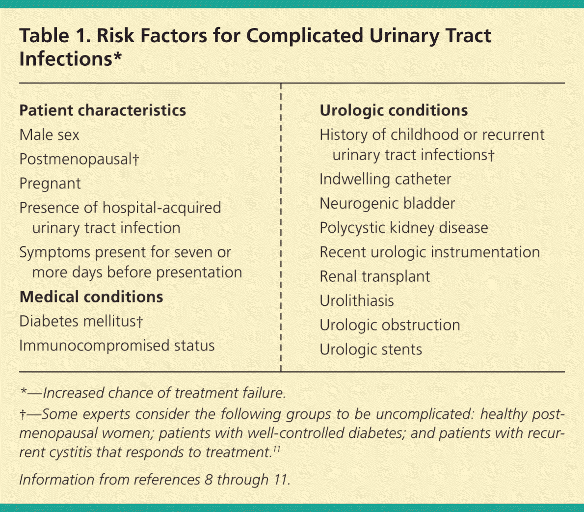
| Patient characteristics |
| Male sex |
| Postmenopausal† |
| Pregnant |
| Presence of hospital-acquired urinary tract infection |
| Symptoms present for seven or more days before presentation |
| Medical conditions |
| Diabetes mellitus† |
| Immunocompromised status |
| Urologic conditions |
| History of childhood or recurrent urinary tract infections† |
| Indwelling catheter |
| Neurogenic bladder |
| Polycystic kidney disease |
| Recent urologic instrumentation |
| Renal transplant |
| Urolithiasis |
| Urologic obstruction |
| Urologic stents |
*—Increased chance of treatment failure.
†—Some experts consider the following groups to be uncomplicated: healthy post-menopausal women; patients with well-controlled diabetes; and patients with recurrent cystitis that responds to treatment.11
Inflammatory, noninfectious conditions that can lead to dysuria include the presence of a foreign body (e.g., stent, bladder stone), noninfectious urethritis (e.g., reactive arthritis, formerly Reiter syndrome), and dermatologic conditions. Noninflammatory conditions can be divided into the following categories: anatomic; endocrine; neoplastic; medication-, food-, or recreational drug–related; iatrogenic; and idiopathic. Any condition that causes hematuria with clots can cause dysuria, including renal neoplasms and nephrolithiasis. Interstitial cystitis (also known as bladder pain syndrome) refers to chronic bladder pain, often with voiding symptoms, lasting six weeks or more without an identifiable cause.12 The differential diagnosis of dysuria is summarized in Table 2.2,4,8,11–19
Table 2. Differential Diagnosis of Dysuria in Adults
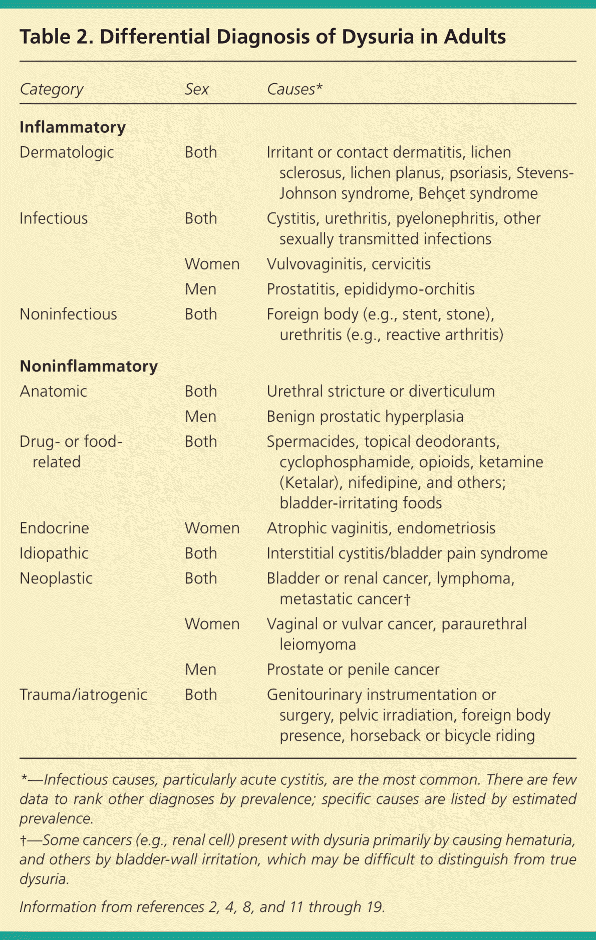
| Category | Sex | Causes* |
|---|---|---|
| Inflammatory | ||
| Dermatologic | Both | Irritant or contact dermatitis, lichen sclerosus, lichen planus, psoriasis, Stevens-Johnson syndrome, Behçet syndrome |
| Infectious | Both | Cystitis, urethritis, pyelonephritis, other sexually transmitted infections |
| Women | Vulvovaginitis, cervicitis | |
| Men | Prostatitis, epididymo-orchitis | |
| Noninfectious | Both | Foreign body (e.g., stent, stone), urethritis (e.g., reactive arthritis) |
| Noninflammatory | ||
| Anatomic | Both | Urethral stricture or diverticulum |
| Men | Benign prostatic hyperplasia | |
| Drug- or food-related | Both | Spermacides, topical deodorants, cyclophosphamide, opioids, ketamine (Ketalar), nifedipine, and others; bladder-irritating foods |
| Endocrine | Women | Atrophic vaginitis, endometriosis |
| Idiopathic | Both | Interstitial cystitis/bladder pain syndrome |
| Neoplastic | Both | Bladder or renal cancer, lymphoma, metastatic cancer† |
| Women | Vaginal or vulvar cancer, paraurethral leiomyoma | |
| Men | Prostate or penile cancer | |
| Trauma/iatrogenic | Both | Genitourinary instrumentation or surgery, pelvic irradiation, foreign body presence, horseback or bicycle riding |
*—Infectious causes, particularly acute cystitis, are the most common. There are few data to rank other diagnoses by prevalence; specific causes are listed by estimated prevalence.
†—Some cancers (e.g., renal cell) present with dysuria primarily by causing hematuria, and others by bladder-wall irritation, which may be difficult to distinguish from true dysuria.
History and Physical Examination
The medical history should characterize the timing, persistence, severity, duration, and exact location of the dysuria. Pain occurring at the start of urination may indicate urethral pathology; pain occurring at the end of urination is usually of bladder origin.1,2,9 Physicians should ask about other bladder symptoms, such as frequency, urgency, incontinence, hematuria, malodorous urine, and nocturia. The history should include the presence of flank pain, nausea, fever, and other systemic symptoms. A history of dysuria, UTIs, STIs, and recent sexual activity are crucial. Additionally, medication use, family history, and procedural history can help identify the cause of dysuria. In women, the history should also include the presence of vaginal discharge or irritation, most recent menstrual period, and type of contraception used.2,8
Specific localization of the discomfort varies between men and women. Women with vaginitis often describe external dysuria, as well as vaginal irritation or discharge. With cystitis, the dysuria is characteristically felt in the bladder or urethra.5,9 In addition to dysuria, men with prostatitis may have deep perineal pain and obstructive urinary symptoms, whereas those with epididymo-orchitis may have localized testicular pain. Lesions from herpes simplex virus of the vulvar or penile area may cause dysuria.2,13 Patients with interstitial cystitis may have suprapubic or abdominal pain related to bladder filling. These patients nearly always report urinary frequency and urgency, whereas dysuria is variable.3,12
A meta-analysis in which approximately 50% of patients had a UTI found that the highest positive predictive value (PV+) of cystitis in women was self-diagnosis of cystitis (86%), followed by the absence of vaginal discharge (82%), presence of hematuria (75%), and urinary frequency (73%). This review found that the combination of dysuria and urinary frequency without vaginal discharge or irritation yielded a very high likelihood of UTI (positive likelihood ratio [LR+] = 24.6). A woman with dysuria and frequency, no risk factors for complicated infection, and no vaginal discharge had a 90% probability of UTI; thus, treatment based on symptoms alone was advocated.9 A study of 196 symptomatic women found that 79% of patients with “considerable” dysuria, suspicion of a UTI, and absence of vaginal symptoms had a UTI.20 In a prospective study of 490 men with symptoms of a UTI, symptoms of dysuria and urgency were significantly associated with a positive urine culture.21 A suggested history for patients with dysuria is provided in eTable A.
eTable A. Elements of a Detailed History for Select Patients with Dysuria
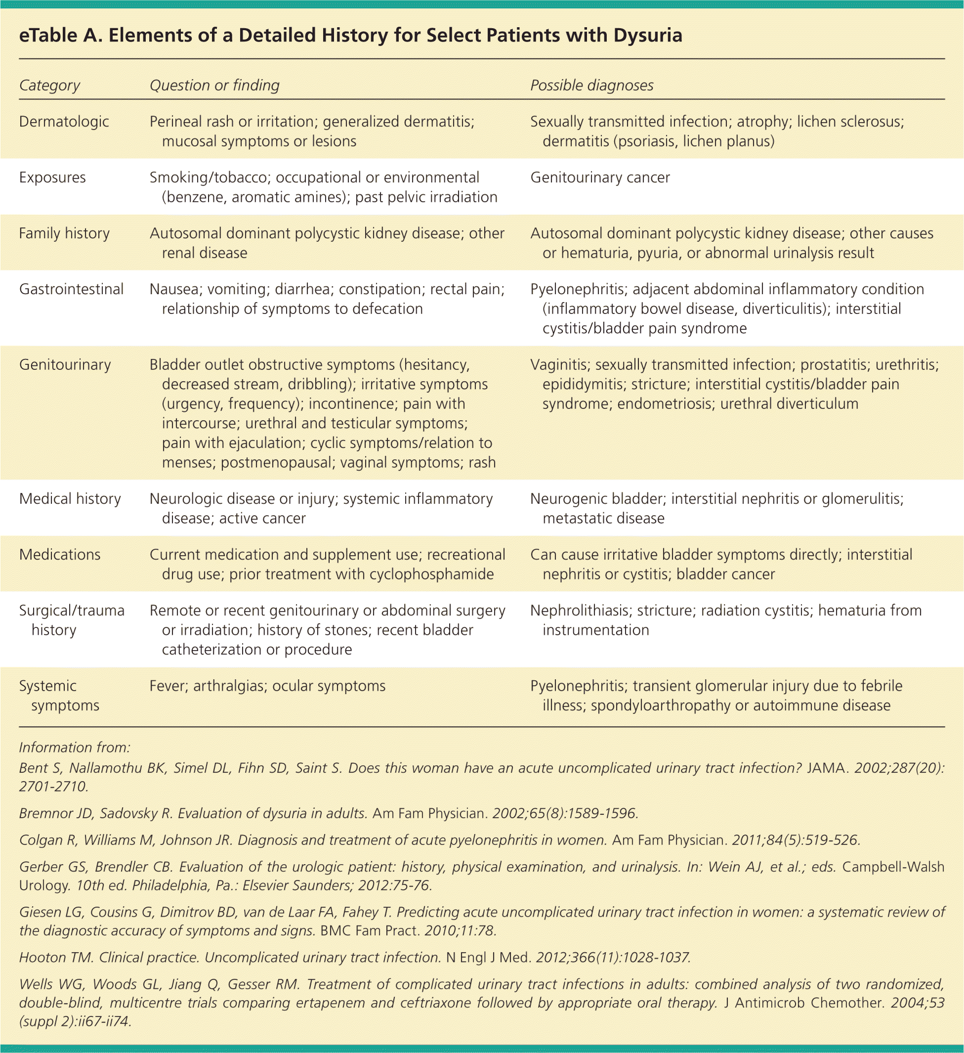
| Category | Question or finding | Possible diagnoses |
|---|---|---|
| Dermatologic | Perineal rash or irritation; generalized dermatitis; mucosal symptoms or lesions | Sexually transmitted infection; atrophy; lichen sclerosus; dermatitis (psoriasis, lichen planus) |
| Exposures | Smoking/tobacco; occupational or environmental (benzene, aromatic amines); past pelvic irradiation | Genitourinary cancer |
| Family history | Autosomal dominant polycystic kidney disease; other renal disease | Autosomal dominant polycystic kidney disease; other causes or hematuria, pyuria, or abnormal urinalysis result |
| Gastrointestinal | Nausea; vomiting; diarrhea; constipation; rectal pain; relationship of symptoms to defecation | Pyelonephritis; adjacent abdominal inflammatory condition (inflammatory bowel disease, diverticulitis); interstitial cystitis/bladder pain syndrome |
| Genitourinary | Bladder outlet obstructive symptoms (hesitancy, decreased stream, dribbling); irritative symptoms (urgency, frequency); incontinence; pain with intercourse; urethral and testicular symptoms; pain with ejaculation; cyclic symptoms/relation to menses; postmenopausal; vaginal symptoms; rash | Vaginitis; sexually transmitted infection; prostatitis; urethritis; epididymitis; stricture; interstitial cystitis/bladder pain syndrome; endometriosis; urethral diverticulum |
| Medical history | Neurologic disease or injury; systemic inflammatory disease; active cancer | Neurogenic bladder; interstitial nephritis or glomerulitis; metastatic disease |
| Medications | Current medication and supplement use; recreational drug use; prior treatment with cyclophosphamide | Can cause irritative bladder symptoms directly; interstitial nephritis or cystitis; bladder cancer |
| Surgical/trauma history | Remote or recent genitourinary or abdominal surgery or irradiation; history of stones; recent bladder catheterization or procedure | Nephrolithiasis; stricture; radiation cystitis; hematuria from instrumentation |
| Systemic symptoms | Fever; arthralgias; ocular symptoms | Pyelonephritis; transient glomerular injury due to febrile illness; spondyloarthropathy or autoimmune disease |
Information from: Bent S, Nallamothu BK, Simel DL, Fihn SD, Saint S. Does this woman have an acute uncomplicated urinary tract infection? JAMA. 2002;287(20): 2701–2710.
Bremnor JD, Sadovsky R. Evaluation of dysuria in adults. Am Fam Physician. 2002;65(8):1589–1596.
Colgan R, Williams M, Johnson JR. Diagnosis and treatment of acute pyelonephritis in women. Am Fam Physician. 2011;84(5):519–526.
Gerber GS, Brendler CB. Evaluation of the urologic patient: history, physical examination, and urinalysis. In: Wein AJ, et al.; eds. Campbell-Walsh Urology. 10th ed. Philadelphia, Pa.: Elsevier Saunders; 2012:75–76.
Giesen LG, Cousins G, Dimitrov BD, van de Laar FA, Fahey T. Predicting acute uncomplicated urinary tract infection in women: a systematic review of the diagnostic accuracy of symptoms and signs. BMC Fam Pract. 2010;11:78.
Hooton TM. Clinical practice. Uncomplicated urinary tract infection. N Engl J Med. 2012;366(11):1028–1037.
Wells WG, Woods GL, Jiang Q, Gesser RM. Treatment of complicated urinary tract infections in adults: combined analysis of two randomized, double-blind, multicentre trials comparing ertapenem and ceftriaxone followed by appropriate oral therapy. J Antimicrob Chemother. 2004;53 (suppl 2):ii67–ii74.
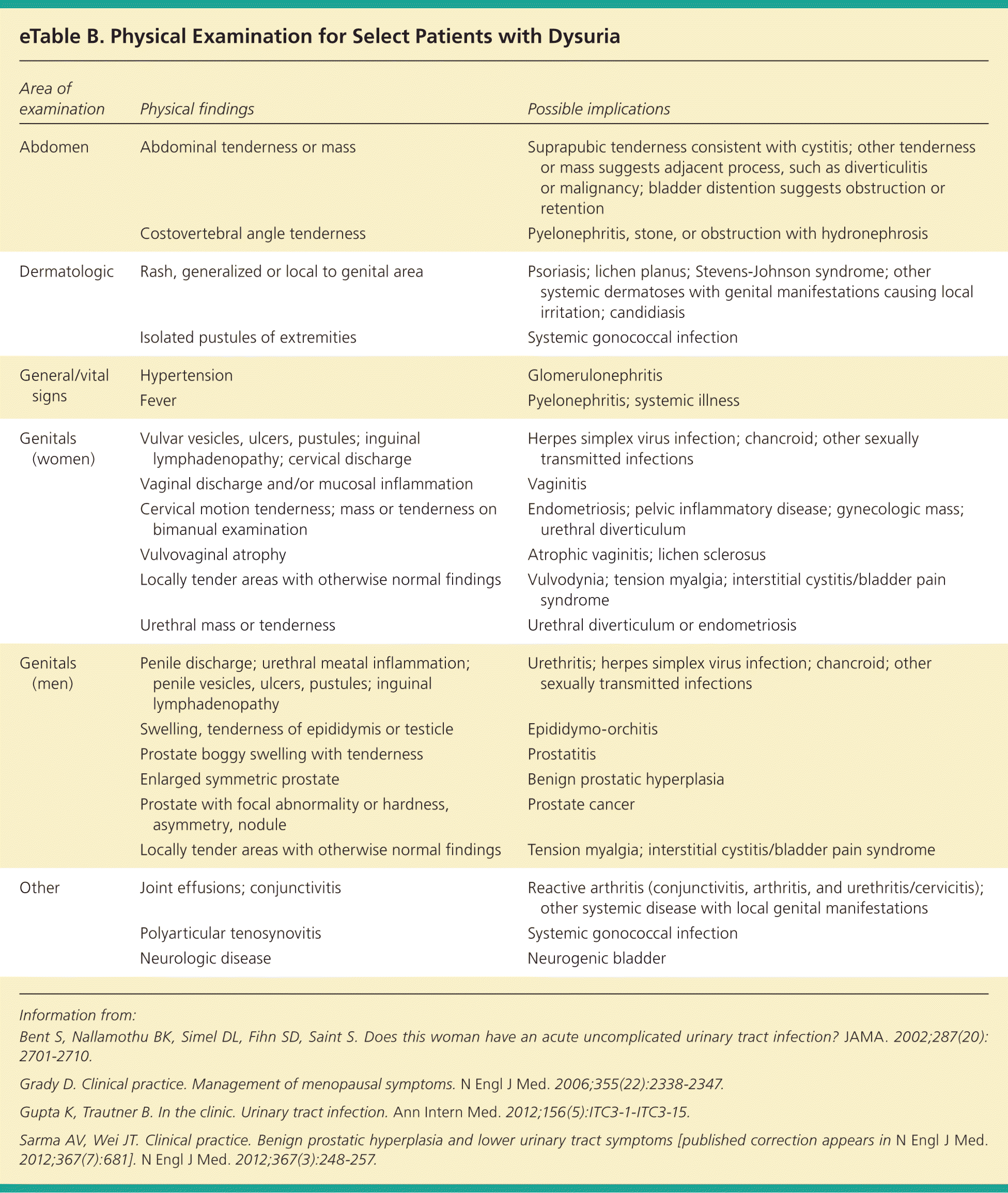
The physical examination, especially when complicated UTI is a consideration, should include vital signs, evaluation for costovertebral angle pain, palpation for abdominal mass or tenderness, and inspection for dermatologic conditions and acute joint effusions. Often the most relevant findings on physical examination are sex-specific, including inspecting for infectious or atrophic vaginitis and STIs in women, and prostatitis and STIs in men.15,18 The presence of costovertebral angle tenderness on examination modestly increases the likelihood of having a UTI in women (LR+ = 1.7).9 Key physical examination findings are discussed in eTable B. Risk factors for complicated UTI and failure to respond to initial treatment are indications for a more detailed history and physical examination.
eTable B. Physical Examination for Select Patients with Dysuria
| Area of examination | Physical findings | Possible implications |
|---|---|---|
| Abdomen | Abdominal tenderness or mass | Suprapubic tenderness consistent with cystitis; other tenderness or mass suggests adjacent process, such as diverticulitis or malignancy; bladder distention suggests obstruction or retention |
| Costovertebral angle tenderness | Pyelonephritis, stone, or obstruction with hydronephrosis | |
| Dermatologic | Rash, generalized or local to genital area | Psoriasis; lichen planus; Stevens-Johnson syndrome; other systemic dermatoses with genital manifestations causing local irritation; candidiasis |
| Isolated pustules of extremities | Systemic gonococcal infection | |
| General/vital signs | Hypertension | Glomerulonephritis |
| Fever | Pyelonephritis; systemic illness | |
| Genitals (women) | Vulvar vesicles, ulcers, pustules; inguinal lymphadenopathy; cervical discharge | Herpes simplex virus infection; chancroid; other sexually transmitted infections |
| Vaginal discharge and/or mucosal inflammation | Vaginitis | |
| Cervical motion tenderness; mass or tenderness on bimanual examination | Endometriosis; pelvic inflammatory disease; gynecologic mass; urethral diverticulum | |
| Vulvovaginal atrophy | Atrophic vaginitis; lichen sclerosus | |
| Locally tender areas with otherwise normal findings | Vulvodynia; tension myalgia; interstitial cystitis/bladder pain syndrome | |
| Urethral mass or tenderness | Urethral diverticulum or endometriosis | |
| Genitals (men) | Penile discharge; urethral meatal inflammation; penile vesicles, ulcers, pustules; inguinal lymphadenopathy | Urethritis; herpes simplex virus infection; chancroid; other sexually transmitted infections |
| Swelling, tenderness of epididymis or testicle | Epididymo-orchitis | |
| Prostate boggy swelling with tenderness | Prostatitis | |
| Enlarged symmetric prostate | Benign prostatic hyperplasia | |
| Prostate with focal abnormality or hardness, asymmetry, nodule | Prostate cancer | |
| Locally tender areas with otherwise normal findings | Tension myalgia; interstitial cystitis/bladder pain syndrome | |
| Other | Joint effusions; conjunctivitis | Reactive arthritis (conjunctivitis, arthritis, and urethritis/cervicitis); other systemic disease with local genital manifestations |
| Polyarticular tenosynovitis | Systemic gonococcal infection | |
| Neurologic disease | Neurogenic bladder |
Information from: Bent S, Nallamothu BK, Simel DL, Fihn SD, Saint S. Does this woman have an acute uncomplicated urinary tract infection? JAMA. 2002;287(20): 2701–2710.
Grady D. Clinical practice. Management of menopausal symptoms. N Engl J Med. 2006;355(22):2338–2347.
Gupta K, Trautner B. In the clinic. Urinary tract infection. Ann Intern Med. 2012;156(5):ITC3-1-ITC3-15.
Sarma AV, Wei JT. Clinical practice. Benign prostatic hyperplasia and lower urinary tract symptoms [published correction appears in N Engl J Med. 2012;367(7):681]. N Engl J Med. 2012;367(3):248–257.
Laboratory Tests
URINALYSIS
Urinalysis is the most useful test in a patient with dysuria; most studies have used dipstick urinalysis. Multiple studies of women with symptoms suggestive of a UTI have demonstrated that the presence of nitrites is highly predictive of a positive culture (PV+ = 75% to 95%); dipstick showing more than trace leukocytes is nearly as predictive (PV+ = 65% to 85%); and the presence of both is almost conclusive (PV+ = 95%).9,20,22–24 Urinary nitrites may be falsely negative in women with a UTI.25 Few studies specifically address the value of urinalysis in men with dysuria, but evidence suggests similar value to the combination of leukocyte esterase, nitrites, and possibly blood.4,21,26,27 Leukocyte esterase or pyuria alone with isolated dysuria suggests urethritis.8,10,28
CULTURES AND CYTOLOGY
Any patient with risk factors for a complicated UTI (Table 18–11 ) or whose symptoms do not respond to initial treatment should have a urine culture and sensitivity analysis. Patients with suspected pyelonephritis should have renal function assessed with serum creatinine measurement, and electrolyte levels should be measured if there is substantial nausea and vomiting. Blood cultures are usually not necessary, but can be obtained in patients with high fever or risk of infectious complications.2,8
In women with vaginal symptoms, secretions should be evaluated with wet mount and potassium hydroxide microscopy or a vaginal pathogens DNA probe. Urethritis should be suspected in younger, sexually active patients with dysuria and pyuria without bacteriuria; in men, urethral inflammation and discharge is typically present. In patients with suspected urethritis, a urethral, vaginal, endocervical, or urine nucleic acid amplification test for Neisseria gonorrhoeae and Chlamydia trachomatis is indicated. Genital ulcerations can be sampled for herpes simplex virus culture or polymerase chain reaction testing, as well as testing for other STIs.28 In men with suspected chronic prostatitis, urine culture after gentle prostatic massage can yield the causative bacterial agent. Prostate-specific antigen level is transiently elevated during acute prostatitis and should not be measured in patients with acute inflammatory symptoms. Urine cytology is helpful if bladder cancer is suspected, such as in older patients with hematuria and a negative culture result.29
IMAGING AND OTHER ADVANCED STUDIES
Imaging is not necessary in most patients with dysuria, although it may be indicated in patients with a complicated UTI, a suspected anatomic anomaly (e.g., abnormal voiding, positive family history of genitourinary anomalies), obstruction or abscess, relapsing infection, or hematuria. Ultrasonography is the preferred initial test for patients with obstruction, abscess, recurrent infection, or suspected kidney stones, because it avoids radiation exposure.30,31 Helical computed tomography urography is used to view the kidneys and adjacent structures, and may be considered to further evaluate patients with possible abscess, obstruction, or suspected anomalies when ultrasonography is not diagnostic.8,30 If urinalysis is unrevealing, cystoscopy can be performed to evaluate for bladder cancer, hematuria, and chronic bladder symptoms. Urodynamic studies can be performed for persistent voiding symptoms with otherwise unrevealing workup,12 although a recent Cochrane review found no evidence that these tests led to reduction in symptoms in men with such indications.32 Further investigation and urology referral should be considered in patients with recurrent UTI, urolithiasis, known urinary tract abnormality or cancer, history of urologic surgery, hematuria, persistent symptoms, or in men with abnormal postvoid residual urine level (greater than 100 mL)8,10,11,29,33 (Table 34,8,9,11,21,23–26,28–30,33,34 ).
Table 3. Diagnostic Tests for Select Patients with Dysuria
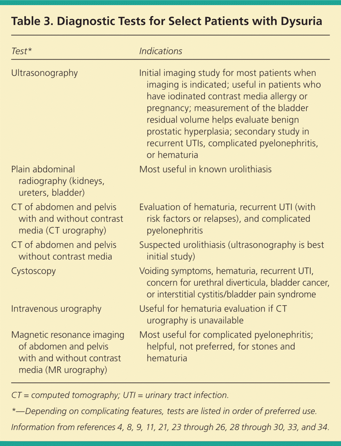
| Test* | Indications |
|---|---|
| Ultrasonography | Initial imaging study for most patients when imaging is indicated; useful in patients who have iodinated contrast media allergy or pregnancy; measurement of the bladder residual volume helps evaluate benign prostatic hyperplasia; secondary study in recurrent UTIs, complicated pyelonephritis, or hematuria |
| Plain abdominal radiography (kidneys, ureters, bladder) | Most useful in known urolithiasis |
| CT of abdomen and pelvis with and without contrast media (CT urography) | Evaluation of hematuria, recurrent UTI (with risk factors or relapses), and complicated pyelonephritis |
| CT of abdomen and pelvis without contrast media | Suspected urolithiasis (ultrasonography is best initial study) |
| Cystoscopy | Voiding symptoms, hematuria, recurrent UTI, concern for urethral diverticula, bladder cancer, or interstitial cystitis/bladder pain syndrome |
| Intravenous urography | Useful for hematuria evaluation if CT urography is unavailable |
| Magnetic resonance imaging of abdomen and pelvis with and without contrast media (MR urography) | Most useful for complicated pyelonephritis; helpful, not preferred, for stones and hematuria |
CT = computed tomography; UTI = urinary tract infection.
*—Depending on complicating features, tests are listed in order of preferred use.
Information from references 4, 8, 9, 11, 21, 23 through 26, 28 through 30, 33, and 34.
Clinical Decision Rules
More than 400 patients were included in an English study to develop and validate a clinical decision rule for women presenting with dysuria and urinary frequency. The scoring system they developed can be used in a two-stage process, with some patients treated empirically based on symptoms alone (Table 4).23 Negative predictive values are poor; many women with UTI will not have all three clinical symptoms that were found to be predictive of UTI (dysuria, nocturia, and offensive urine odor), and about one-fourth of patients with urinary symptoms and a normal dipstick result have UTI, so appropriate follow-up is important.
Table 4. Clinical Decision Rule for UTI in Women Without Signs of Complicated Disease
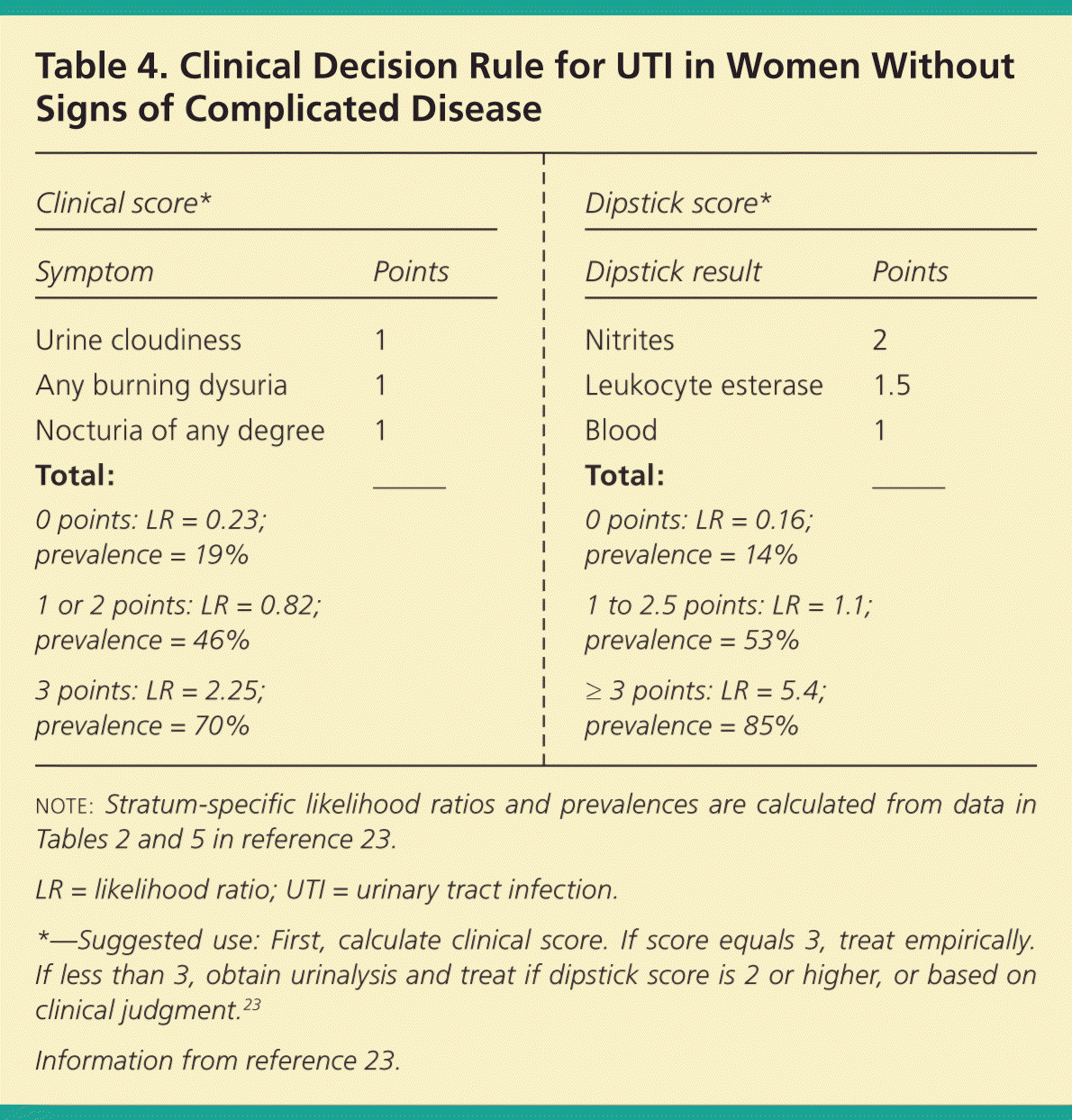
| Clinical score* | Dipstick score* | ||
|---|---|---|---|
| Symptom | Points | Dipstick result | Points |
| Urine cloudiness | 1 | Nitrites | 2 |
| Any burning dysuria | 1 | Leukocyte esterase | 1.5 |
| Nocturia of any degree | 1 | Blood | 1 |
| Total: | ____ | Total: | ____ |
| 0 points: LR = 0.23; | 0 points: LR = 0.16; | ||
| prevalence = 19% | prevalence = 14% | ||
| 1 or 2 points: LR = 0.82; | 1 to 2.5 points: LR = 1.1; | ||
| prevalence = 46% | prevalence = 53% | ||
| 3 points: LR = 2.25; | ≥ 3 points: LR = 5.4; | ||
| prevalence = 70% | prevalence = 85% | ||
note: Stratum-specific likelihood ratios and prevalences are calculated from data in Tables 2 and 5 in reference 23.
LR = likelihood ratio; UTI = urinary tract infection.
*—Suggested use: First, calculate clinical score. If score equals 3, treat empirically. If less than 3, obtain urinalysis and treat if dipstick score is 2 or higher, or based on clinical judgment.23
Information from reference 23.
A Dutch study included 490 outpatient men with dysuria, frequency, or urgency, while excluding men with symptoms suggestive of an STI or a complicated UTI. The authors found that the combination of age (60 years or older) and either a positive leukocyte esterase or nitrite test result had the best positive and negative predictive values for UTI (83% and 60%, respectively).21
Another study developed a symptoms-history-urinalysis score that included symptoms of frequency, nocturia, dysuria, hematuria, and offensive-smelling urine; history of a previous UTI; and urinalysis results (protein, blood, and nitrites). The study found that a score of 0 or greater on the 13-item score sheet (range of possible scores from –19 to +31) identified 85% of women with infection; 25% of women without infection also had a score of 0 or greater.26
All of the decision rules caution that in a patient with dysuria, these combinations of variables can reassure physicians that a UTI is likely present, but are not very useful in ruling out a UTI when they are absent. It is important to address the other clinical features discussed here to narrow the differential diagnosis.
Approach to the Patient
Many studies advise that, in the right clinical setting, symptoms alone can identify patients with a high likelihood of UTI who are candidates for empiric therapy. Women with an uncomplicated history who present with acute dysuria, urinary urgency or frequency, and no vaginal discharge can be treated for acute cystitis without other evaluation.9,23–25,31,35 Several studies suggest that this approach is effective in reducing costs while improving patient satisfaction, with no increase in adverse outcomes.5,35,36 This approach is reflected in the algorithms presented here for the evaluation and follow-up of dysuria. These algorithms are based on evaluation of the existing evidence. Figure 12,4,5,10,20,22,24,25,31,37,38 addresses the initial presentation of a woman with acute dysuria, and Figure 22,4,8,9,12,29,33 addresses the follow-up evaluation. Figure 3 presents the approach to a man with dysuria.4,18,21,26–29,39 For patients with initially normal dip-stick urinalysis and culture results, Table 5 lists common conditions that may be causing dysuria, the typical presentation, and management recommendations.4,12–19,28 It is important to use clinical judgment and to be aware of the inclusion and exclusion criteria for the studies on which these algorithms are based. For example, a sexually active adolescent with dysuria is more likely to have an STI than cystitis, and urinalysis results may be negative.40–42 Similarly, an older patient who experiences dysuria shortly after having an indwelling catheter and who has a negative urinalysis result still has a high likelihood of having a UTI.38
Figure 1. Initial Approach in a Woman with Acute Dysuria
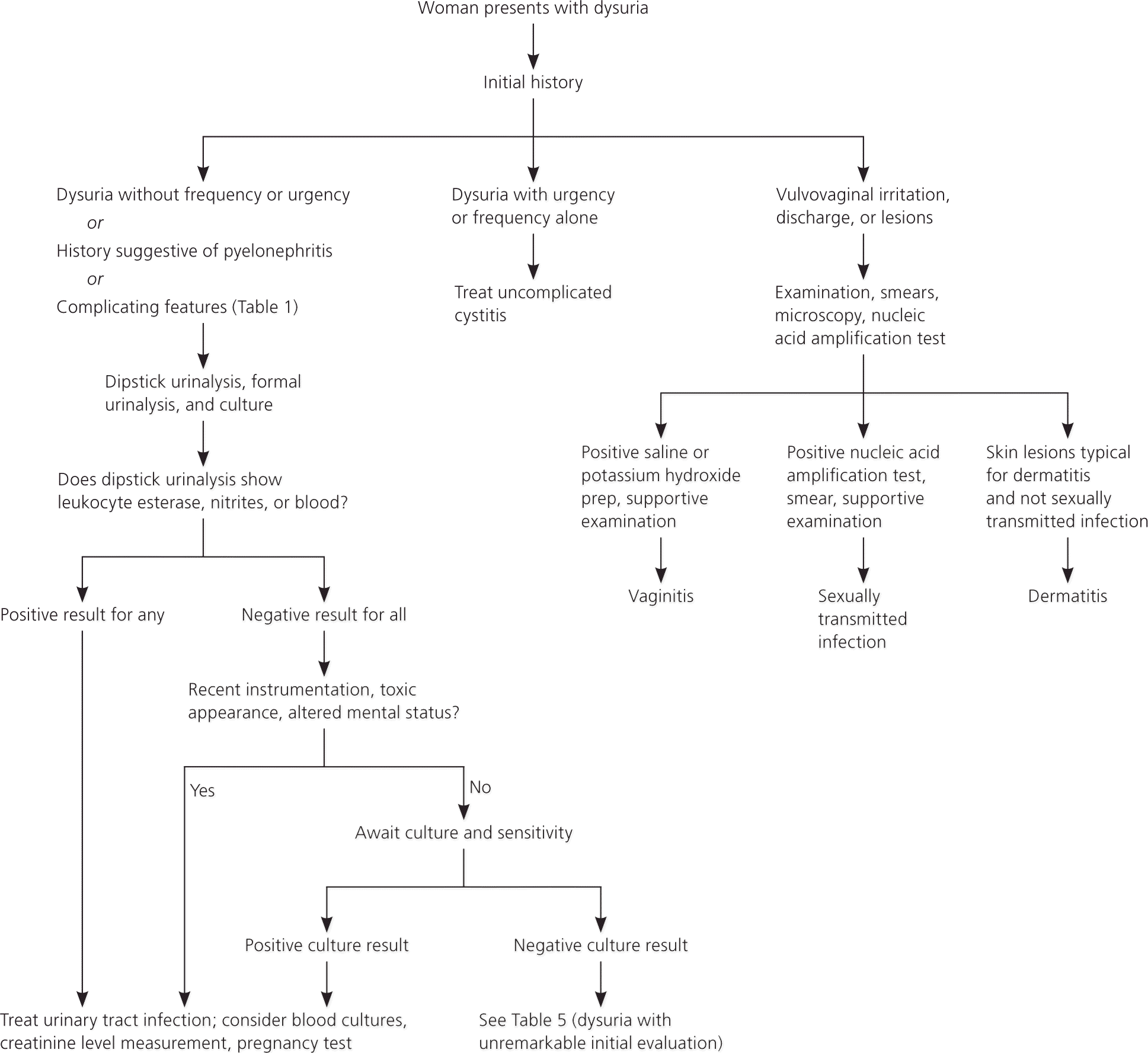
Algorithm for the initial approach in a woman with acute dysuria.
Information from references 2, 4, 5, 10, 20, 22, 24, 25, 31, 37, and 38.
Figure 2. Follow-up Evaluation in a Woman with Acute Dysuria
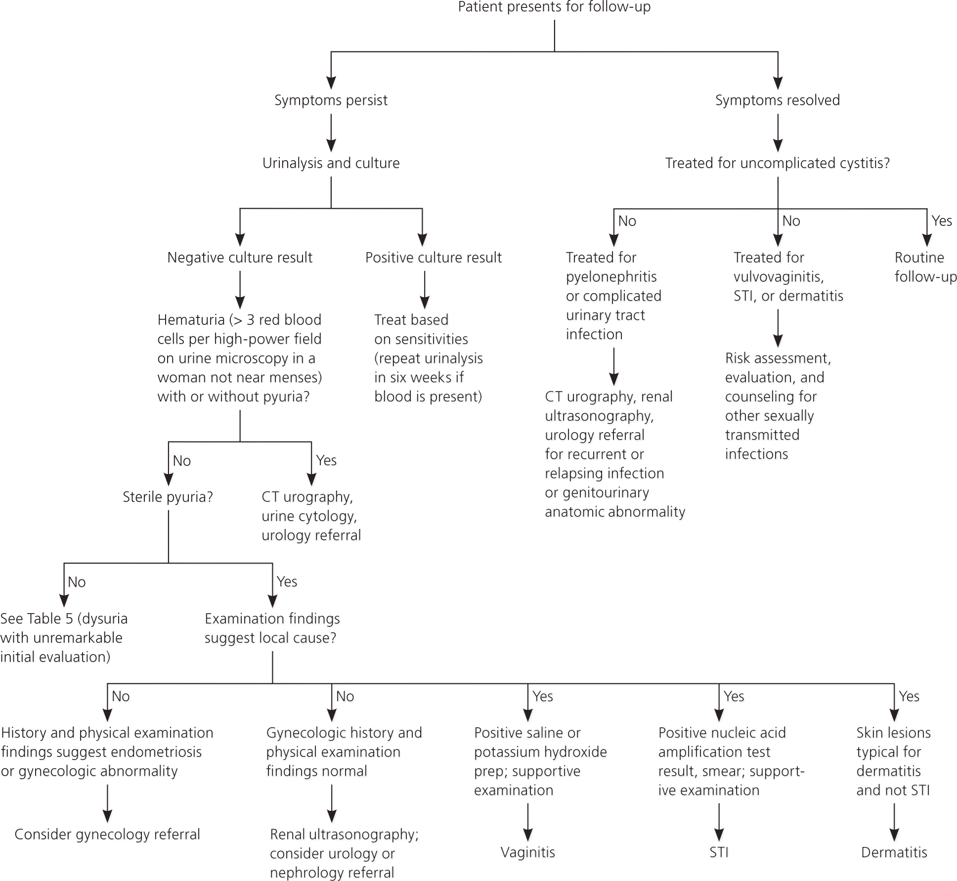
Algorithm for the follow-up evaluation in a woman with acute dysuria. (CT = computed tomography; STI = sexually transmitted infection.)
Information from references 2, 4, 8, 9, 12, 29, and 33.
Figure 3. Approach in a Man with Acute Dysuria
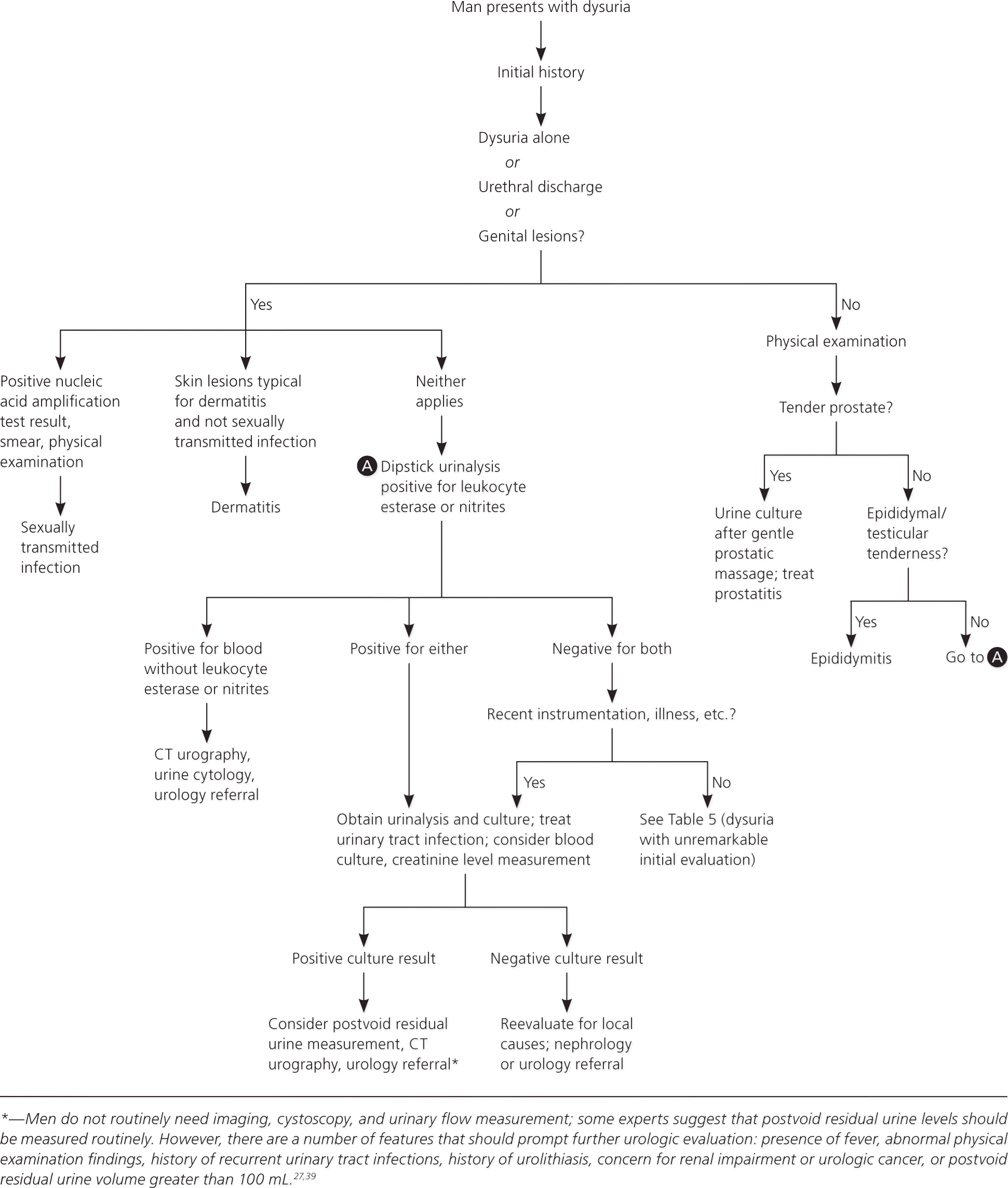
Algorithm for the approach in a man with acute dysuria. (CT = computed tomography.)
Information from references 4, 18, 21, 26 through 29, and 39.
Table 5. Considerations in Patients with Dysuria and Unremarkable Initial Evaluation
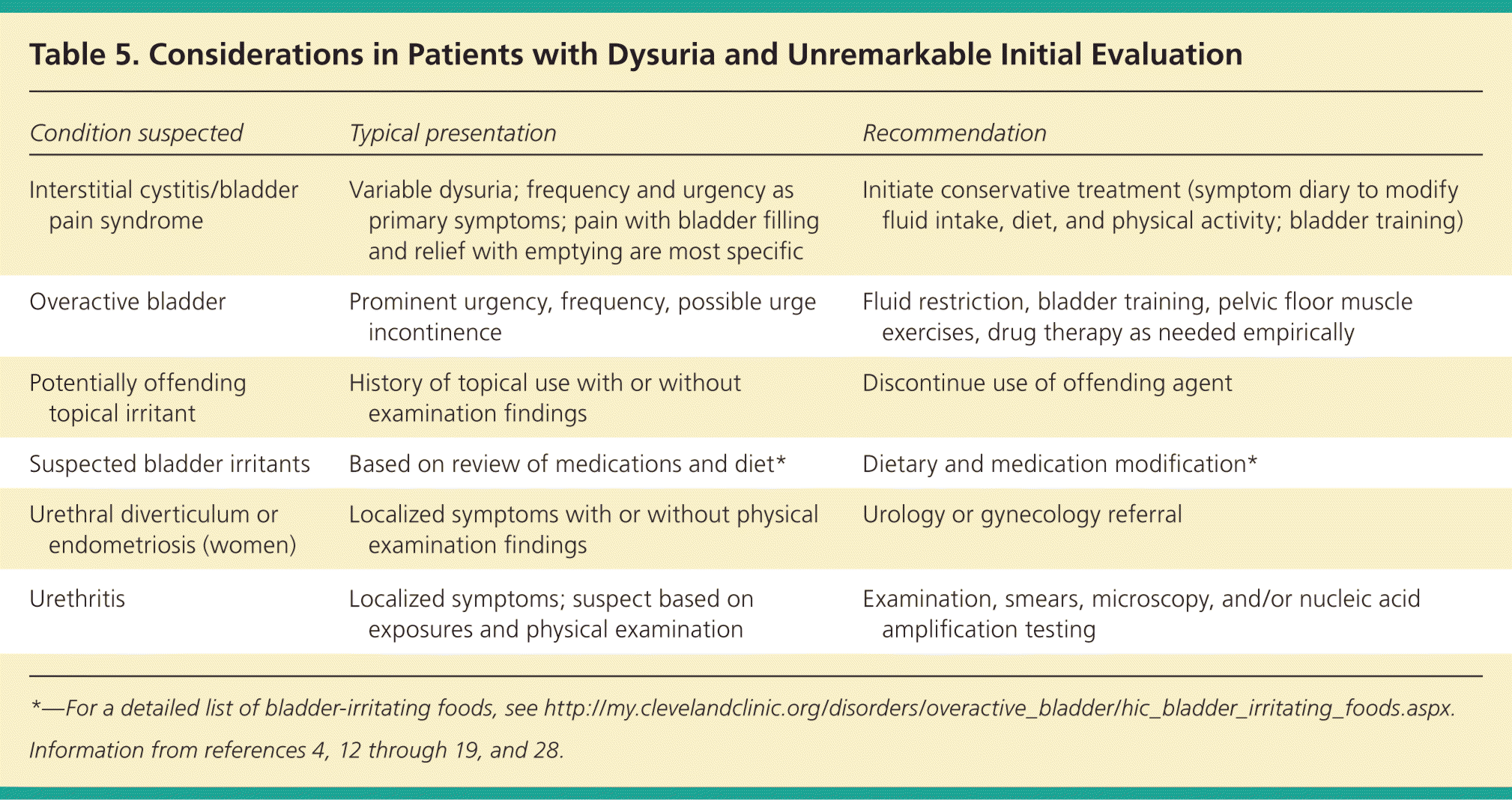
| Condition suspected | Typical presentation | Recommendation |
|---|---|---|
| Interstitial cystitis/bladder pain syndrome | Variable dysuria; frequency and urgency as primary symptoms; pain with bladder filling and relief with emptying are most specific | Initiate conservative treatment (symptom diary to modify fluid intake, diet, and physical activity; bladder training) |
| Overactive bladder | Prominent urgency, frequency, possible urge incontinence | Fluid restriction, bladder training, pelvic floor muscle exercises, drug therapy as needed empirically |
| Potentially offending topical irritant | History of topical use with or without examination findings | Discontinue use of offending agent |
| Suspected bladder irritants | Based on review of medications and diet* | Dietary and medication modification* |
| Urethral diverticulum or endometriosis (women) | Localized symptoms with or without physical examination findings | Urology or gynecology referral |
| Urethritis | Localized symptoms; suspect based on exposures and physical examination | Examination, smears, microscopy, and/or nucleic acid amplification testing |
*—For a detailed list of bladder-irritating foods, see http://my.clevelandclinic.org/disorders/overactive_bladder/hic_bladder_irritating_foods.aspx.
Data Sources: A PubMed search was completed in Clinical Queries using the key terms dysuria, urinary tract infection (acute, recurrent, elderly), and hematuria, as well as for the specific disease entities considered (e.g., gonococcal urethritis). The search included meta-analyses, randomized controlled trials, clinical trials, and reviews. The following databases and summaries were also used: Cochrane Database of Systematic Reviews, BMJ Clinical Evidence, National Guideline Clearinghouse, Essential Evidence Plus, and UpToDate. Search dates: January through August 2014, and May through July 2015.
The opinions expressed herein are those of the authors and are not necessarily representative of those of the U.S. Army or Department of Defense.
