
Am Fam Physician. 2017;95(1):37-38
Author disclosure: No relevant financial affiliations.
A 20-year-old U.S Marine presented with a rash on his left upper arm. He reported getting a mild sunburn nine days earlier. Three days after the sunburn, he received routine vaccinations before overseas deployment. The vaccines included anthrax, hepatitis A and B, Japanese encephalitis virus, typhoid, and vaccinia virus (smallpox). He first noted a small, slightly erythematous, raised rash on his left upper deltoid area two days before presentation. Within 24 hours, the rash had become several times larger. The patient reported discomfort in the area. He did not have a fever or history of similar episodes.
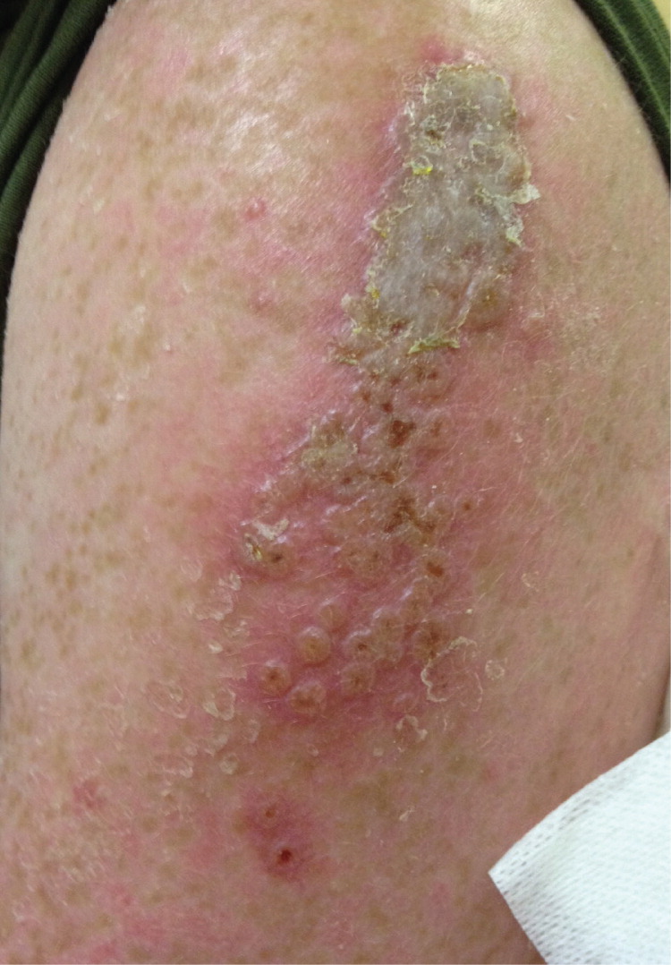
On physical examination, the patient was comfortable and in no acute distress. His skin examination revealed a 5- × 2-cm erythematous plaque on the left upper lateral deltoid area that continued distally with an 8- × 4-cm area of coalescing umbilicated papules (Figure 1).
Question
Discussion
The answer is E: vaccinia reaction. The patient's skin findings of a large, coalesced plaque with transmitted lesions distal to the inoculated site are consistent with a robust vaccinia uptake and autoinoculation. When smallpox was eradicated in 1979, routine vaccination ended.1,2 After the 2001 anthrax attacks, vaccination of certain populations resumed, including members of the U.S. military before deployment to combat zones, first responders, and civilian public health care professionals.3,4
The vaccine is administered via scarification, which is more effective than intramuscular injection used in other vaccines. In this process, the needle penetrates the patient's epidermis 15 times in the deltoid area. After vaccination, patients will have a skin reaction that lasts an average of 14 to 21 days. Within two to three days, several papules and vesicles appear at the vaccination site. Typically, at seven to nine days, a localized pustule manifests, crusting over within a few days. Ultimately, the scab detaches and leaves a residual scar.
There is some risk with the vaccinia virus vaccine because it uses a live virus. Robust uptake leads to development of a large erythematous plaque and induration of at least 10 cm. Autoinoculation (accidental inoculation) typically occurs via transmission by the hands from the inoculation site to another area, typically the face. In a generalized vaccinia reaction, the lesions are papulovesicular and become pustules; they are typically extensive following vaccination. A progressive vaccinia reaction is rare but often severe and can result in the destruction of local skin and underlying soft tissue structures.5
Eczema vaccinatum occurs in patients who have a history of eczema. Eczema typically presents as dry, itchy skin with erythema, scaling, and lichenification. After vaccination, patients may develop an extensive vesiculopustular eruption, mostly in areas of active dermatitis. Lesions may appear umbilicated and in crops.6
Erythema multiforme is a mucocutaneous hypersensitivity reaction resulting from infections or medications. It is classically characterized by target lesions that look like a bull's eye. The target lesions erupt over 24 to 48 hours and remain for up to two weeks.
Molluscum contagiosum is a viral infection that occurs in children and adults. In children, it spreads through close skin contact. In adults, it is typically sexually transmitted. It has umbilicated papules similar to a vaccinia reaction, but the appearance of the scab over the lesion would be unusual with molluscum contagiosum.
A secondary bacterial infection arises after trauma or injury to the skin. The area develops an indurated skin lesion that is tender and warm to the touch. Systemic signs of infection might also be present. Common causes include streptococci and staphylococci.
| Condition | History | Characteristics |
|---|---|---|
| Eczema vaccinatum | Eczema | Vesiculopustular eruption after vaccination, mostly in areas of active dermatitis; lesions may appear umbilicated and in crops |
| Erythema multiforme | Mucocutaneous hypersensitivity reaction resulting from infections or medications | Classic erythematous target lesions that look like a bull's eye; target lesions erupt over 24 to 48 hours and remain for up to two weeks |
| Molluscum contagiosum | Viral infection; in children, it spreads through close skin contact; in adults, it is typically sexually transmitted | Umbilicated papules, without scabbing over the lesion |
| Secondary bacterial infection | Infection arises after trauma or injury to the skin | Indurated skin lesion that is tender and warm to the touch; systemic signs of infection may be present |
| Vaccinia reaction | Recent vaccination; may have autoinoculation transmitted by the hand from the vaccination site to another area, typically the face | Large erythematous and indurated lesion at the site of vaccination from robust uptake |
